
Method and apparatus for displaying body sounds and performing diagnosis based on body sound analysis

a body sound and analysis method technology, applied in the field of non-invasive diagnostic systems and techniques, can solve the problems of inability to record a given sound (e.g., a particular inspiration or expiration), time-consuming and laborious, and achieve the effect of facilitating the diagnosis of certain diseases

Inactive Publication Date: 2009-04-21
STETHOGRAPHICS
View PDF26 Cites 23 Cited by
  • Summary
  • Abstract
  • Description
  • Claims
  • Application Information

AI Technical Summary

Benefits of technology

"The present invention is a system for recording, displaying, and analyzing body sounds to diagnose various diseases. The system includes a plurality of transducers that detect the sound or vibration of the body at different sites around the patient's chest. The detected data is then conditioned and converted into digital format and stored on a computer station. A computer program organizes the data and can display it in real-time or print it for review by a physician. The program can also analyze the data and provide a possible diagnosis based on pre-defined criteria or guidelines. The system can also localize the origin of abnormal sounds and allow patients to input sounds for diagnosis. Overall, the invention provides a richer, more fully coordinated set of data for accurately detecting body sound abnormalities."

Problems solved by technology

First, the intelligent stethoscope has only a single microphone, so that obtaining recordings at multiple locations is time-consuming.
A single microphone also makes it impossible to record a given sound (e.g., a particular inspiration or expiration) from more than one point on the chest.
Due to these limitations, the intelligent stethoscope is not that likely to provide accurate diagnoses.

Method used


Image

  • Method and apparatus for displaying body sounds and performing diagnosis based on body sound analysis
  • Method and apparatus for displaying body sounds and performing diagnosis based on body sound analysis
  • Method and apparatus for displaying body sounds and performing diagnosis based on body sound analysis

Examples


Embodiment Construction

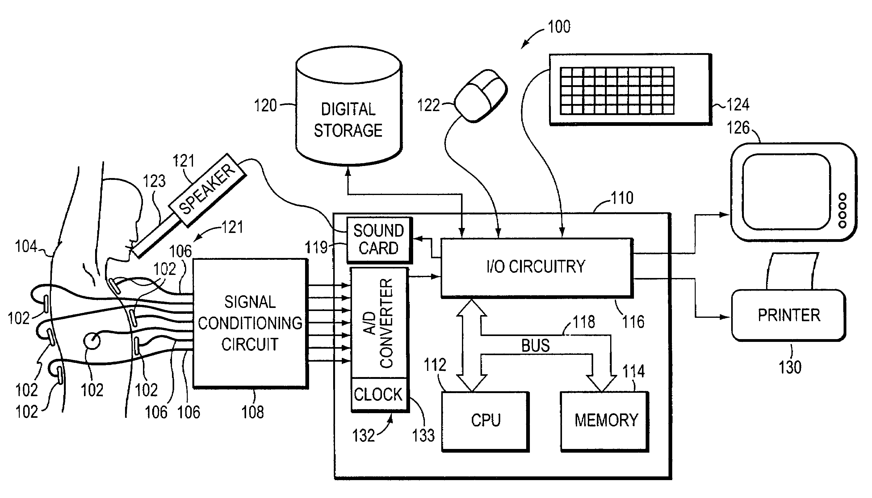
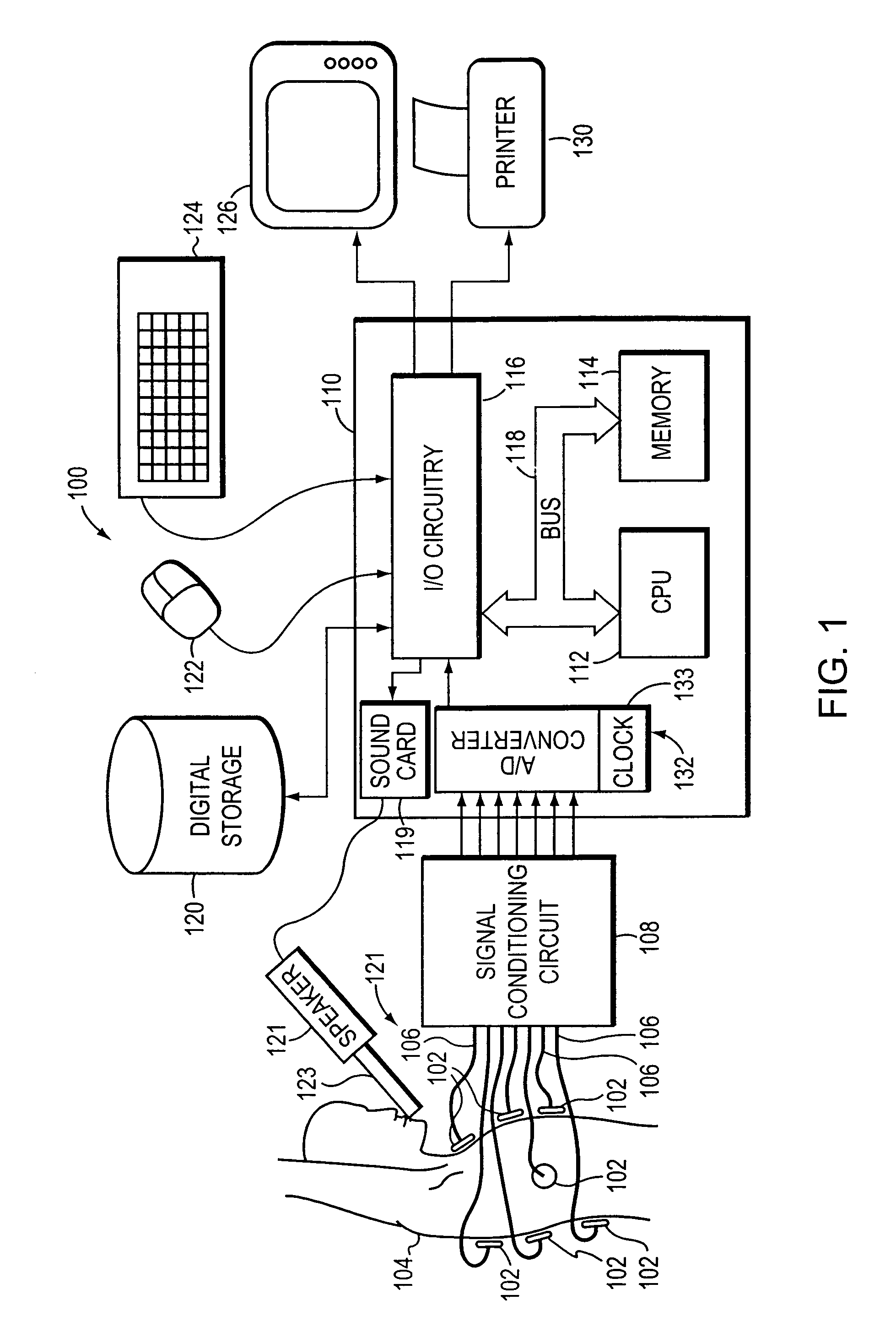
[0040]FIG. 1 is a block diagram of the lung sound recording and analysis system 100 of the present invention. The system 100 includes a sensor system 101 which includes a plurality of sound transducers, such as analog microphones 102, that may be placed at various sites around the chest or other area of a patient 104. In the preferred embodiment of the invention, the system 100 uses sixteen different sites, of which, fifteen are located around the chest and one is located at the patient's trachea. More specifically, there is one site on the left side, one site on the right side, two sites on the upper front chest separated by the spinal column (proximate to the top portion of the lungs), one site on the lower right front chest, two sites on the upper back (proximate to the top portion of the lungs), four sites in the middle back (proximate to the mid portion of the lungs), four sites at the lower back (proximate to the bottom of the lungs) and one site at the trachea. It should be u...


PUM


Abstract

A lung sound diagnostic system for use in collecting, organizing and analyzing lung sounds associated with the inspiration(s) and expiration(s) of a patient. The system includes a plurality of transducers that may be placed at various sites around the patient's chest. The microphones are coupled to signal processing circuitry and A / D converters which digitize the data and preferably provides the digital data to a computer station. A data collection and organization program, executing on the computer station, organizes and formats the data into a combination display for display or printing. The combinational display includes at least two display elements. In a first display element, the data is shown for both inspiration and expiration combined in a first time scale. In a second display element, the data for inspiration and expiration are shown individually in a second time scale that is time-expanded relative to the first time scale. The system may also include application programs for detecting and classifying abnormal sounds. The resulting information may be displayed in a variety of formats to facilitate diagnosis. Additionally, the system may include an analysis program for comparing selected criteria corresponding to the detected abnormal sounds with predefined thresholds in order to provide a likely diagnosis.

Description

[0001]This application is a divisional of application Ser. No. 10 / 080,209 titled METHOD AND APPARATUS FOR DISPLAYING LUNG SOUNDS AND PERFORMING DIAGNOSIS BASED ON LUNG SOUND ANALYSIS, filed on Feb. 21, 2002, now U.S. Pat. No. 6,790,183 which is a continuation-in-part of application Ser. No. 09 / 699,546, filed Oct. 30, 2000, now U.S. Pat. No. 6,394,967, which is a continuation of application Ser. No. 09 / 172,343, filed Oct. 14, 1998, now U.S. Pat No. 6,139,505.BACKGROUND OF THE INVENTION[0002]1. Field of the Invention[0003]The present invention relates generally to non-invasive diagnostic systems and techniques, and more specifically, to a method and apparatus for diagnosis based upon the review and analysis of body sounds.[0004]2. Background Information[0005]Since the time of its invention in the early 1800's, the stethoscope has been used routinely by physicians to amplify sounds in the human body. The physician typically places the chest piece of the stethoscope against the patient'...

Claims


Application Information

Patent Timeline
Patent Type & Authority Patents(United States)
IPC IPC(8): A61B5/00A61B7/00A61B8/00
CPCA61B5/061A61B7/003A61B8/00A61B5/7264
Inventor MURPHY, RAYMOND L. H.
Owner STETHOGRAPHICS
Learn More