Apparatus and method for assessing retinal damage

a retinal damage and retinal technology, applied in the field of retinal damage assessment, can solve the problems of glaucoma primarily destructive of the patient's visual field, optic nerve head atrophia and visual field defects, and it is not practical to test any given point in the grid with a wide range of stimulus intensities, so as to reduce the duration of perimetry testing, the effect of increasing the retinal area and prolonging the tes

Inactive Publication Date: 2009-04-28
THE RES FOUND OF STATE UNIV OF NEW YORK
View PDF16 Cites 18 Cited by
  • Summary
  • Abstract
  • Description
  • Claims
  • Application Information

AI Technical Summary

Problems solved by technology

Atrophy of the optic nerve head and visual field defects may arise in the setting of an IOP that is within normal limits.
Because glaucoma leads to destruction of retinal nerve fibers carrying the modulated output of retinal photoreceptor cells (as opposed to destruction of the lens or cornea, which form the refractive media of the eye), glaucoma is primarily destructive of a patient's visual fields.
Since it is impractical to test every point in the visual field, each strategy uses a grid of test points that covers a circular central area of the visual field that is most likely to show glaucomatous defects.
Just as it is not practical to test each point in the visual field, it is not practical to test any given point in the grid with a wide range of stimulus intensities.
If the patient misses a significant number of these stimuli, then the automated perimeter is considered to have detected a defect that warrants further testing.
Automated perimetry is beset with a number of disadvantages arising from the subjectivity of the patients' responses and the excessive duration of the test.
Patients dislike the test because it is long and tedious.
Test anxiety, attention deficits, fatigue and boredom interfere with perimetry in both the young and the elderly.
A desire to influence the outcome of the test favorably can result in false positives (pressing the button to signal visualization of a light stimulus when no light has been activated).
Attention deficits can result in false negatives (failing to press the button to signal visualization of a light stimulus having a suprathreshold luminance).
The results of automated perimetry are often unreliable.
However, the patient's response to a larger-area threshold test in perimetry may be mediated by undamaged retinal areas near damaged retinal areas.
Thus, a large-area threshold test in perimetry is likely to be associated with a substantial risk of a falsely normal test result.
The pupillary response to spatially-localized luminance increments has been used as an indicator of glaucomatous retinal damage, but the small-area stimuli used in pupil perimetry may target small retinal areas that only weakly stimulate a PLR, and may fail to stimulate a PLR if the small retinal area that is being stimulated by light has been damaged by glaucoma.
Large variations in pupil response amplitude among patients and the changes in sensitivity of the pupil response with the retinal location of the small-area light stimulus have also limited the usefulness of such measurements.
Additionally, prevailing pupil perimetry takes a relatively long time to perform.

Method used


Image

  • Apparatus and method for assessing retinal damage
  • Apparatus and method for assessing retinal damage
  • Apparatus and method for assessing retinal damage

Examples


Embodiment Construction

3.1 Glaucoma

[0061]As used herein the term glaucoma comprises all pathophysiological states of the human eye presenting with an elevation in IOP and either a visual field defect or optic atrophy, and includes but is not limited to acute angle-closure glaucoma, angle-recession glaucoma, drug-induced glaucoma, hemolytic glaucoma, hemosiderotic glaucoma, juvenile glaucoma, low-tension glaucoma, malignant glaucoma, narrow-angle glaucoma, neovascular glaucoma, normotensive glaucoma, phacolytic glaucoma, phacomorphic glaucoma, pigmentary glaucoma post-surgical glaucoma, primary open-angle glaucoma, primary infantile glaucoma, pseudoexfoliative glaucoma, pupillary block glaucoma uveitic glaucoma, aniridia, aqueous misdirection syndrome, irido-corneal-endothelial syndrome and plateau iris syndrome.
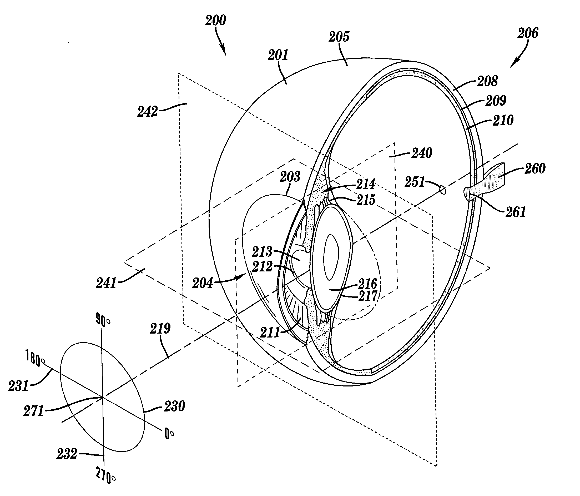
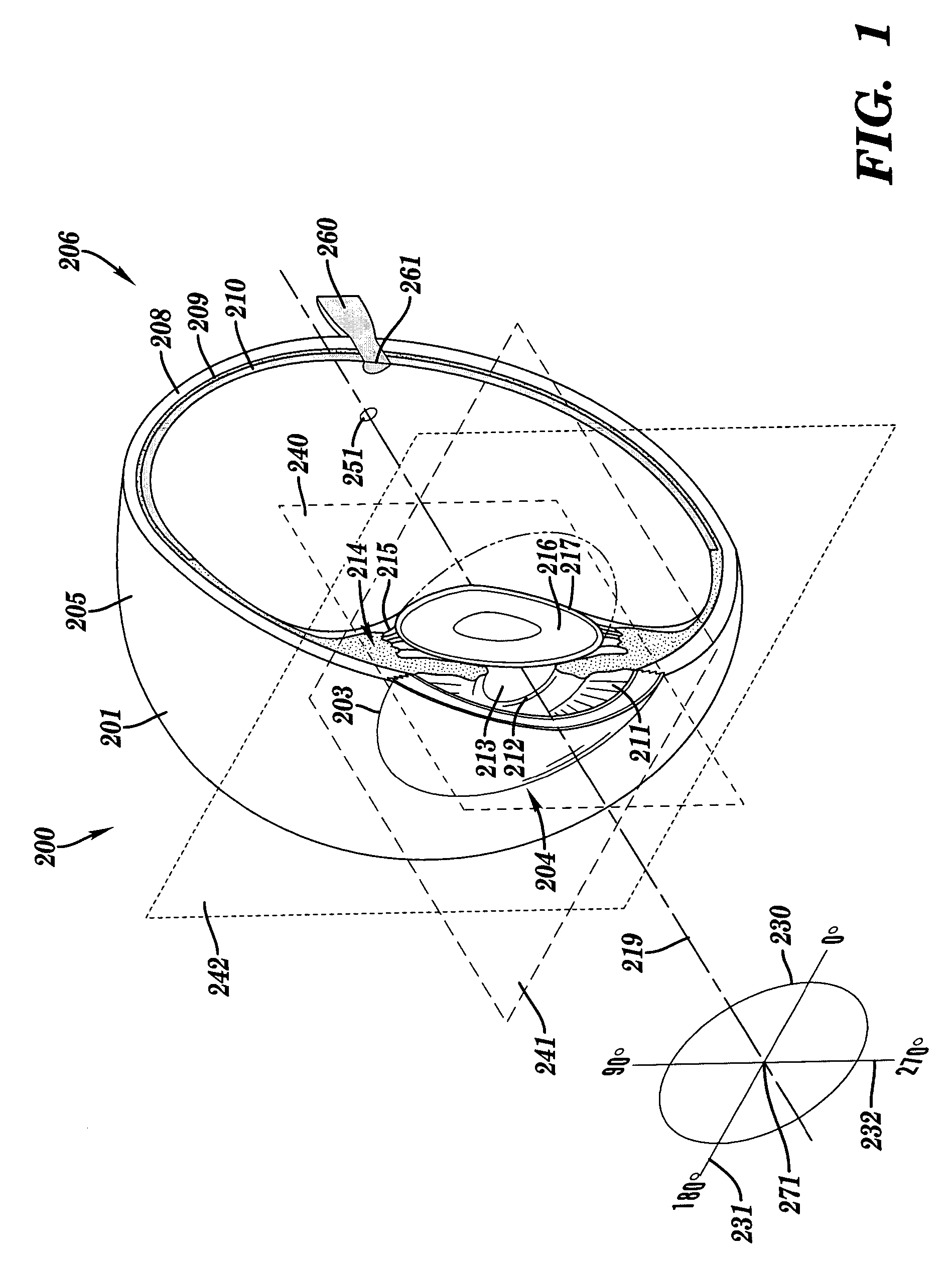
3.2 Ocular Anatomy

[0062]FIG. 1 is a schematic perspective illustration of a human eye 200, set against an anatomical sagittal plane 240, an anatomical axial plane 241 and an anatomical coronal plan...


PUM


Abstract

The invention administers an objective clinical test to an eye that measures the visual sensitivity of the superior retina and the inferior retina, by alternately presenting a stimulus pair comprising a shaped superior light stimulus and a shaped inferior light stimulus that are horizontal mirror images of one another and have shapes encompassing visual field defects. The shaped superior and inferior light stimuli stimulate pupillary responses whose amplitudes are measured. A cycle-averaged pupillary response balance and a luminance ratio are computed for each presentation of a stimulus pair. A stimulus pair response curve is computed by fitting cycle-averaged pupillary response balances to a sigmoid function of the luminance ratios. A balanced luminance ratio at which the cycle-averaged pupillary response balance is equal to about zero is computed from the sigmoid function. The balanced luminance ratio is indicative of the presence and location of retinal nerve damage.

Description

RELATED APPLICATION[0001]The present invention claims priority to U.S. Provisional No. 60 / 551,649 filed on Mar. 9, 2004, which is incorporated herein by reference in its entirety.BACKGROUND OF THE INVENTION[0002]1. Technical Field[0003]The present invention generally relates to an apparatus and method for assessing retinal damage to an eye and in particular to an apparatus and method for assessing glaucomatous retinal damage to an eye.[0004]2. Related Art2.1 Glaucoma Epidemiology[0005]Glaucoma is the second most common cause of blindness in the United States, and the single most important cause of blindness among black Americans. About 80,000 Americans are blind from primary open-angle glaucoma (“POAG”). At least 2 million Americans have POAG, but about half are unaware of it. About 5 to 10 million Americans are ocular hypertensives having an elevated intraocular pressure (“IOP”) that places them at increased risk for the development of POAG. The rate at which such ocular hypertensi...

Claims


Application Information

Patent Timeline
Patent Type & Authority Patents(United States)
IPC IPC(8): A61B3/10A61B3/00A61B3/11
CPCA61B3/112A61B3/024
Inventor WYATT, HARRY J.
Owner THE RES FOUND OF STATE UNIV OF NEW YORK
Why Eureka
Try Eureka