System and method supporting imaging and monitoring applications

a technology for monitoring applications and systems, applied in applications, tomography, instruments, etc., can solve the problems of occupying a large space, restricting the movement of doctors, and bulky external displays, and achieve the effect of being suitable for mobile applications

Active Publication Date: 2009-06-23
FUJIFILM SONOSITE
View PDF29 Cites 188 Cited by
  • Summary
  • Abstract
  • Description
  • Claims
  • Application Information

AI Technical Summary

Benefits of technology

[0008]The present invention is directed to systems and methods providing a portable diagnostic medical device that processes information from multiple sources. An embodiment of the invention is a unified device that supports dual-mode operation providing imaging and monitoring applications. The unified device preferably provides for imaging and the transfer of image information from one point to another. The unified device of embodiments also may preferably be used for patient monitoring, such as to monitor the patient's vital signs or other information about the patient. The unified device of embodiments is small, portable and suitable for mobile applications.

Problems solved by technology

Many medical personnel may need to be present during the performance of a clinical procedure; however, routine monitoring or imaging examinations may only demand nurses or imaging technicians, such as sonographers, to be present to perform these examinations.
Accordingly, with respect to monitoring and imaging, a physician's movement is limited.
These external displays are typically bulky, taking up a large amount of space, and are generally not positioned close to one another.

Method used


Image

  • System and method supporting imaging and monitoring applications
  • System and method supporting imaging and monitoring applications
  • System and method supporting imaging and monitoring applications

Examples


Embodiment Construction

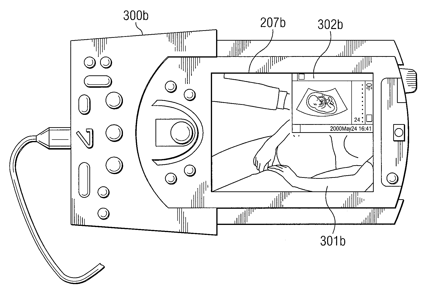
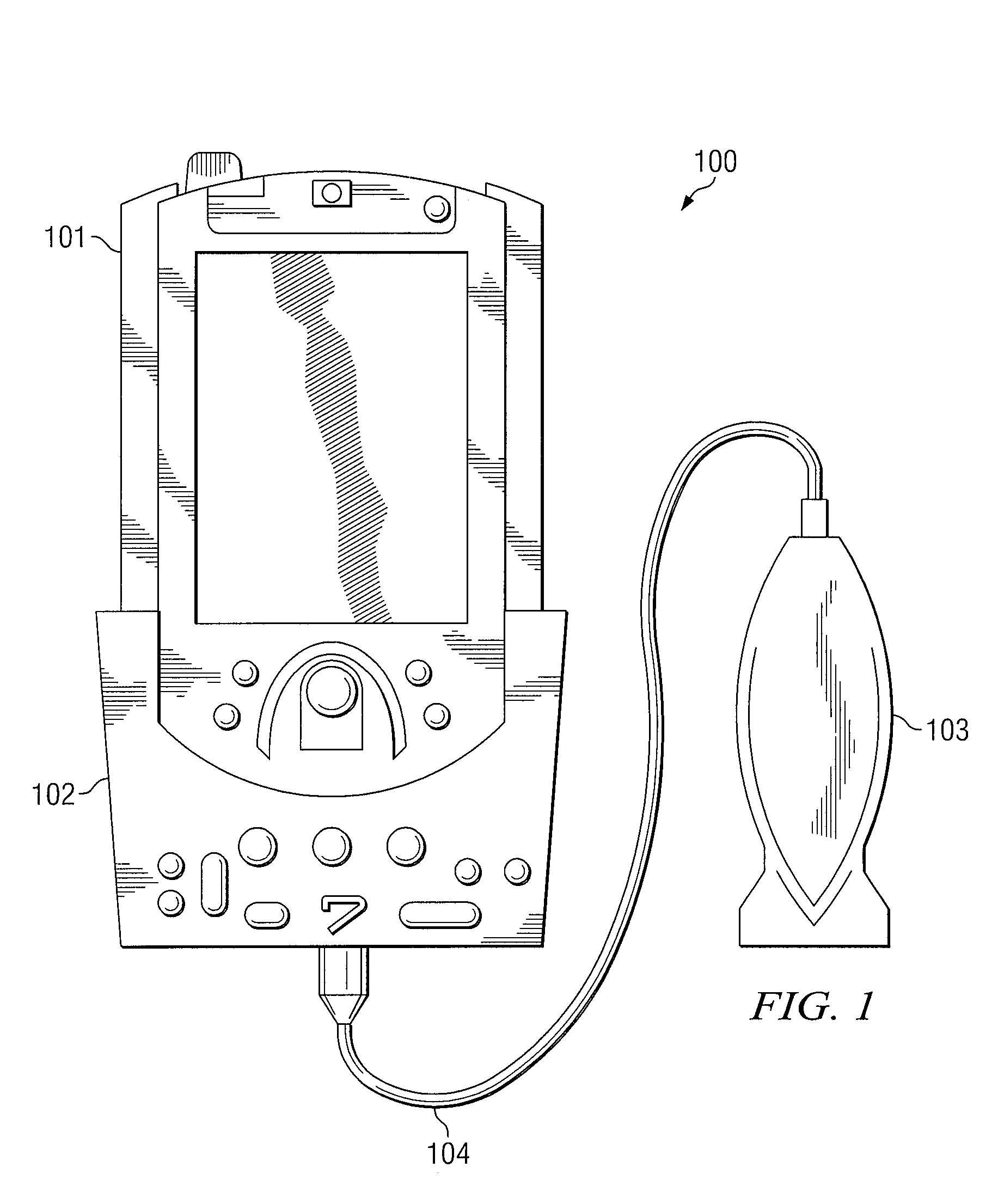
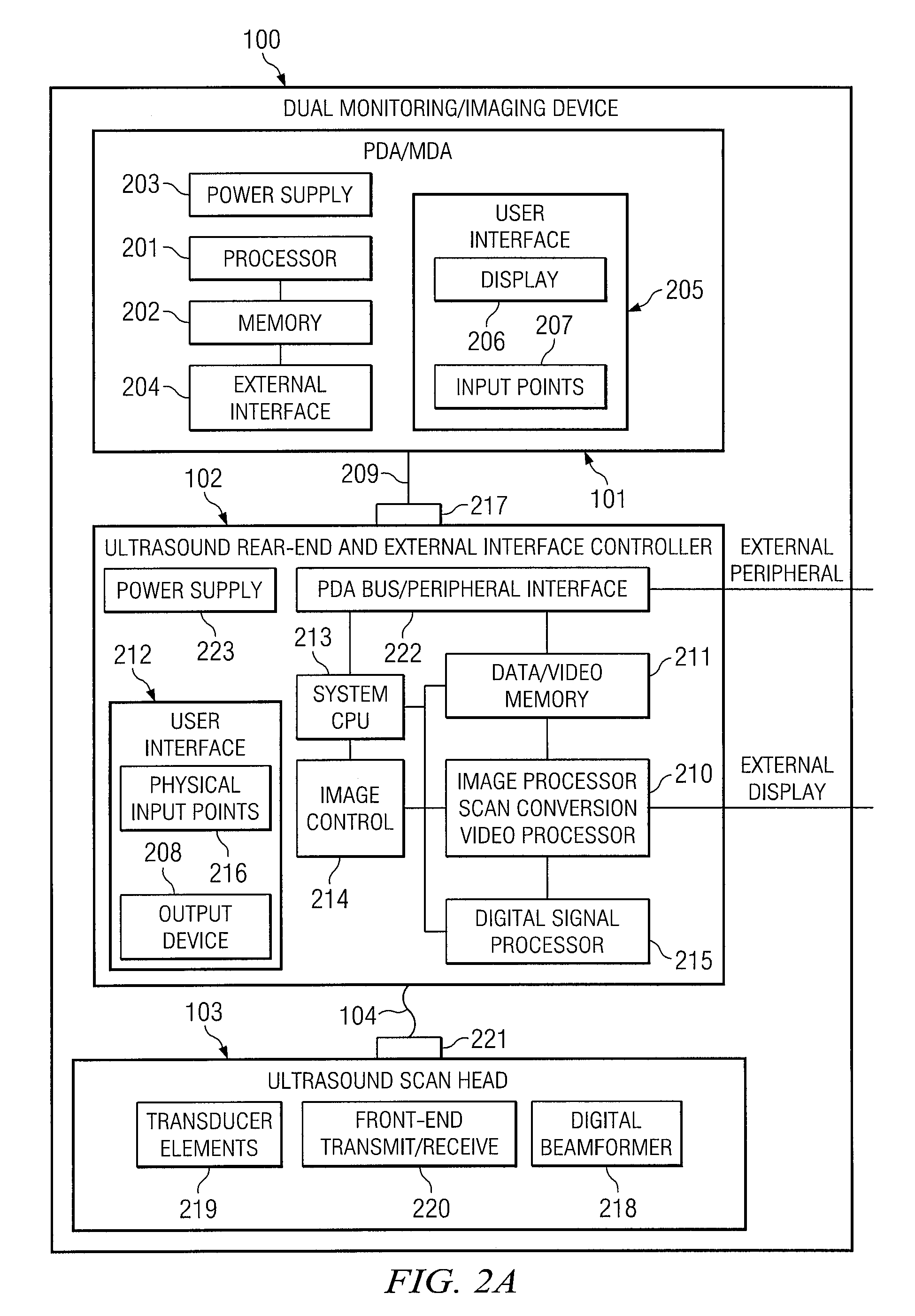
[0029]In a first embodiment, a unified device for processing information, such as monitoring and imaging, from multiple sources is provided. The device is comprised of an external controller or sleeve for receiving a personal digital assistant (PDA) or medical data assistant (MDA), and an interconnect to allow the external controller and / or PDA / MDA to communicate with an ultrasound scan head. FIG. 1 depicts an embodiment of dual monitoring / imaging device 100 comprising PDA / MDA 101, external controller 102 for receiving PDA / MDA 101, scan head 103, and interconnect 104 for attaching scan head 103 to external controller 102. Interconnect 104, which may be wireless or wireline, allows for communication between scan head 103 and external controller 102 as well as to allow communication of ultrasound signals generated by scan head 103 to external controller 102. External controller 102 processes the signals into images for eventual display on PDA / MDA 101 and / or external display systems (n...


PUM


Abstract

The present invention is directed to a system and method in which a portable medical device supports simultaneous imaging and monitoring applications. This portable device includes an external controller for receiving a PDA/MDA via a communication bus. An ultrasound scan head also is preferably attached to the external controller via an interconnect. In operation, real-time video feed of the patient may be acquired using the device and then displayed on the PDA/MDA. Using the ultrasound scan head in conjunction with the external controller, images may be acquired and displayed on the PDA/MDA, and vital sign monitoring of a single patient or multiple patients may be performed. Video feed, images and/or monitoring information may be transferred to a remote expert for review and analysis on the remote expert's communication device.

Description

CROSS-REFERENCE TO RELATED APPLICATION[0001]The present application claims priority to U.S. Provisional Patent Application No. 60 / 491,919, entitled “Ultrasound Conversion Kit for a PDA / MDA,” filed Jul. 31, 2003, the disclosure of which is hereby incorporated herein by reference.TECHNICAL FIELD[0002]Embodiments of this disclosure are directed to medical diagnostic and / or treatment systems and more particularly to medical devices for supporting imaging and monitoring applications.BACKGROUND OF THE INVENTION[0003]Clinical space at a hospital, such as a surgical suite, is typically crowded with various medical personnel and large pieces of equipment occupying the space. Many medical personnel may need to be present during the performance of a clinical procedure; however, routine monitoring or imaging examinations may only demand nurses or imaging technicians, such as sonographers, to be present to perform these examinations. While the physician in charge of the patient may not need to b...

Claims


Application Information

Patent Timeline
Patent Type & Authority Patents(United States)
IPC IPC(8): A61B8/00A61B5/00G16H30/20G16H30/40G16H40/67
CPCA61B8/14A61B8/4411A61B8/4472A61B8/56A61B8/565G06F19/3418H04N19/162A61B2560/0456G06F19/321G06F19/3406G16H40/63G16H30/20G16H40/67G16H30/40
Inventor HWANG, JUINJET
Owner FUJIFILM SONOSITE
Why Eureka
Try Eureka