

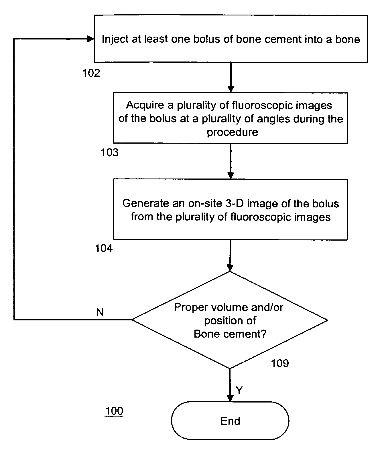
Intra-operative 3-D reconstruction of bone cement boli using X-rays
a 3-d reconstruction and bone cement technology, applied in the field of three-dimensional imaging, can solve the problems of impracticality of ct scans, extra cost and radiation exposure for patients, and achieve the effects of preventing secondary damage to the vertebral body, and reducing the risk of fractur
- Summary
- Abstract
- Description
- Claims
- Application Information
AI Technical Summary
Benefits of technology
Problems solved by technology
Method used
Image

Examples
example
[0046]Six cadaveric spinal segments (T9-L4; Male, ages 63-88) from a coinciding study were analyzed. They were placed two at a time in a water bath and scanned in a clinical CT scanner with 1 mm transverse slices. Each specimen was then prepared by cutting off the L1-L3 segment and removing the intervertebral discs from each end. Vertebroplasty was then performed on each of the six 3-level segments; every L1 and L3 vertebra was then injected with 10 cc of PMMA, in a bi-pedicular injection scheme (two discrete injections, one through each lateral pedicle).
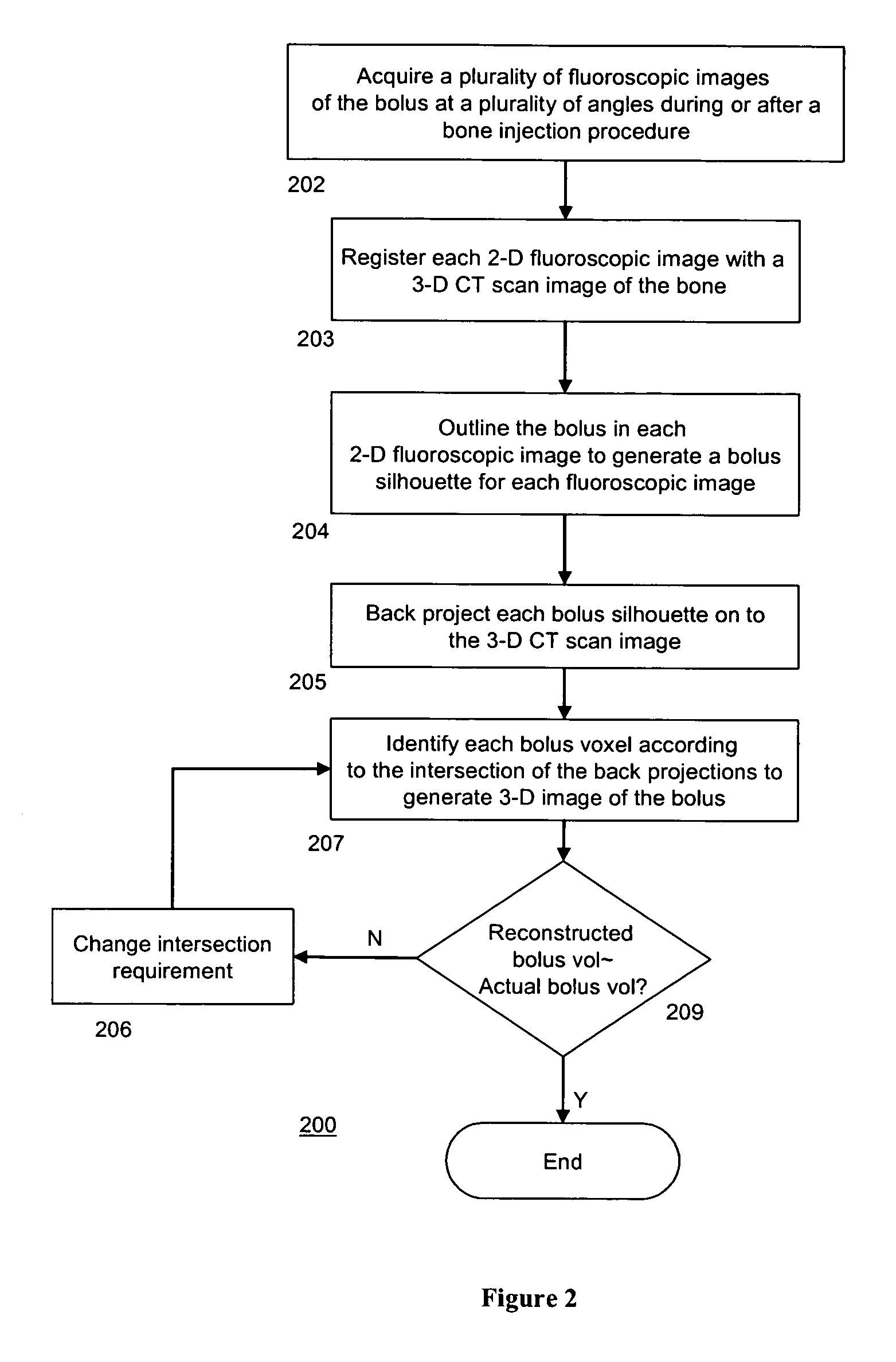
[0047]After treatment, each specimen was transferred to a cylindrical container with a stand and demarcated angles. The container was placed on a treatment table, and a fluoroscope projector was placed directly above in a fixed position. By rotating the container in fixed increments, 12 fluoroscopic X-ray images were taken evenly spaced angles of 15° over a 180° range, as opposite projections provide similar information. Each X-ray ...
PUM
 Abstract
Description
Claims
Application Information