Method and system for near-infrared fluorescence contrast-enhanced imaging with area illumination and area detection

a near-infrared fluorescence contrast and imaging technology, applied in the field of biomedical imaging, can solve problems such as difficult to solve underdetermined inverse problems, and achieve the effect of efficient excited and more data

Active Publication Date: 2009-10-06
TEXAS A&M NUNIV SYST THE
View PDF73 Cites 26 Cited by
  • Summary
  • Abstract
  • Description
  • Claims
  • Application Information

AI Technical Summary

Benefits of technology

[0007]Embodiments of the invention provide a number of technical advantages. Embodiments of the invention may include all, some, or none of these advantages. An approach employed herein involves area illumination and area detection on the same tissue-like surface. This approach overcomes the disadvantages associated with point illumination and point detection given that (1) fluorophores within a large tissue volume are more efficiently excited via area illumination and (2) significantly more data is provided via area detection. In addition, non-contact imaging is possible. Some applications may arise in medical imaging of diseased tissues and intraoperative 3D identification of diseased (and tumor) lesion margin for effective and complete resection of diseased tissue, among others.

Problems solved by technology

Disadvantages with point illumination and point detection include (1) possible failure to sufficiently excite the contrast-enhanced target and (2) the production of sparse data sets, which make the underdetermined inverse problem difficult to solve.

Method used


Image

  • Method and system for near-infrared fluorescence contrast-enhanced imaging with area illumination and area detection
  • Method and system for near-infrared fluorescence contrast-enhanced imaging with area illumination and area detection
  • Method and system for near-infrared fluorescence contrast-enhanced imaging with area illumination and area detection

Examples


Embodiment Construction

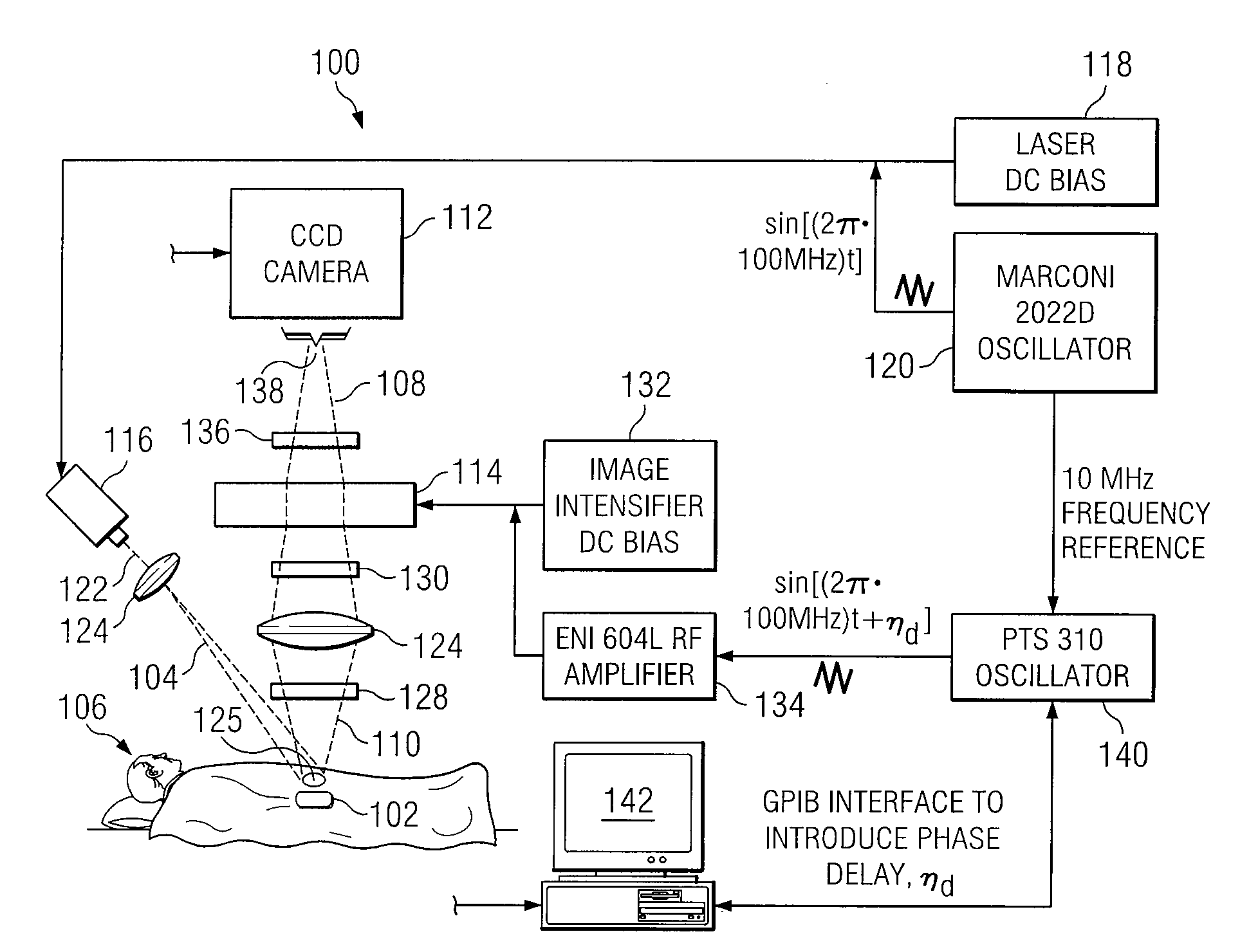
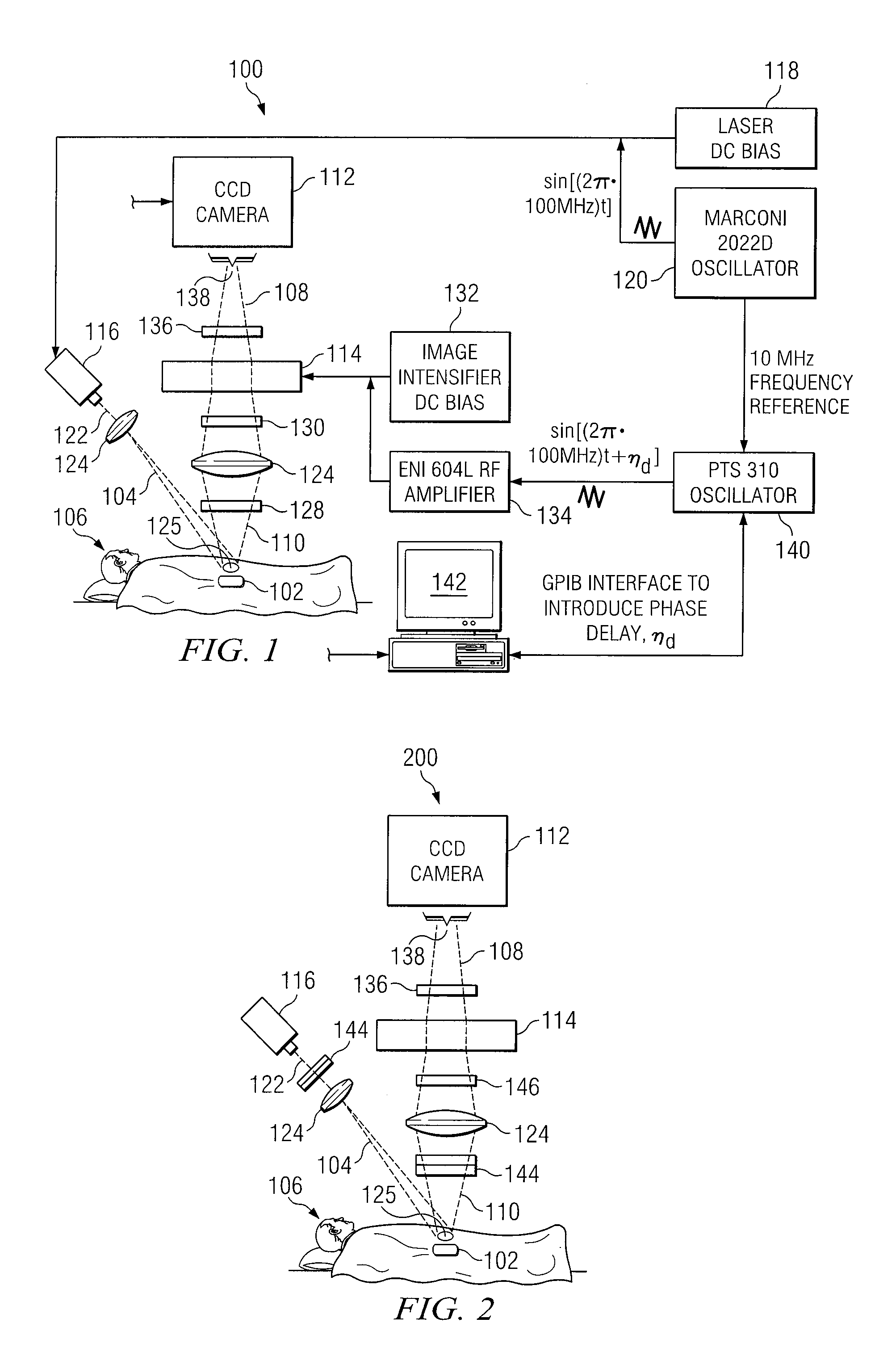
[0015]FIG. 1 is a schematic illustrating a system for excitation area illumination and area detection using near-infrared (“NIR”) frequency-domain photon migration (“FDPM”) imaging techniques according to one embodiment of the present invention. In particular, system 100 generates three-dimensional (“3D”) images of fluorescent target 102 within a light scattering material (e.g., tissue) based on emission light detected at a two-dimensional (“2D”) sensor surface. At a high level, system 100 delivers an intensity-modulated excitation light 104 into the tissue of a body 106 to cause stimulated emission of fluorescent target 102. Emission light 108 from the fluorescent target 102 is detected by intensified charge-coupled camera (ICCD) 112 via gain modulated image intensifier 114. A 3D image of fluorescent target 102 is generated based, at least in part, on excitation light 104 and emission light 108. System 100 provides several advantages over the prior art such as, for example, the abi...


PUM

PropertyMeasurementUnit
surface areaaaaaaaaaaa
sizeaaaaaaaaaa
fluorescent lifetimeaaaaaaaaaa

Abstract

According to one embodiment of the invention, a method for biomedical imaging includes directing time-varying excitation light at a surface area of a light scattering material, the material comprising a fluorescent target. Time-varying emission light from the fluorescent target is detected, substantially at a two-dimensional sensor surface, in response to the time-varying excitation light stimulating the fluorescent target. The time-varying emission light is filtered to reject excitation light re-emitted from the material. A three-dimensional image of the fluorescent target is generated based on the detection substantially at the sensor surface.

Description

RELATED APPLICATIONS[0001]This application claims the benefit under 35 U.S.C. § 119(e) of U.S. Provisional Application Ser. No. 60 / 480,526 filed Jun. 20, 2003.GOVERNMENT RIGHTS[0002]This invention was made with Government support from the National Institute of Health, Contract No. R01 CA67176, and R01 EB002107. The government may have certain rights in this invention.TECHNICAL FIELD OF THE INVENTION[0003]The present invention relates generally to biomedical imaging and, more particularly, to a method and system for near infrared fluorescence contrast-enhanced imaging with area illumination and area detection.BACKGROUND OF THE INVENTION[0004]Fluorescent contrast agents have been developed and / or employed that enhance the optical detection of diseased tissues. These agents excite and re-emit at near-infrared (NIR) wavelengths, which deeply penetrate tissues. Localization of a contrast-enhanced target in three dimensions necessitates: (1) rapid and precise acquisition of fluorescence m...

Claims


Application Information

Patent Timeline
Patent Type & Authority Patents(United States)
IPC IPC(8): A61B5/00G01N21/64
CPCA61B5/0059G01N21/6456G01N21/6428G01N21/6408A61B5/0073
Inventor SEVICK-MURACA, EVA M.THOMPSON, ALAN B.RANADHIR, ROY
Owner TEXAS A&M NUNIV SYST THE
Why Eureka
Try Eureka