

Diagnostic and therapeutic methods based on the L1 adhesion molecule for ovarian and endometrial tumors
- Summary
- Abstract
- Description
- Claims
- Application Information
AI Technical Summary
Benefits of technology
Problems solved by technology
Method used
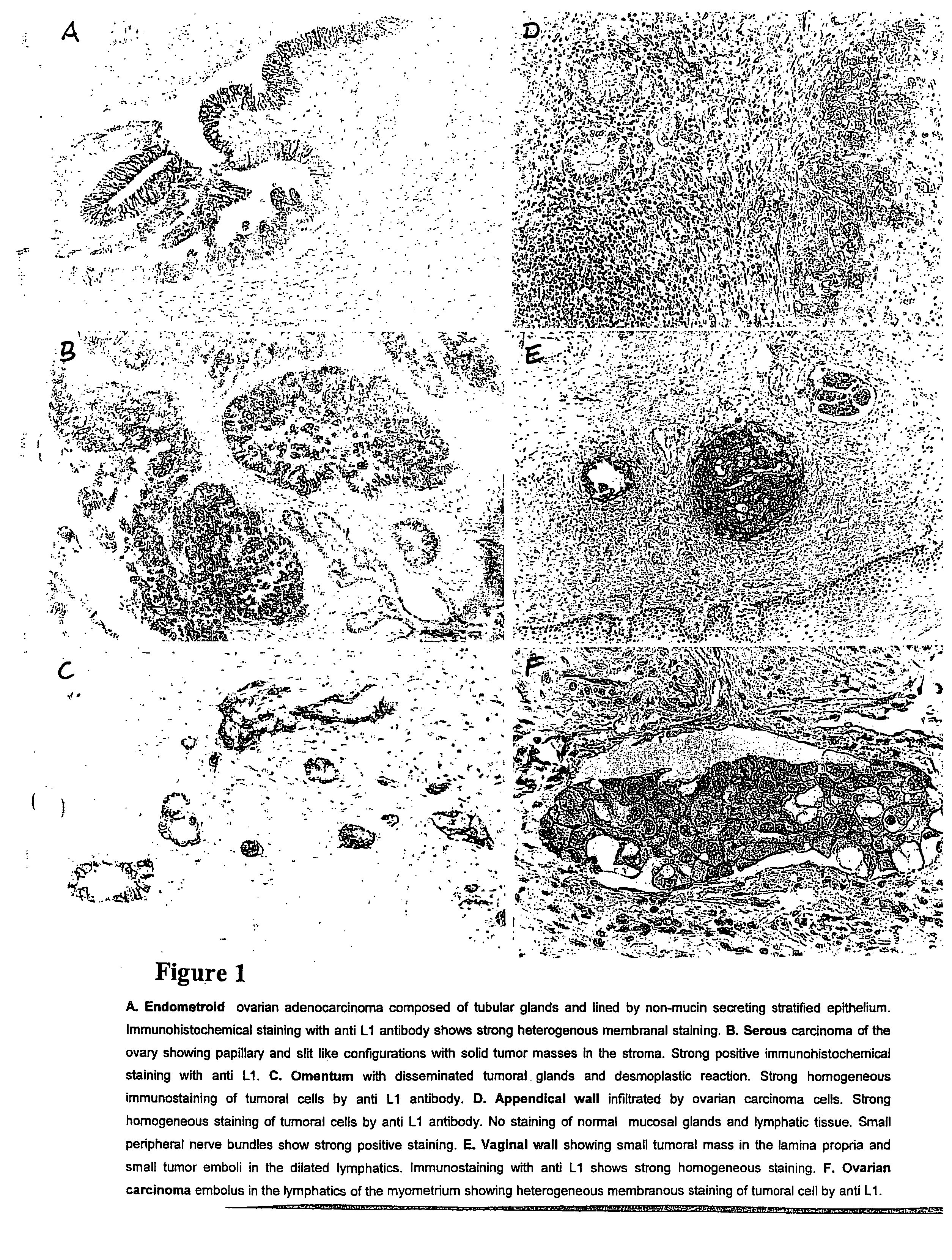
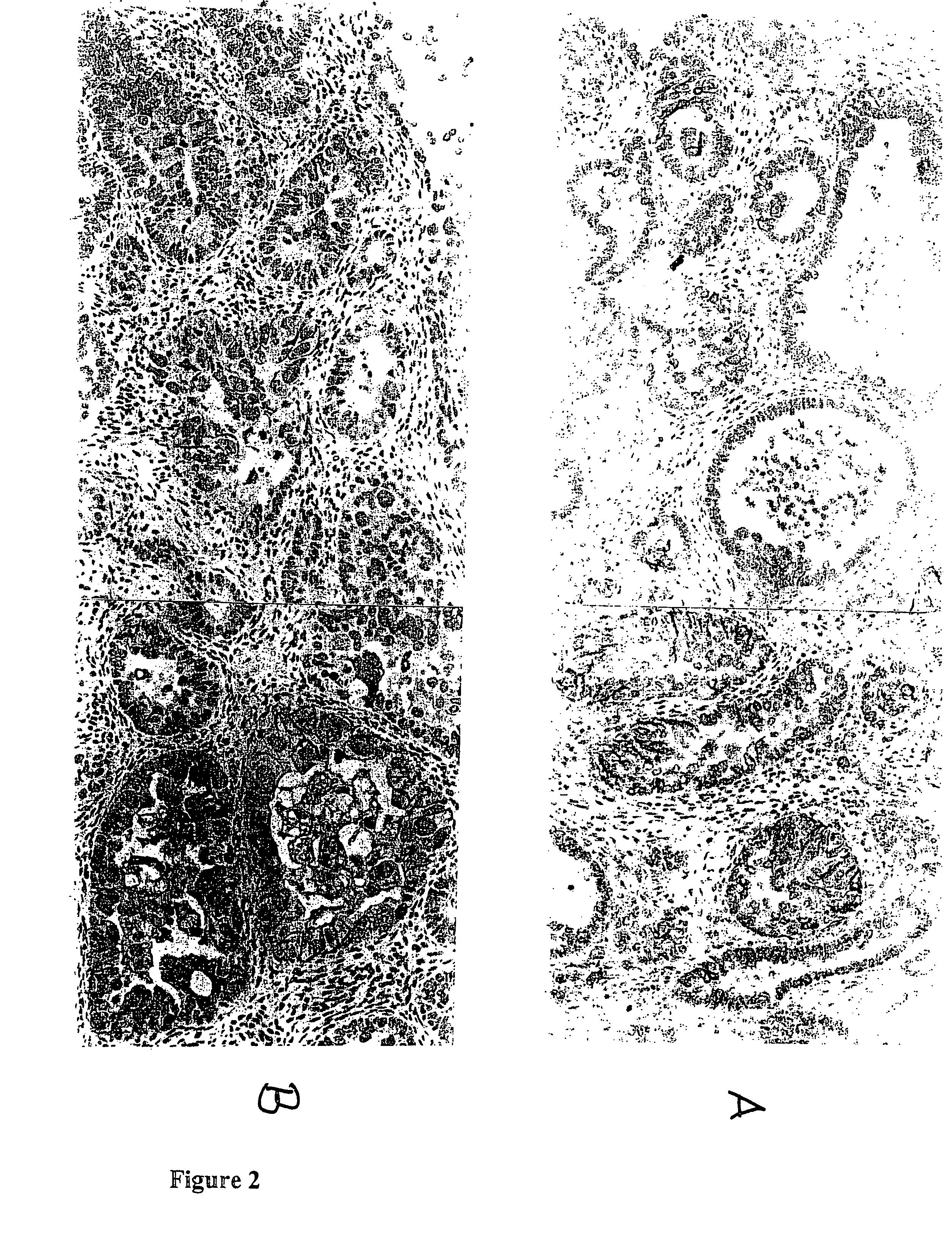
Image

Examples
example 2
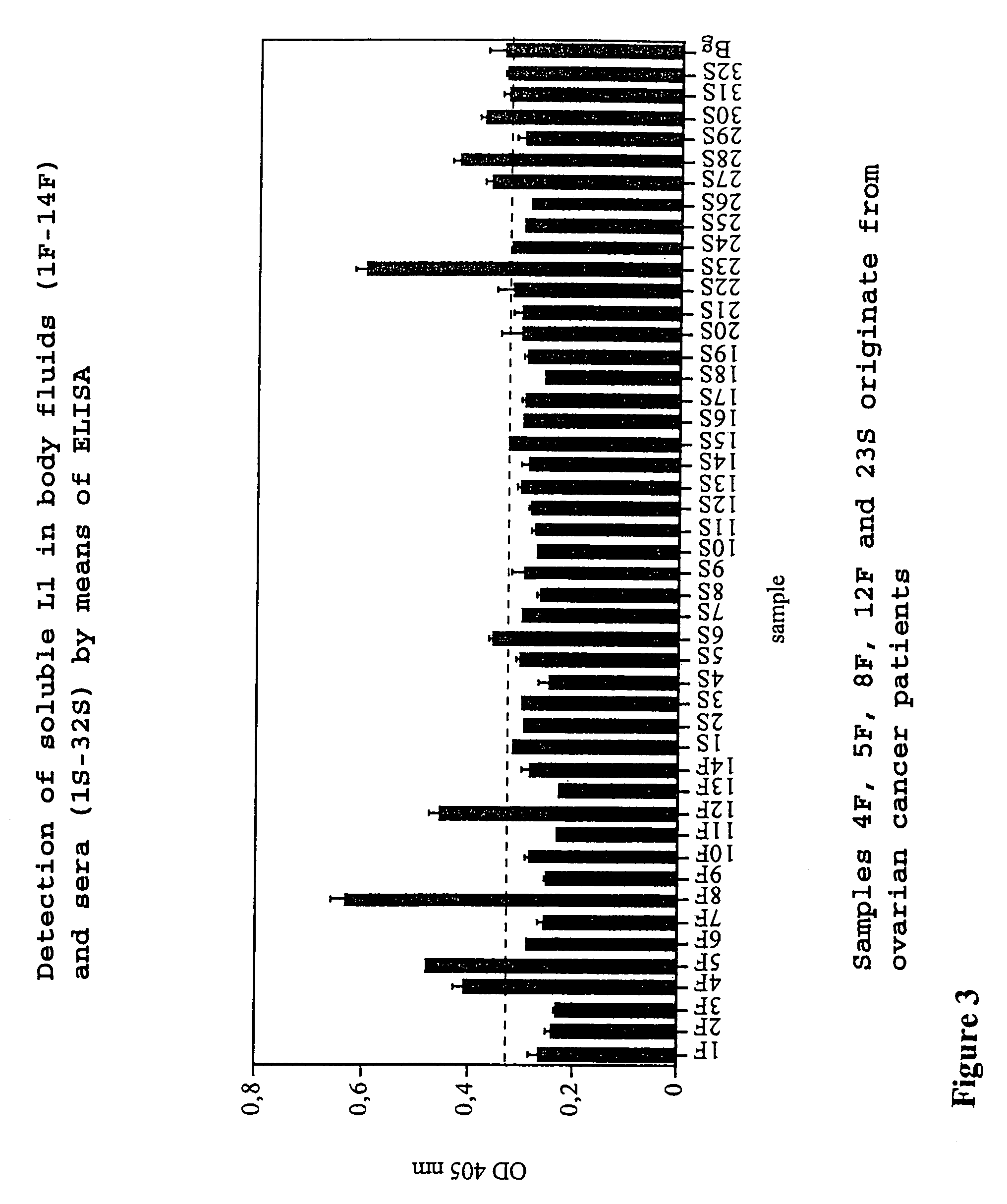
ELISA Shows the Presence of Soluble L1 in sera and Ascitic Fluid of Tumor Patients, a Correlation between the Presence of L1 and the Tumor Type (Ovarian and Endometrial Tumor) being Observed.
[0052]Samples of body fluids (ascites, serum) were tested for the presence of soluble L1 using a “capture” ELISA. For this purpose, microtitration plates were coated with the human anti L1 antibody (concentration: 1 μg / ml) described in Example 1 and then a blocking step was carried out with 3% BSA in PBS (45 min., room temperature) to eliminate the non-specific bonding to the plate. The body fluid was added at differing concentrations (1:2 and 1:10 of the fluid in 3% BSA in PBS), and incubation was carried out at room temperature for 1 hour. Therefore, four wash steps followed in Tris-buffered common salt solution (TBS, pH 8.0, in the presence of 0.02% Tween-20). Bound soluble L1 was determined by the addition of human biotin-conjugated anti L1 antibody. For this purpose, the biotinylated antibo...
example 3
A New ELISA Format for the Detection of Soluble L1
[0053]The ELISA described above in Example 2 used the coating of the microtiter plate with L1 mAb 1 (capturing monoclonal antibody) and the detection of soluble L1 with a biotinylated (or otherwise labelled) L1 mAb 2 (detecting monoclonal antibody). This type of ELISA is termed G / K format. We have now developed another format in which we use both for capturing and detection the same mAb to L1 (K / K format). In FIG. 5A and B a comparison of both types of ELISA is shown using a positive serum (CA526, ovarian tumor patient) and seval control sera from unrelated tumors. It is obvious that the new ELISA format in FIG. 5B gives a better signal to noise ratio (appr. 5-8 fold) than the previous format shown in FIG. 5A. As the K / K format can only work when the antibody epitope for detection is not blocked by the antibdy used for capturing, these results implicate that in the serum sample the soluble L1 is dimeric or multimeric. The control dat...
example 4
PCR Analysis for Detecting L1 mRNA in Tumor Samples
[0054]Primer sequences for PCR analysis are deduced from the chimeral human L1 cDNA sequence (EMBL / GenBank accession number M 74387; SEQ ID NO: 16). For the detection of human mRNA encoding L1 in tumor tissue or patient serum samples mRNA is isolated using commercial kits (Roche Molecular Biochemicals) and transcribed into cDNA. A nested PCR approach is used with the following primer combinations: First amplification primer 1: ACTGAGGGCTGGTTCATC (SEQ ID NO: 8)(sense), primer 2: CTTGCACTGTACTGGCCA (SEQ ID NO: 9)(antisense)(one cycle 45 sec, 94° C., 30 cycles of 1 min at 94° C., 1 min at 56° C., 1 min at 72° C.). For the second PCR 1 μl of the first PCR reaction is used with primer 1: ACTCAGTGAAGGATAAGGAG (SEQ ID NO: 10)(sense), primer 2: TTGAGCGATGGCTGCTGCT (SEQ ID NO: 11)(antisense). In an alternative protocol the following primers are used: primer 1: AGGTCCCTGGAGAGTG (SEQ ID NO: 12) (sense); primer 2: TTGAGCGATGGCTGCTGCT (SEQ ID NO...
PUM
| Property | Measurement | Unit |
|---|---|---|
| pH | aaaaa | aaaaa |
| concentration | aaaaa | aaaaa |
| adhesion | aaaaa | aaaaa |
Abstract
Description
Claims
Application Information