
Process for producing cartilage cells for transplantation

a technology of cartilage cells and cartilage, which is applied in the direction of skeletal/connective tissue cells, prosthesis, biocide, etc., can solve the problems of inability to obtain a sufficient amount of human chondrocytes using the currently available methods, and the difficulty of applying human chondrocytes to transplantation therapy in practice,

Inactive Publication Date: 2010-04-27
YANAGA KATSU +1
View PDF4 Cites 0 Cited by
  • Summary
  • Abstract
  • Description
  • Claims
  • Application Information

AI Technical Summary

Benefits of technology

This method allows for the rapid production of a large quantity of normal human chondrocytes suitable for transplantation, providing a safe and effective cartilage therapy material that can be used to treat various cartilage-related defects without the risks associated with nonhuman feeder cells.

Problems solved by technology

In humans, however, only a small amount of cartilage can be collected, therefore, only a small number of chondrocytes can be used at the initiation of the culture.
Thus, it is highly difficult to apply human chondrocytes to transplantation therapy in practice.
Namely, there is a problem that although a large amount of normal chondrocytes is required for transplantation in humans, it has been impossible to obtain a sufficient amount of human chondrocytes using the currently available methods.
The use of nonhuman animal feeder cells is, however, accompanied with problems of unexpected bacterial or viral infections and complicated treatments are needed to prevent these infections.

Method used


Image

  • Process for producing cartilage cells for transplantation
  • Process for producing cartilage cells for transplantation
  • Process for producing cartilage cells for transplantation

Examples


example 1

Chondrocyte culture

[0050]Composition of culture medium: To the DME(H) medium, 10% of FBS, 10 ng / ml of human FGF (Kaken Pharmaceutical Research Institute), 40 ng / ml hydrocortisone (Sigma) and 5 ng / ml human IGF-I (GIBCO) were added.

[0051]Collected cartilage: From human rear auricular cartilage, a sample piece (about 1×1 cm2) having perichondrium bonded to one face was collected.

[0052](a) Chondrocyte Fraction

[0053]The cartilage piece obtained above was disinfected with penicillin G (800 u / ml), kanamycin (1 mg / ml) and Fungizone (2.5 ug / ml). It was then diced with a scalpel and then allowed to stand in F-12 medium containing 0.3% of type II collagenase (Worthington Biochemical) at 4° C. overnight. On the next day, the culture medium was shaken at 37° C. for 4 hours and centrifuged. The precipitate thus obtained was employed as a cell fraction at the initiation of the culture.

[0054](b) Primary Culture

[0055]The cell fraction as described above was seeded by using the above-described medium...

example 2

Transplantation of Chondrocytes

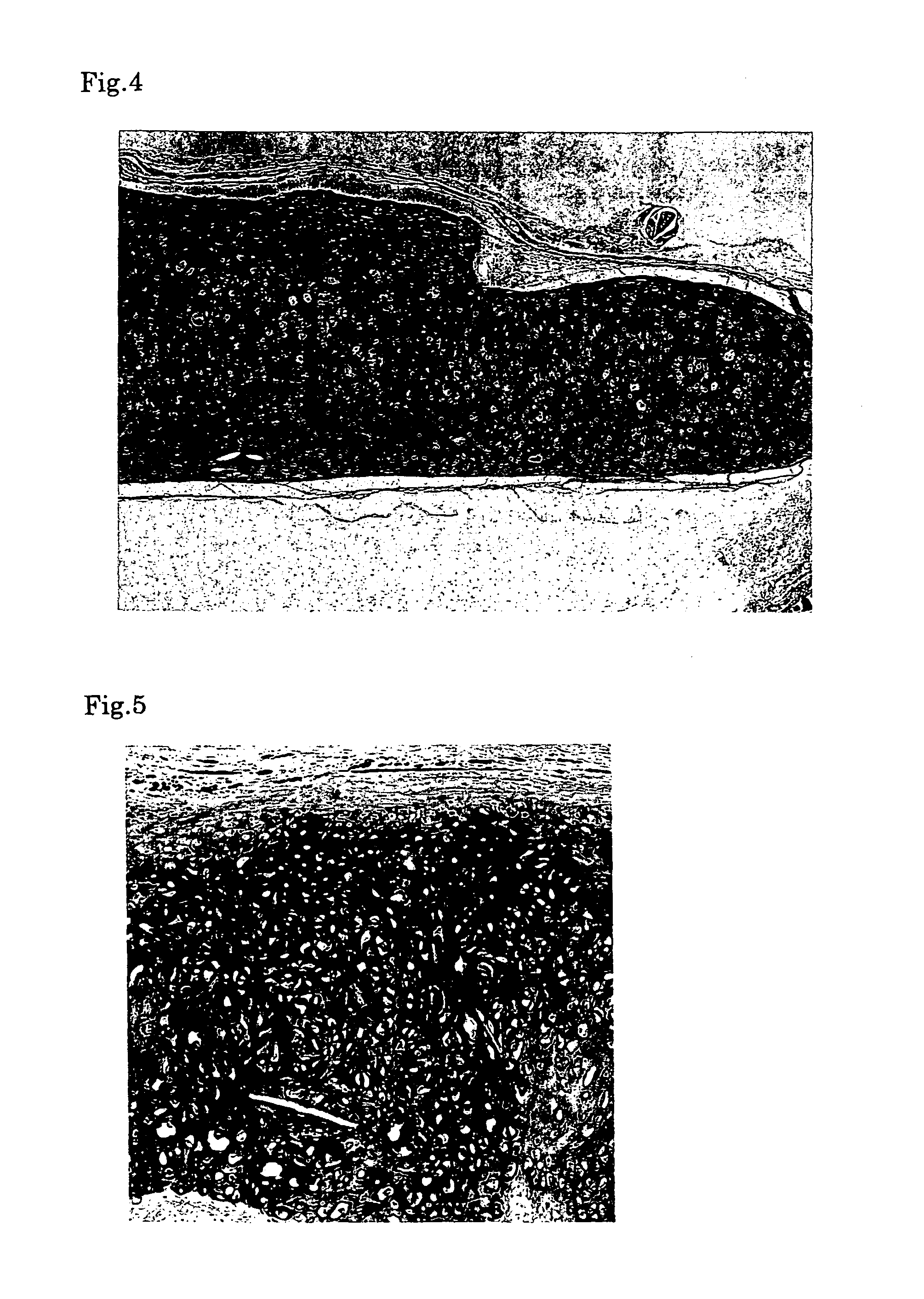
[0061]First, the medium was removed from the culture flask and the cells were harvested with a cell lifter. Then the harvested cells were collected with a syringe. Next, the chondrocytes collected into the syringe were injected into a defect in a cartilage of a nude mouse by using an injection needle. Six months after the transplantation, a specimen was sampled from the transplantation site and histologically examined to confirm the success in engraftment. When the sampled tissue was stained with hematoxylin-eosine (HE), namely, it was observed that cells were multilayered and bonded to each other via the matrix (FIG. 4). When the cells were immunologically stained for type II collagen serving as a molecular marker of cartilage tissue, the extracellular matrix was stained, indicating that the extracellular matrix was a cartilage-specific matrix (FIG. 5). Moreover, metachromacia was shown in toluidine blue staining, indicating the presence of aggrecan s...


PUM

PropertyMeasurementUnit
areaaaaaaaaaaa
culture timeaaaaaaaaaa
areaaaaaaaaaaa

Abstract

This invention is intended to provide a method to quickly obtain a large amount of normal human chondrocytes or a mass thereof without fear of bacterial or viral infection. Namely, a method of producing human chondrocytes characterized by comprising co-culturing chondrocytes obtained from a cartilage having perichondrium, for example, auricular cartilage together with the perichondrium; and a method of producing human chondrocytes characterized by comprising monolayer or multilayer seeding the cultured cells once or more and culturing to give a chondrocyte mass.

Description

[0001]This application is a U.S. National Stage under 35 U.S.C §371 of International Application No. PCT / JP04 / 05071 (filed Apr. 8, 2004), which claims priority under 35 U.S.C. §119(a)-(d) to Application No. JP2003-109707 filed on Apr. 15, 2003 and JP2003-176351 filed Jun. 20, 2003.TECHNICAL FIELD[0002]This invention relates to a method of producing normal human chondrocytes and the normal human chondrocytes obtained by the method. It also relates to a cartilage therapy material using the thus obtained normal human chondrocytes.BACKGROUND ART[0003]In a cartilage tissue, chondrocytes exist in the state of being embedded in the matrix. These chondrocytes can be separated from the matrix by treating the cartilage with an enzyme such as collagenase. Attempts have been made to utilize these separated chondrocytes in transplantation therapy, in particular, autotransplantation of chondrocytes for treating cartilage-related diseases. It has been experimentally confirmed that transplantation ...

Claims


Application Information

Patent Timeline
Patent Type & Authority Patents(United States)
IPC IPC(8): A01N65/00A61L27/00A61L27/38A61L27/44C12N5/07C12N5/077
CPCA61L27/3817C12N5/0655A61L27/3852A61L2430/06C12N5/0602A61L27/00
Inventor YANAGA, HIROKO
Owner YANAGA KATSU
Learn More