Implantable auricular cartilage composite stent

A composite stent, implantable technology, applied in nose implants, medical science, prostheses, etc., can solve the problems of insufficient support, poor stability, poor hardness, etc., to improve stability, increase support, increase The effect of its own strength and hardness

Active Publication Date: 2020-02-11
朱伟伟 +1
View PDF21 Cites 0 Cited by
  • Summary
  • Abstract
  • Description
  • Claims
  • Application Information

AI Technical Summary

Problems solved by technology

[0006] However, in the above technical solutions of the prior art, simply using cartilage tissue as a scaffold is prone to insufficient support strength, resulting in technical problems of low strength, poor hardness, and poor stability of the cartilage scaffold.

Method used


Image

  • Implantable auricular cartilage composite stent
  • Implantable auricular cartilage composite stent
  • Implantable auricular cartilage composite stent

Examples


Embodiment 1

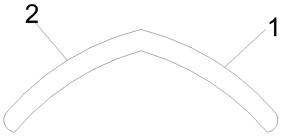
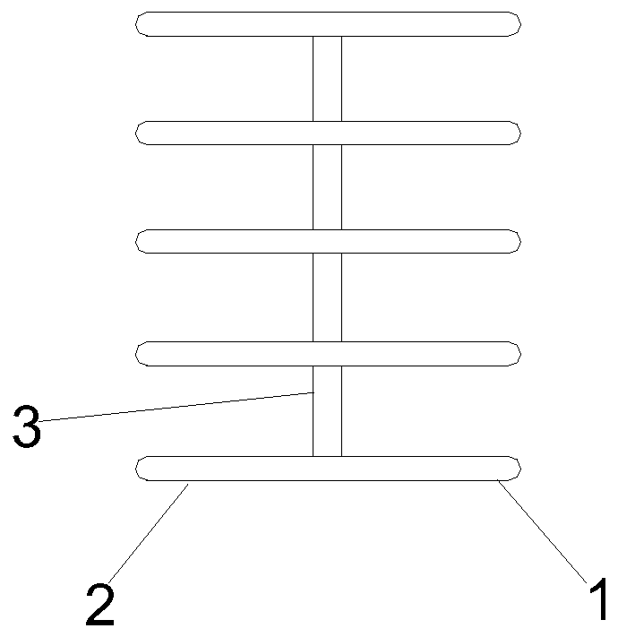
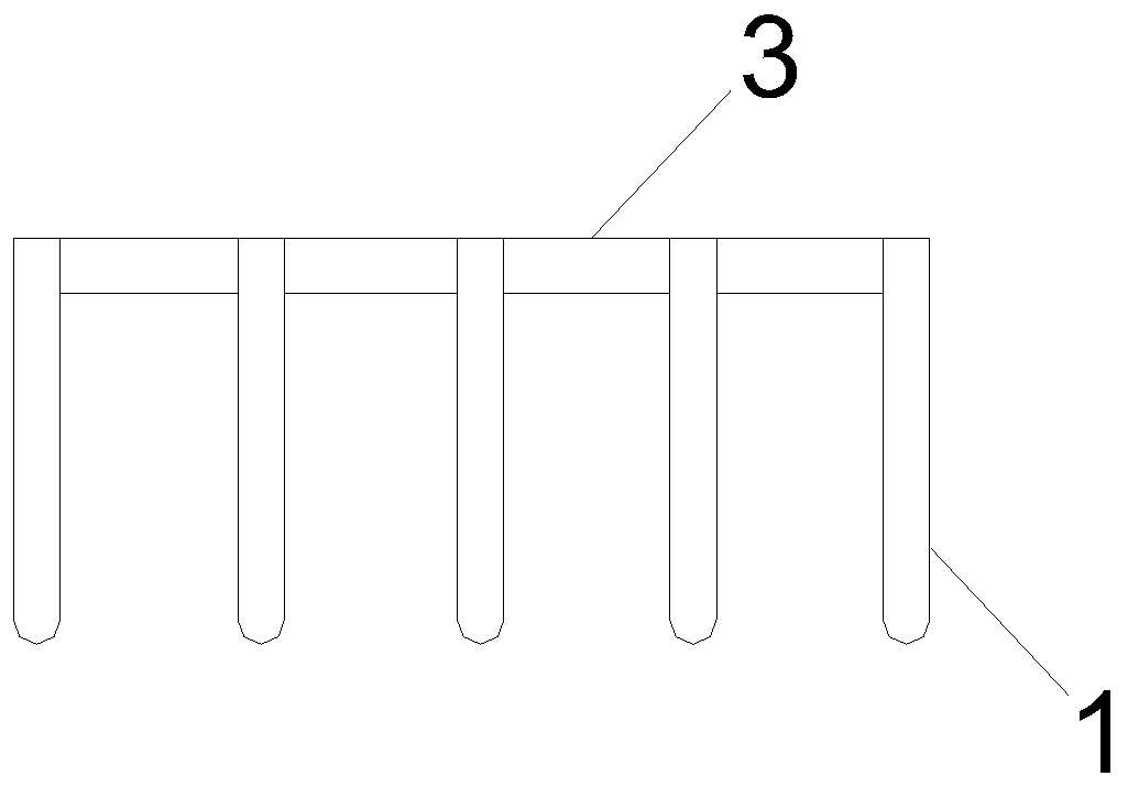
[0052] Such as Figure 1-4 As shown, an implantable ear cartilage composite bracket; includes a first bracket 1 and a second bracket 2; one end of the first bracket 1 and the second bracket 2 is connected with a connecting frame 3, and the first bracket 1 and the second bracket 2 are connected to each other. The second support 2 forms a "human" structure through the connection frame 3; the first support 1, the second support 2 and the connection frame 3 all include a line carving fiber core 4 and an ear cartilage support layer 5, and the line carving line The outside of the fiber core 4 is wrapped with an ear cartilage support layer 5 .

[0053] The thread-carving fiber core 4 has a helical structure. The acute angle included in the "human" shape of the first bracket 1 and the second bracket 2 ranges from 35° to 50°.

[0054] In addition, during the preparation of the ear cartilage scaffold, the removed ear cartilage tissue was minced, washed three times with phosphate hydro...

Embodiment 2

[0059] The preparation method of the implantable ear cartilage composite bracket comprises the following steps:

[0060] S1: Add the thread carving thread into the N,N-dimethylformamide solvent and stir it, after ultrasonic treatment, let it stand for a period of time, add alkaline solution to solidify, stretch while solidifying, wash, dry, and get the thread carving thread fiber Core 4;

[0061] S2: Take the hyaline ear cartilage tissue, cut it into pieces, add active agent and protease inhibitor for centrifugation, remove the supernatant, and keep the precipitated part of the solution;

[0062] S3: add active agent and protease inhibitor buffer solution to the precipitation solution obtained in S2, oscillate and mix well, and wash the precipitated part repeatedly after centrifugation to obtain cartilage particles for later use;

[0063] S4: spread the cartilage particles obtained in S3 evenly on a culture dish, and then vacuum freeze-dry them in a freeze dryer to obtain car...

Embodiment 3

[0072] The difference from Examples 1 and 2 is that in order to further increase the stability of the ear cartilage scaffold, the specific surface area A of the ear cartilage cell particles is 850-1920 cm 2 / g; PH value is 7-8 to increase the adhesion of ear chondrocytes, and at the same time increase the contact area with the cross-linking agent, which is beneficial to the proliferation and growth of ear chondrocytes. The specific surface area A of the ear chondrocyte particles and the pH value satisfy: A·PH is greater than or equal to 5950 and less than or equal to 15360.

[0073] The cell content M of the extracellular matrix of the ear cartilage, the specific surface area A of the ear chondrocytes and the pH value satisfy the following relationship:

[0074] A=μ·(PH) 3 / M 1 / 2 ;

[0075] In the above formula, μ is the specific surface area coefficient, and the value range is 3.24-6.65; only numerical calculations are performed here.

[0076] In order to improve the bioa...


PUM


Abstract

The invention discloses an implantable auricular cartilage composite stent. The implantable auricular cartilage composite stent comprises a first stent and a second stent, wherein a connecting frame is connected to one end of the first stent and one end of the second stent; an inverted V-shaped structure is formed by the first stent and the second stent through the connecting frame; the first stent, the second stent and the connecting frame comprise line engraving line fiber cores and auricular cartilage stent layers; and outer sides of the line engraving line fiber cores are coated with auricular cartilage stent layers correspondingly. According to the implantable auricular cartilage composite stent disclosed by the invention, the stent is manufactured by the auricular cartilage tissue; the line engraving line fiber cores are implanted in the center of the auricular cartilage tissue, so that the supporting force of the cartilage stent is increased, the intensity and hardness of the auricular cartilage stent are improved, and the stability of the auricular cartilage stents are further promoted; and through the inverted V-shaped structure, the implantable auricular cartilage composite stent crosses the nasal septum, so that the mechanical property is increased, and the stability of the cartilage stent is further improved.

Description

technical field [0001] The invention belongs to the technical field of biomaterials used in medicine, and in particular relates to an implanted ear cartilage composite bracket. Background technique [0002] The clinical application of cartilage scaffolds is common in plastic surgery and orthopedics, but with the increase of applications, there are also many shortcomings. For example, in plastic surgery, ear cartilage is used to pad the tip of the nose by taking out the cartilage of the pinna and placing it on the tip of the nose. , to achieve the purpose of nose tip shaping. There are two main methods of ear cartilage pad pen tip, one is to build a nose tip bracket, and then lift the nose tip up, and the other is to make the ear cartilage into a shield shape, pad it under the skin of the nose tip, and reach the tip of the nose purpose of shaping. [0003] At present, ear cartilage scaffolds are used for support. Ear cartilage alone is not strong enough to support the nasal...

Claims


Application Information

Patent Timeline
Patent Type & Authority Applications(China)
IPC IPC(8): A61F2/18
CPCA61F2/186A61F2/30756A61F2002/30108A61F2002/30764
Inventor 朱伟伟柳晓燕
Owner 朱伟伟
Why Eureka
Try Eureka