Method of assessing tissue viability using near-infrared spectroscopy
a tissue viability and near-infrared technology, applied in the field of tissue viability assessment using near-infrared spectroscopy, can solve the problems of compromising the communication of fluid from the main body portion to the tissue portion, and achieve the effect of ensuring tissue viability, improving the likelihood of tissue survival, and determining the viability of the tissu
- Summary
- Abstract
- Description
- Claims
- Application Information
AI Technical Summary
Benefits of technology
Problems solved by technology
Method used
Image
Examples
example i
Principles of Optical Near-Infrared Tissue Spectroscopy
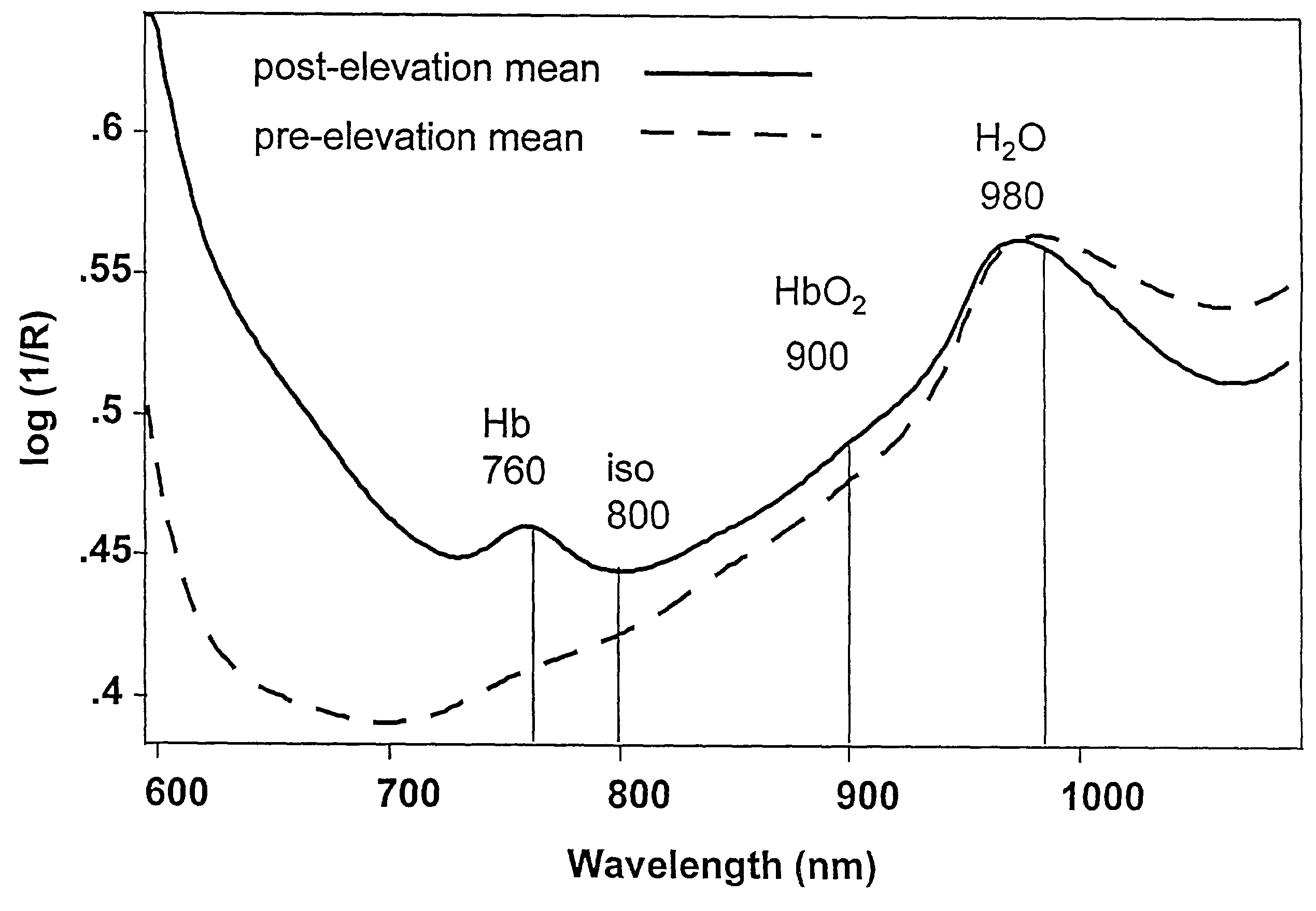
[0063]Infrared spectroscopy deals with the interaction of IR radiation with matter. The IR region of the electromagnetic spectrum is generally considered to lie in the wavelength range from 700-250,000 nm and is further subdivided into three subregions: The near-IR region, (from 700-2500 nm), the mid-IR region (from 2500-50000 nm) and the far-IR region (beyond 50000 nm). Visible and near-IR spectra in the 600 to 1100 nm range are presented in this study. The spectroscopy of tissues involves measuring the attenuation of the light intensity by the tissue relative to the incident light intensity.
[0064]Herein, the relative change and distribution of the levels of oxyhemoglobin (HbO2), and deoxyhemoglobin (Hb) in tissue, following flap elevation is examined and these data are used to predict tissue viability. The near-IR and visible absorption spectra of Hb, HbO2 and water are well understood and the differential absorption by these ...
example ii
Anaesthesia and Surgical Procedures
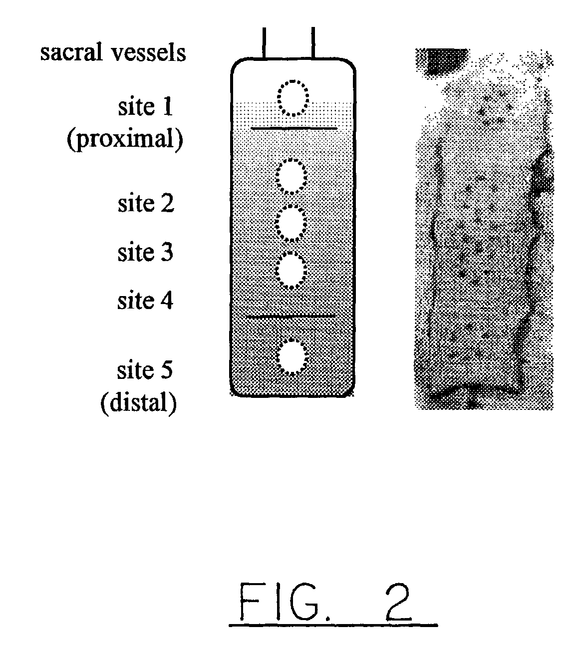
[0072]3×10 cm reverse McFarlane flaps raised on the dorsa of 9 Sprague Dawley rats weighing between 380-410 grams were used. The rats were acclimatized for a minimum period of 2 weeks prior, and the individual surgeries were performed over a three month period. All the procedures were done under 1.2-2% isoflurane inhalational anaesthesia. Five days prior to surgery, rats were shaved and a depilatory agent (Nair™) was applied on the rat dorsum. At that time, reproducible anatomic points for the near-IR spectroscopy measurements were marked on the skin, as shown in FIG. 2.
[0073]Surgery was performed 3 days (72 h) after pre-elevation measurements, which were to be used as control values in the analyses. Prior to surgery, the flap location was defined and marked based on the location of the sacral vessels, following which pre-elevation images and spectra were acquired. Twenty minutes prior to surgery, the rats were premedicated with 0.05 mg / kg of atrop...
example iii
Near-Infrared Measurements
[0075]Near infrared radiance images of 256×256 pixels were collected at 650, 700, 760, 800, 850, 900 and 980 nm using a Photometrics Series 200 CCD camera fitted with a Nikon Macro AF60 lens using 10 nm bandpass (FWHH) Lyot type filters (OCLI, Santa Rosa, Calif.). Visible and near-IR spectra were collected with a NIRSystems 6500 (Perstop, Silver Springs, Mass.) spectrometer using a custom bifurcated fiber optic bundle (CeramOptic, Enfield, Conn.). A 99% Spectralon® reflectance standard (LabSphere Inc.) was used as a reference to convert raw data into reflectance spectra. Each reflectance spectrum consisted of 32 co-added scans collected between 600-1100 nm at 10 nm resolution.
[0076]Pilot studies indicated that approximately the distal half of the flap failed while the half proximal to the vascular pedicle remained viable. Five dorsal monitoring sites were located along the length of the flap, as shown in FIG. 2. In order to have reproducible anatomical poin...
PUM
 Abstract
Description
Claims
Application Information
 - R&D
- Intellectual Property
- Life Sciences
- Materials
- Tech Scout
- Unparalleled Data Quality
- Higher Quality Content
- 60% Fewer Hallucinations
Browse by: Latest US Patents, China's latest patents, Technical Efficacy Thesaurus, Application Domain, Technology Topic, Popular Technical Reports.
© 2025 PatSnap. All rights reserved.Legal|Privacy policy|Modern Slavery Act Transparency Statement|Sitemap|About US| Contact US: help@patsnap.com


