Check patentability & draft patents in minutes with Patsnap Eureka AI!

Combined apparatus for imaging the inner part of a body, and method thereof

Active Publication Date: 2010-06-08
ESAOTE
View PDF14 Cites 11 Cited by
  • Summary
  • Abstract
  • Description
  • Claims
  • Application Information

AI Technical Summary

Benefits of technology

[0010]The invention has an object of providing an apparatus for imaging the inner part of a body, particularly for diagnostic imaging thereof and especially for a so called 3D imaging, which obviates the drawbacks of prior art apparatuses, while allowing to obtain an improved imaging apparatus which has better global imaging functions, a smaller global size, a higher versatility and lower costs.

Problems solved by technology

In order to image with sufficient clearness and accuracy tissues or flows which have an unfavorable imaging behavior, with reference to a specific type of imaging method, arrangements or constructions of imaging apparatuses are often needed which increase the cost and hinder the installation thereof.
Further, there exists a tendency to provide a separate apparatus for each type of imaging method with considerable purchase costs.
The peculiar characteristic of these apparatuses is a reduced size of the magnetic structure which allows to considerably limit the weight and dimensions, hence both purchase and installation costs thereof.
In these conditions, imaging of tissues which have unfavorable characteristics with respect to the Nuclear Magnetic Resonance imaging technique in use is even more difficult.
This document does not deal with the hardware integration of the two system but limit its disclosure to the techniques for combining the data obtained by the two different imaging systems.
Hence, while one of the imaging devices is enabled, the other is temporarily disabled and vice versa.
In this case, the different slice planes along which ultrasound imaging occurs may not be equally spaced and especially may also not be parallel to each other due to orientation inaccuracies caused by radial displacements.
In this case, Nuclear Magnetic Resonance imaging may still be performed while the ultrasonic probe is enabled because, if the marking elements provide strong signals, the risk that artifacts may be generated while locating the orientation of the image slice planes of the probe is not relevant.
Typically, high quality Nuclear Magnetic Resonance imaging sequences, i.e., having a high resolution, a high contrast and an optimized signal-to-noise ratio are relatively long, whereby it is difficult to determine the moment in which the contrast agents for Nuclear Magnetic Resonance imaging reach the region under examination of the body.
The result of a long imaging sequence may be that, when it ends, the contrast agent may be no longer present in a sufficient amount in the region under examination.

Method used


Image

  • Combined apparatus for imaging the inner part of a body, and method thereof
  • Combined apparatus for imaging the inner part of a body, and method thereof
  • Combined apparatus for imaging the inner part of a body, and method thereof

Examples


Embodiment Construction

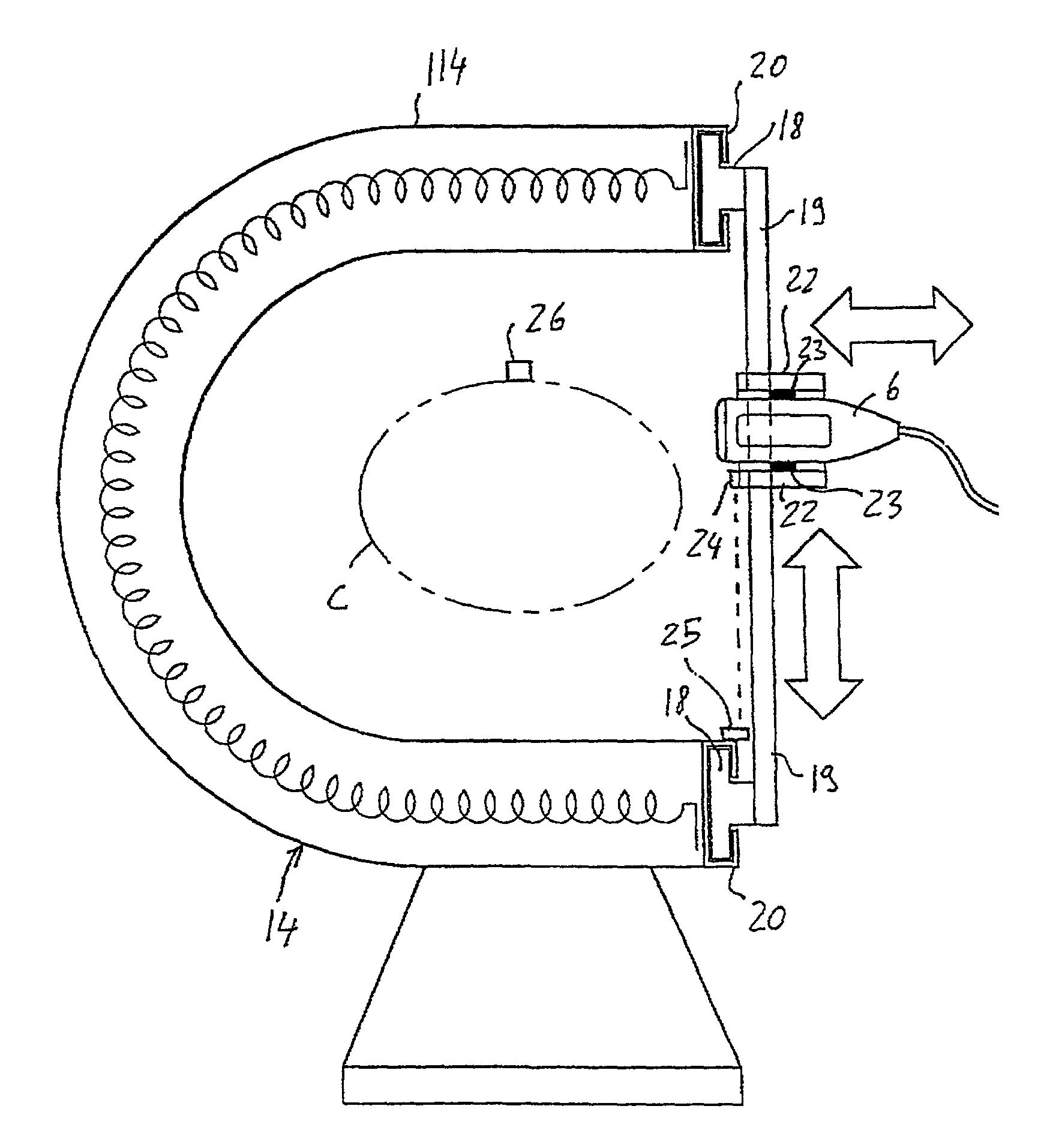
[0075]With reference to FIGS. 1 and 2, an integrated apparatus for ultrasound imaging and Nuclear Magnetic Resonance imaging comprises a Nuclear Magnetic Resonance imaging device and an ultrasound imaging device. The Nuclear Magnetic Resonance imaging device comprises a magnetic structure generally denoted as 1, which includes a permanent, resistive or superconducting magnet for generating a static field in a cavity 101 designed to receive a part of the patient body; coils for transmitting radio-frequency pulses for exciting nuclear spins, gradient coils for applying selecting and phase and frequency encoding gradients and receiving coils. All these elements are known per se and are not shown in detail.

[0076]In FIGS. 1 and 2, the magnetic structure 1 is contained in a separate case and has a peripherally closed annular shape, with two opposite open sides. A patient table 2, in the form of a table / chair is associated to the magnetic structure 1. Particularly, the table is shorter tha...


PUM


Abstract

A combined apparatus for diagnostic imaging an inner part of a body, the apparatus including at least a device for imaging and displaying images according to at least two types of beams, waves or signals transmitted toward a body or a part of the body to be examined, the device including a Nuclear Magnetic Resonance imaging and displaying device and an ultrasound imaging and displaying device in an integrated configuration within the same apparatus.

Description

CROSS-REFERENCE TO RELATED APPLICATIONS[0001]This application claims priority under 35 U.S.C. §§119 to IT SV2001A00020, filed in Italy on Jun. 8, 2001; the entire contents of which are hereby incorporated by reference.BACKGROUND OF THE INVENTION[0002]1. Field of the Invention[0003]The invention relates to a combined apparatus for imaging the internal part of a body, particularly for diagnostic imaging thereof, comprising at least means for imaging and displaying images according to at least one type of beams, waves or signals transmitted by a body or a part thereof to be examined.[0004]2. Description of Related Art[0005]Imaging apparatuses are well-known and include ultrasound imaging apparatuses, Nuclear Magnetic Resonance imaging apparatuses, X-Ray apparatuses, etc. In such type of conventional apparatuses, each apparatus always uses a single type of wave or beam for imaging. From the diagnostic point of view, human body tissues and human body fluid flows provide better or worse d...

Claims


Application Information

Patent Timeline
IPC IPC(8): A61B5/05A61B8/00A61B5/055A61B5/352G01R33/28
CPCA61B5/742G01R33/4814A61B8/00A61B8/5238A61B5/7475A61B5/0456A61B5/0035A61B5/352
Inventor BIGLIERI, EUGENIOPUGLIESE, OSVALDOBESSI, LORENZOSATRAGNO, LUIGISCHIAVINI, LUCIANO
Owner ESAOTE
Features
  • R&D
  • Intellectual Property
  • Life Sciences
  • Materials
  • Tech Scout
  • Unparalleled Data Quality
  • Higher Quality Content
  • 60% Fewer Hallucinations
Learn More