Diagnostic scanning microscope for information-enriched qualitative histopathology
a scanning microscope and information-enriched technology, applied in the field of light optical microscopy, can solve the problems of increasing resolution, corresponding loss of field, and under-utilization of prior-art mechanized procedures by the profession, and achieves the effect of effective continuous linear coverage and highly advantageous increase in imaging speed
- Summary
- Abstract
- Description
- Claims
- Application Information
AI Technical Summary
Benefits of technology
Problems solved by technology
Method used
Image
Examples
example 1
[0066]FIG. 25 shows a histopathologic section of urothelium taken from a region judged to be histologically normal at some distance from a papillary carcinoma lesion in a bladder. Nine nuclei in the image (identified by highlighted boundaries in the figure) were selected as suitable for analysis for their in-focus position. Upon digital analysis, two of these nuclei showed a nuclear signature resembling that of papillary carcinoma, even though visually the section was normal. Utilizing a computer graphic marker in the form of an outlining box, the diagnostic information is revealed directly on the image, thereby providing a visual, quantitative diagnostic clue not otherwise detectable from the image.
example 2
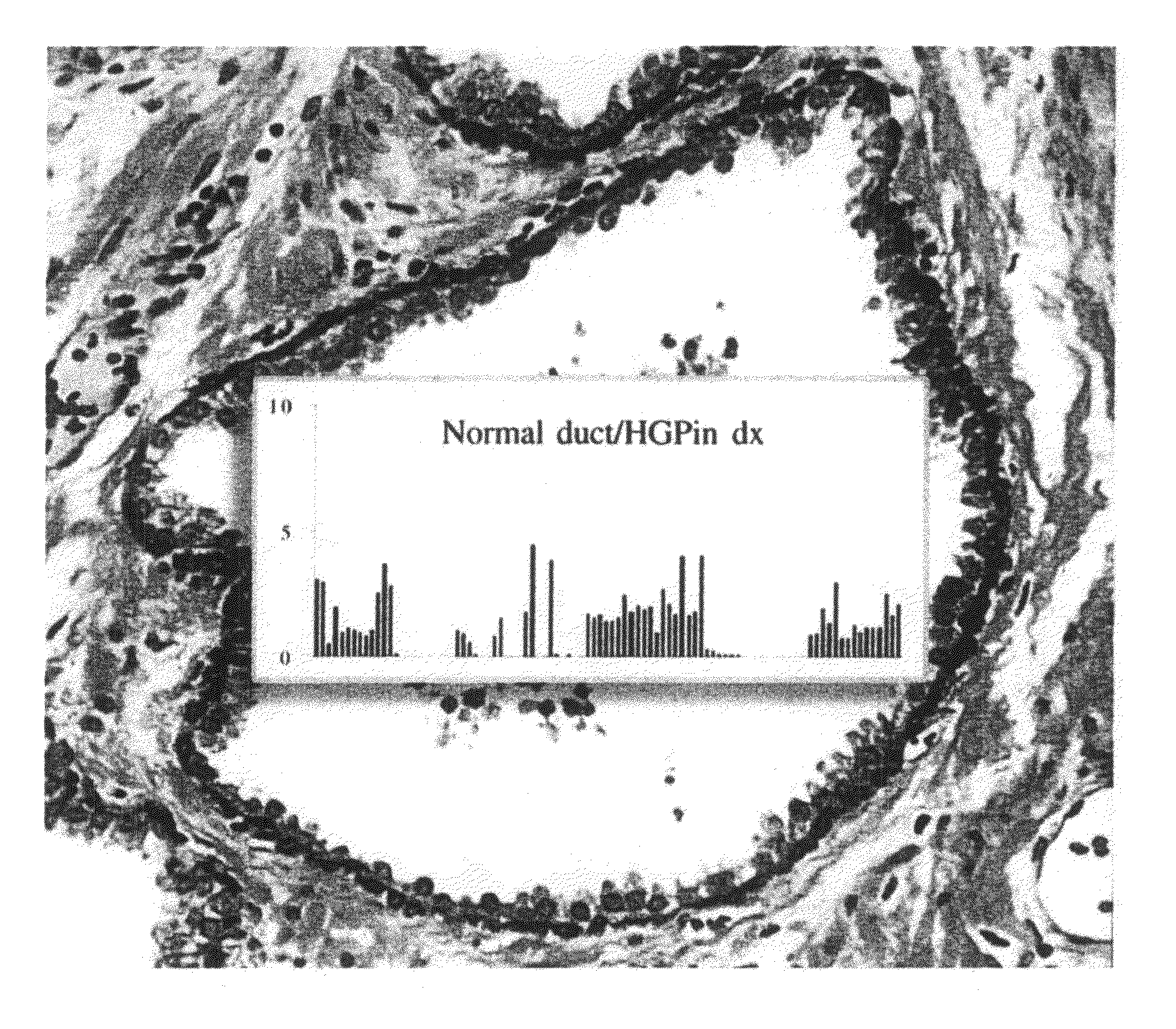
[0067]A tissue sample from a prostate gland duct was sectioned, fixed and stained with hematoxylin / eosin. The resulting slide was scanned with a high numerical aperture microscope objective to produce an image of the sample, as shown in FIG. 26. The image exhibits all criteria for a normal gland, such as a single-layered epithelium and a basal cell layer without gaps. However, a numeric analysis of the chromatin pattern of the nuclei revealed an overall signature known to be typical of a “normal appearing gland” in the vicinity of a prostatic intraepithelial neoplasia (PIN) lesion. FIG. 27 illustrates the signature of the imaged sample (seen in the middle of the figure) in comparison with the signatures of normal tissue (above) and cancerous tissue (below).
[0068]According to one approach, the signature profile of the epithelium is displayed superimposed on the image of the stained sample to reveal the diagnostic clue that is not otherwise visually perceptible, as illustrated in FIG....
PUM
 Abstract
Description
Claims
Application Information
 - R&D
- Intellectual Property
- Life Sciences
- Materials
- Tech Scout
- Unparalleled Data Quality
- Higher Quality Content
- 60% Fewer Hallucinations
Browse by: Latest US Patents, China's latest patents, Technical Efficacy Thesaurus, Application Domain, Technology Topic, Popular Technical Reports.
© 2025 PatSnap. All rights reserved.Legal|Privacy policy|Modern Slavery Act Transparency Statement|Sitemap|About US| Contact US: help@patsnap.com


