Method for post-processing a three-dimensional image data set of vessel structure

a three-dimensional image and data set technology, applied in image enhancement, instruments, angiography, etc., can solve the problems of large loss of local resolution, difficult segmentation correctly, and low local resolution of three-dimensional image data sets generated with currently used imaging modalities

Inactive Publication Date: 2011-03-08
SIEMENS HEALTHCARE GMBH
View PDF7 Cites 20 Cited by
  • Summary
  • Abstract
  • Description
  • Claims
  • Application Information

AI Technical Summary

Benefits of technology

[0018]The registration can either be achieved by the so-called 2D-3D registration method in which for example different projections are calculated from the 3D image data set and compared to the 2D DSA. In the special case in which the 3D image data set was obtained with the same angiography systems as the 2D DSA, the registration can be determined more easily from the known system geometry.
[0020]Optionally the 3D image data set can be scaled up before step (f) to a higher local resolution, so that the more precise location information of the 2D DSA is not lost in the unsharp pixels of the 3D image data set.
[0025]If the 3D position of the differing vessel section in the 3D image data set is then known the segmentation threshold values for the 3D image data set can be changed so that the differences are reduced. In this case the segment boundaries between the vessel section and the background are shifted through pixel-by-pixel changing of a segmentation threshold value of the 3D image data set. This means that the segmentation parameters of the 3D image data set are adapted locally (not globally) to guarantee an optimum 3D display. Optionally the voxel-by-voxel segmentation threshold values can be smoothed to avoid abrupt transitions.

Problems solved by technology

The three-dimensional image data sets generated with currently used imaging modalities do not however possess any high local resolution.
An x-ray image does in principle have a high local resolution; however a large part of local resolution is lost through the reconstruction of many x-ray images into one three-dimensional image data set, e.g. in CT or in 3D rotation angiography.
In the area of the neck of an aneurysm in particular it is very difficult to perform a segmentation correctly.
If the segmentation is thus optimized to a specific area (e.g. the aneurysm neck), under some circumstances this leads to a less than optimum segmentation of other areas (e.g. feeding vessels, middle of the aneurysm).

Method used


Image

  • Method for post-processing a three-dimensional image data set of vessel structure
  • Method for post-processing a three-dimensional image data set of vessel structure
  • Method for post-processing a three-dimensional image data set of vessel structure

Examples


Embodiment Construction

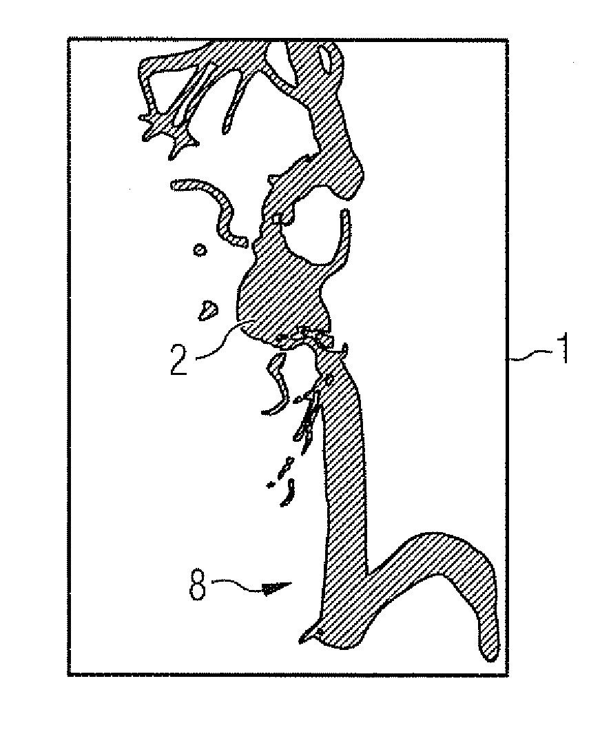
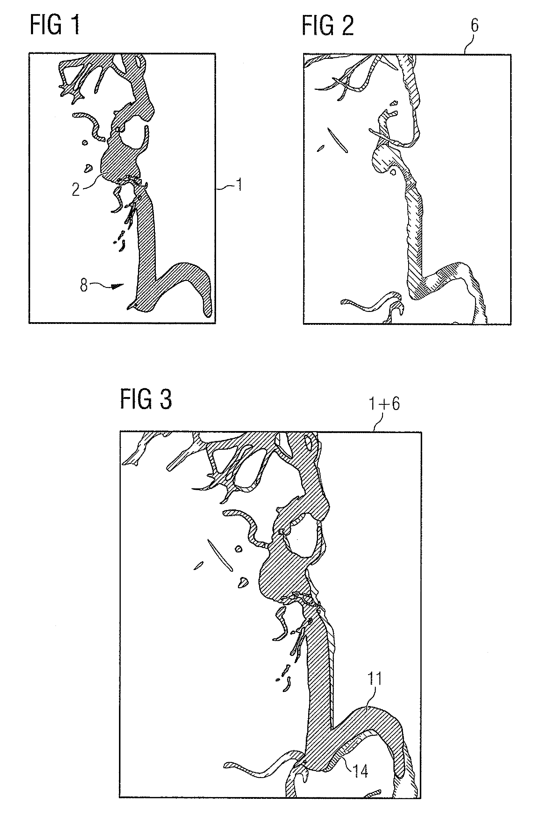
[0036]FIG. 1 shows a typical 2D DSA 1 of a vessel structure 2 with an aneurysm 2a. As one might suspect, the 2D DSA has outstanding local resolution but there is an absence of any depth information.

[0037]FIG. 2 on the other hand shows a three-dimensional display 3 of the same vessel tree which has been obtained by a 3D rotation pass with a C-arm x-ray device. The 3D image data set has been segmented with a global segmentation threshold value and only the values lying above the threshold value are shown. A so-called “volume rendering” display has been selected in which the vessel structure in the 3D volume has been provided with computed shadow and light effects in order to create a visual three-dimensional impression.

[0038]It is precisely with 3D image data sets that the selected global segmentation threshold value is very important, since an aneurysm in particular is displayed markedly differently with different threshold values.

[0039]In accordance with the invention, as in FIG. 3,...


PUM


Abstract

The invention relates to a method for post-processing a 3D image data set of a vessel structure of a human or animal body, in which a 2D DSA (Digital Subtraction Angiography) of the vessel structure is recorded and registered with the 3D image data set. The 2D DSA is compared with a corresponding projection image computed from the 3D data set and this is changed, e.g. by changing the segmentation parameters, to adapt it to the 2D DSA. This enables the outstanding local resolution of the 2D DSA to be used for improving the 3D image data set.

Description

CROSS REFERENCE TO RELATED APPLICATIONS[0001]This application claims priority of German application No. 10 2006 045 423.5 filed Sep. 26, 2006, which is incorporated by reference herein in its entirety.FIELD OF THE INVENTION[0002]The invention relates to a method for post-processing a three-dimensional (3D) image data set of a vessel structure of the body of a human or an animal, with the three-dimensional image data set having been recorded with a medical imaging modality.BACKGROUND OF THE INVENTION[0003]To show blood vessels there are currently recording options available with which three-dimensional image data records can be created. Imaging modalities can be used for this purpose, e.g. computer tomography (CT), magnetic resonance tomography (MR) or 3D rotation angiography.[0004]Important applications in such case are the diagnosis of vessel diseases such as aneurysms and stenoses and the planning of therapies. The planning is a matter of predicting the probability of a rupture of...

Claims


Application Information

Patent Timeline
Patent Type & Authority Patents(United States)
IPC IPC(8): G06K9/00
CPCA61B5/02007A61B5/055A61B6/032A61B6/481A61B6/504A61B19/52G06T5/50G06T7/0024A61B2017/00778A61B2019/5238A61B2019/5289G06T2207/10072G06T2207/10116G06T2207/20148G06T2207/30101Y10S378/901A61B90/36A61B2090/364A61B2090/376G06T7/30G06T7/136
Inventor PFISTER, MARCUSREDEL, THOMAS
Owner SIEMENS HEALTHCARE GMBH
Why Eureka
Try Eureka