Method of automatic extraction of the pulmonary artery tree from 3D medical images

a 3d medical image and automatic extraction technology, applied in the field of medical imaging, can solve the problems of difficult to distinguish pulmonary arteries from veins, inconvenient clinical practice, and inconvenient us

Inactive Publication Date: 2011-06-28
KONINK PHILIPS ELECTRONICS NV
View PDF6 Cites 19 Cited by
  • Summary
  • Abstract
  • Description
  • Claims
  • Application Information

AI Technical Summary

Benefits of technology

[0013]The present invention has the advantage over the prior art that it provides advantageously suppression of pulmonary veins from the pulmonary vessel tree, in applications and visualizations modes that merely concern the pulmonary artery tree.

Problems solved by technology

When extracting the pulmonary vessel tree from Multi-Slice CT data, difficulties arise when distinguishing pulmonary arteries from veins.
Hence, these methods are not well suited for clinical practice.
Hence, this method is based on a mental act of the individual observer an consequently it is prone to errors.

Method used


Image

  • Method of automatic extraction of the pulmonary artery tree from 3D medical images
  • Method of automatic extraction of the pulmonary artery tree from 3D medical images
  • Method of automatic extraction of the pulmonary artery tree from 3D medical images

Examples


Embodiment Construction

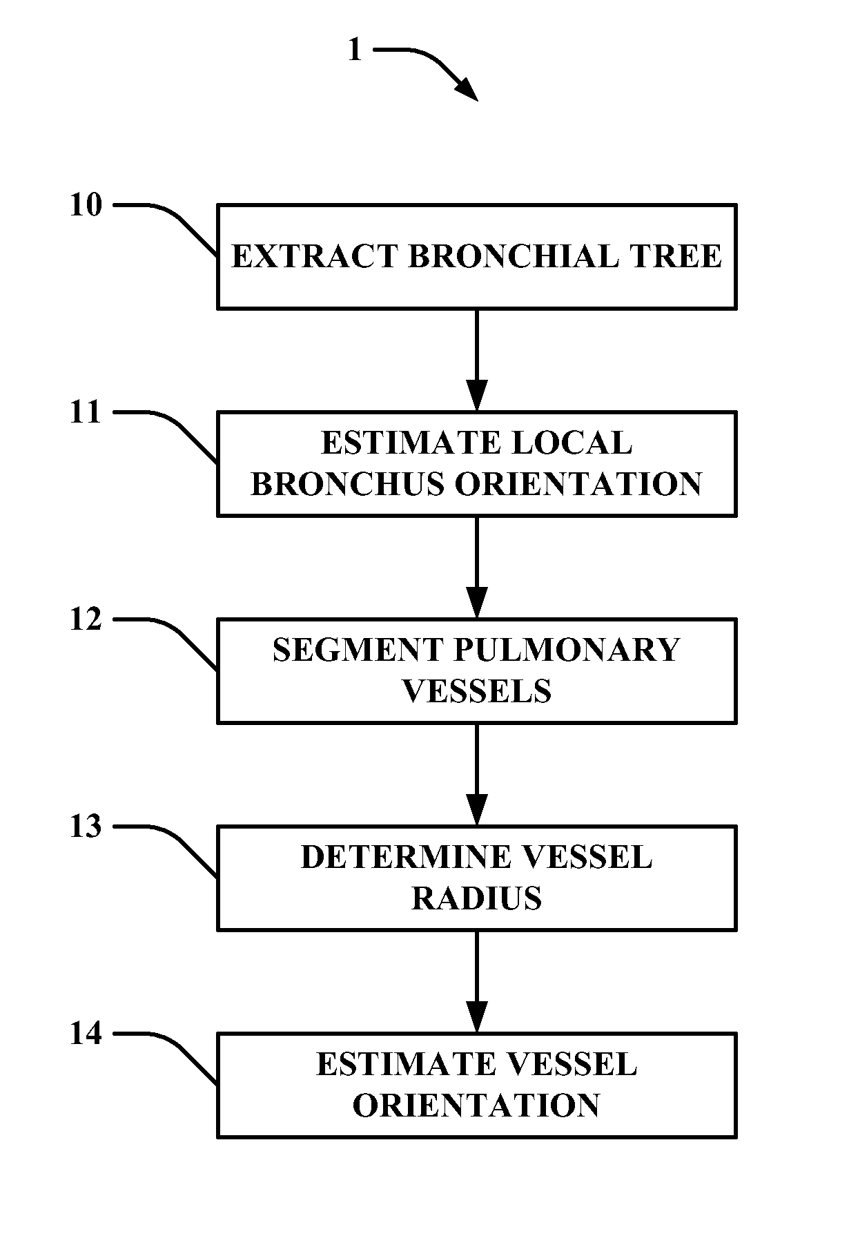
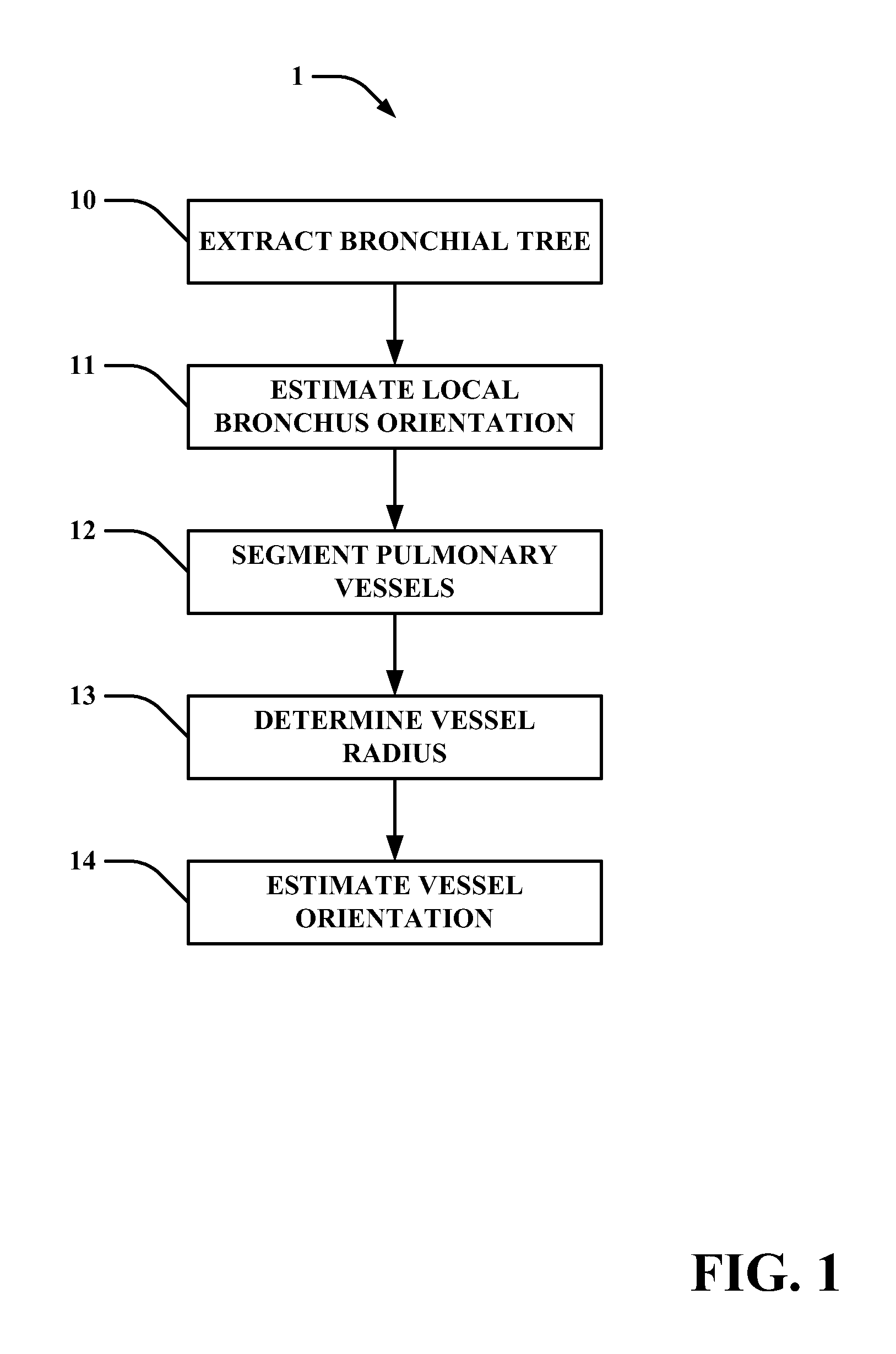
[0029]The following description focuses on an embodiment of the present invention applicable to a Computer Tomography (CT) system and in particular to Multi-Slice CT Data. However, it will be appreciated that the invention is not limited to this application but may be applied to many other imaging systems, including for example Magnetic Resonance Imaging (MRI) systems, 3D Ultrasonic (3D-US) systems, etc.

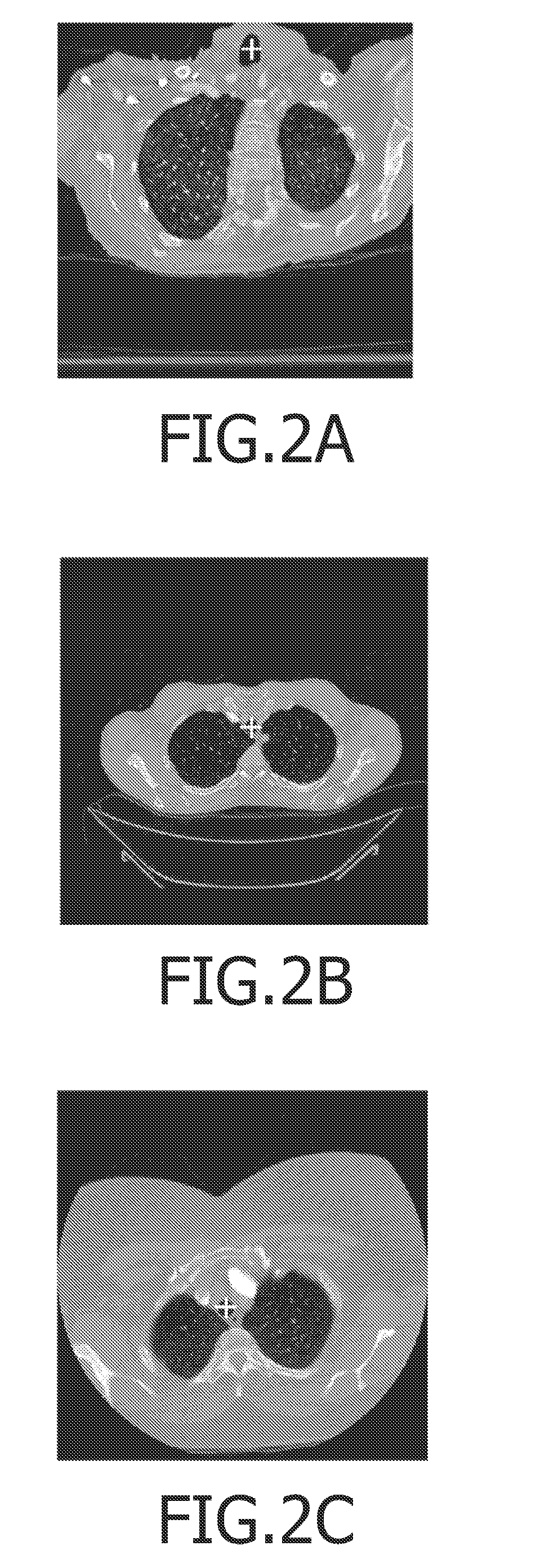
[0030]Below, an automated method for the extraction of the pulmonary artery tree from multi-slice CT data, making use of the tracheobronchial tree, is described. Having the bronchial tree and the accompanying arterial tree available these trees may be used for a joint visualization, e.g., in a virtual bronchoscopy application. Bronchial and arterial diameters are measured fully automatically along both trees. Positions where the ratio of these radii exhibits unusual values may be marked in a display and suggested for further assessment by the radiologist (see e.g. white dots / markings...


PUM


Abstract

An automated method (1) for the automatic extraction of a pulmonary vessel tree from a 3D medical image, such as multi-slice CT data, is disclosed. A segmented pulmonary vessel is identified as either an artery or a vein by determining a measure for arterialness for the vessel. The measure is based on a relation of the orientation of a local bronchus to the orientation of the segmented pulmonary vessel of the local bronchus. When a vessel is identified as a pulmonary artery, it is added to the pulmonary artery tree. Radii of the pulmonary artery and bronchus are measured automatically and positions where a ratio of these radii exhibits unusual values are presented in a display, preferably for suggesting further assessment by a radiologist, which for instance is useful for pulmonary embolism detection.

Description

FIELD OF THE INVENTION[0001]This invention pertains in general to the field of medical imaging. More particularly the invention relates to a method of automatic extraction of the pulmonary artery tree from 3D medical images, for instance for pulmonary embolism detection.BACKGROUND OF THE INVENTION[0002]Multi-detector row computed tomography (MDCT) allows the acquisition of high-resolution data of the entire thorax. The quality of the data allows the assessment of the bronchi as well as the pulmonary artery tree. Extraction of the pulmonary artery tree is an important pre-processing step, e.g., for embolism detection. Also, in the quantitative assessment of the bronchial tree the accompanying artery tree contains important additional information that can be extracted from MDCT data. Hence, for diagnosis and treatment of asthmatic and emphysematic patients, the simultaneous assessment of the tracheobronchial tree and the accompanying pulmonary artery tree is crucial. E.g., the ratio o...

Claims


Application Information

Patent Timeline
Patent Type & Authority Patents(United States)
IPC IPC(8): G06K9/00
CPCG06T7/0081G06T7/0089G06T7/0091G06T2200/04G06T2207/30101G06T2207/20044G06T2207/20156G06T2207/30061G06T2207/10081G06T7/11G06T7/149G06T7/155
Inventor BUELOW, THOMASWIEMKER, RAFAELBLAFFERT, THOMASLORENZ, CRISTIANRENISCH, STEFFEN
Owner KONINK PHILIPS ELECTRONICS NV
Why Eureka
Try Eureka