System and method for quality assurance in pathology

a pathology and quality assurance technology, applied in the field of pathology and microscopy, can solve the problems of inaccuracy of interpretation, lack of standardization of microscopes, compromising spatial details and color fidelity, etc., and achieve the effect of improving diagnosis and improving quality assurance in pathology

Active Publication Date: 2012-04-24
LEICA BIOSYST IMAGING
View PDF17 Cites 8 Cited by
  • Summary
  • Abstract
  • Description
  • Claims
  • Application Information

AI Technical Summary

Benefits of technology

[0011]Accordingly, described herein are systems and methods for improving quality assurance in pathology using digital slide tools for assessing slide quality to trigger (i) preparation of new glass slides, (ii) re-scanning of glass slides, or (iii) enhancement of digital slides prior to interpretation by the pathologist. Digital slides created by slide scanning instruments contribute to improved diagnosis by pathologists by providing an automatic, systematic, objective, and consistent means for digital slide creation and analysis, enabling the use of computer implemented image analysis and image correction tools.
[0012]A digital pathology system (slide scanning instrument and software) assesses and improves the quality of a digital slide. The improved digital slide has a higher image quality that results in increased efficiency in the interpretation of such digital slides when they are viewed on a monitor by a pathologist. These improved digital slides yield a more objective diagnosis than reading the corresponding glass slide under a microscope.

Problems solved by technology

While use of tissue processing instruments and automatic staining equipment has increased the quality and consistency of slide preparations, a significant portion of glass slides that are read by pathologists are still suboptimal and may contribute to inaccurate interpretations.
One of the possible contributing factors to the subjectivity of reading pathology slides is the lack of standardization of the microscope, a tool that has been in use for hundreds of years and which is not recognized as an approved medical device.
Many pathologists' microscopes are not maintained in optimal working condition or furnished with optimal objective lenses, thus compromising spatial details and color fidelity that may be essential to making more accurate diagnoses.
This conventional process suffers from the inability of the histotechnologist to carefully review every area of a glass slide (some labs process hundreds or thousands of slides every day).
The conventional process additionally suffers from the increasing shortage of qualified histotechnologists, and pressure on laboratories to continually improve productivity.
Furthermore, the overall aging of the population and the associated increased incidence of cancer (and surgical biopsies) only exacerbate the challenges of quality-assuring glass slides before they are read by a pathologist due to the significant increase in the number of glass slides that are prepared.

Method used


Image

  • System and method for quality assurance in pathology
  • System and method for quality assurance in pathology
  • System and method for quality assurance in pathology

Examples


Embodiment Construction

[0021]Certain embodiments as disclosed herein provide for systems and methods for quality assurance in pathology. After reading this description it will become apparent to one skilled in the art how to implement the invention in various alternative embodiments and alternative applications. However, although various embodiments of the present invention will be described herein, it is understood that these embodiments are presented by way of example only, and not limitation. As such, this detailed description of various alternative embodiments should not be construed to limit the scope or breadth of the present invention as set forth in the appended claims.

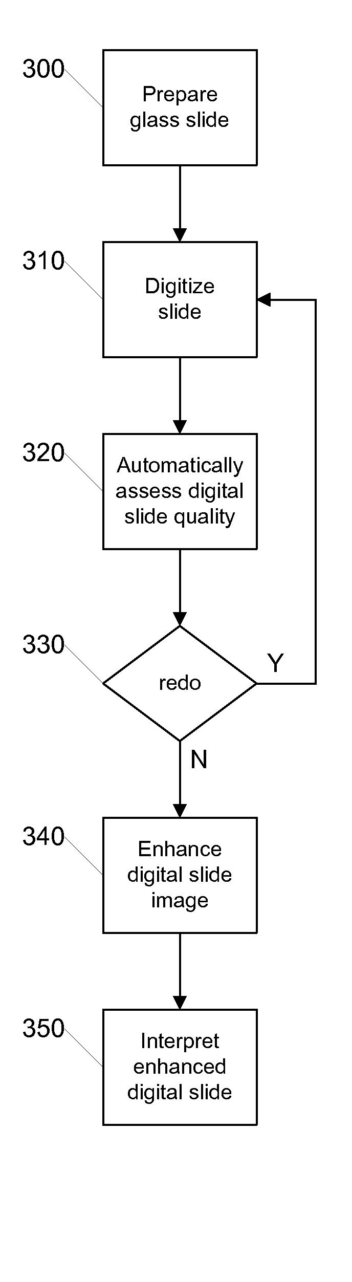
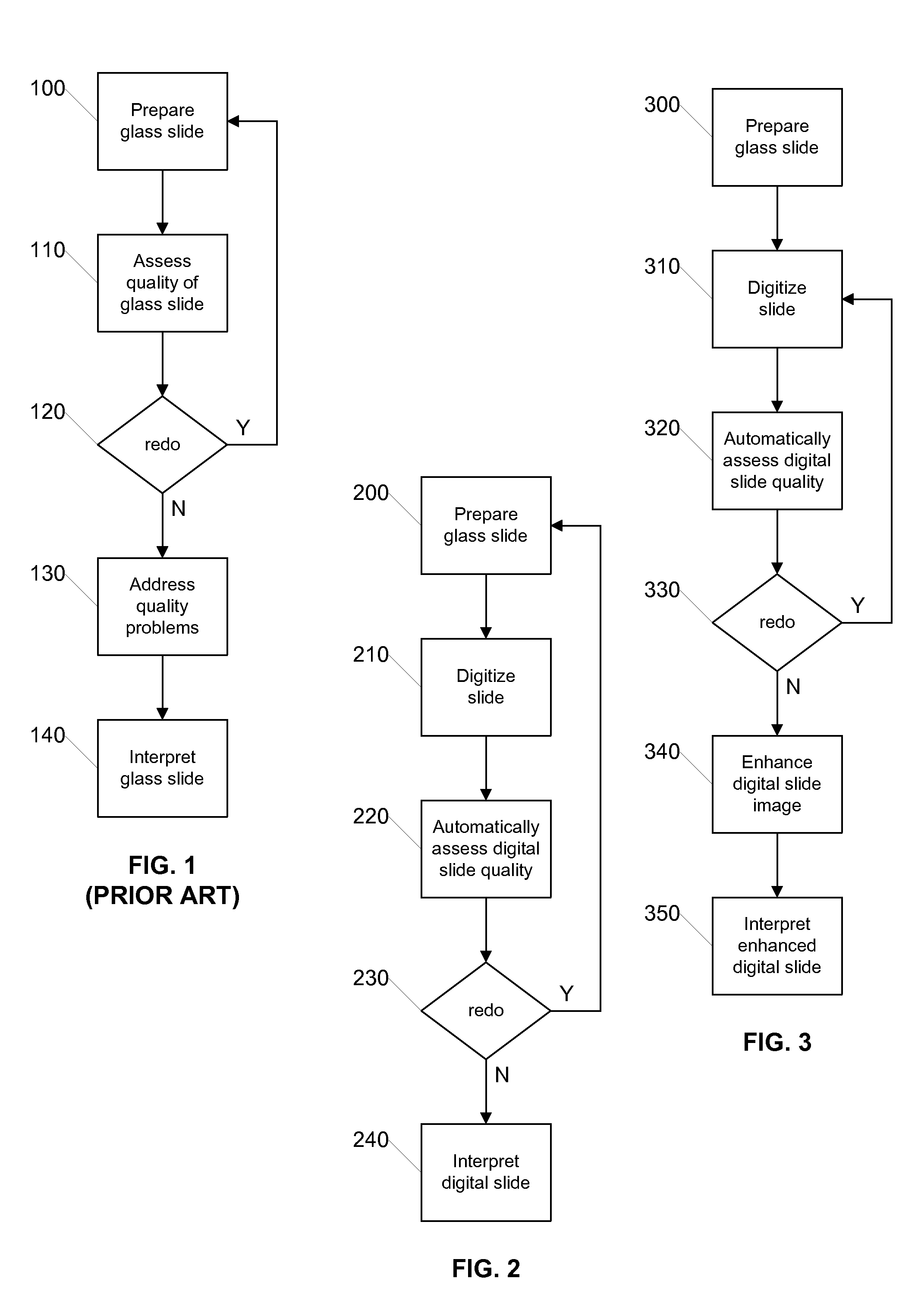
[0022]FIG. 2 is a flow diagram illustrating a process for quality assurance using digital slides according to an embodiment of the invention. The illustrated process can be carried out by a digital pathology system such as that later described with respect to FIG. 5. Initially, in step 200 the glass slide is prepared in the conventi...


PUM


Abstract

Systems and methods for improving quality assurance in pathology using automated quality assessment and digital image enhancements on digital slides prior to analysis by the pathologist are provided. A digital pathology system (slide scanning instrument and software) creates, assesses and improves the quality of a digital slide. The improved digital slide image has a higher image quality that results in increased efficiency and accuracy in the analysis and diagnosis of such digital slides when they are reviewed on a monitor by a pathologist. These improved digital slides yield a more objective diagnosis than reading the corresponding glass slide under a microscope.

Description

RELATED APPLICATION[0001]The present application claims priority to U.S. provisional patent application Ser. No. 60 / 916,252 filed May 4, 2007, which is incorporated herein by reference in its entirety.BACKGROUND[0002]1. Field of the Invention[0003]The present invention generally relates to pathology and microscopy and more particularly relates to improvements in quality assurance for pathology using digital microscopy.[0004]2. Related Art[0005]The diagnosis of glass microscope slides by a pathologist is known to be subjective. Many factors contribute to this lack of objectivity, including the training and skill of the pathologist and the quality of the glass slides from which the diagnosis was made. While use of tissue processing instruments and automatic staining equipment has increased the quality and consistency of slide preparations, a significant portion of glass slides that are read by pathologists are still suboptimal and may contribute to inaccurate interpretations.[0006]One...

Claims


Application Information

Patent Timeline
Patent Type & Authority Patents(United States)
IPC IPC(8): G06K9/00
CPCG02B21/365G06F19/30G06K9/00134G06K9/033G06T5/001G06T7/0002G06K9/00127G06Q50/22G06T2207/10056G06T2207/30024G16H30/20G16H40/20G06V20/693G06V10/987G06V20/69G06T7/0012G06T2207/10024
Inventor SOENKSEN, DIRKZIRKER-SMITH, KATHY
Owner LEICA BIOSYST IMAGING
Why Eureka
Try Eureka