
Joint placement methods and apparatuses

a joint and placement technology, applied in the field of joint placement, can solve the problems of limited range of motion, post-surgical pain, dislocation, etc., and achieve the effects of improving the alignment of modifying the range of motion, and improving the bony coverage to the cup

Inactive Publication Date: 2012-05-15
IGO TECH
View PDF6 Cites 45 Cited by
  • Summary
  • Abstract
  • Description
  • Claims
  • Application Information

AI Technical Summary

Benefits of technology

"The invention provides a method and apparatus for accurately placing prosthetic components in a joint. The method involves determining the range of motion of the joint and planning the placement of the components based on that range of motion and the stability of the components. The apparatus includes a primary cylinder for placement over an exposed neck of a prosthetic femoral stem and a first alignment receptacle for receiving a spatially tracked probe. The invention helps to improve the stability and range of motion of the joint and optimize the placement of the components."

Problems solved by technology

Intervention to correct problems of a joint suffers from difficulty in finding an optimal implant placement including position and orientation.
Problems arising from incorrect placement may involve, among other symptoms, post surgical pain, limited range of motion, dislocation, post surgical fracture, premature implant failure, and adverse biologic response consequent to any of the preceding problems.
These points of impingement cause a lever effect and potentially result in dislocation.
Besides being painful and stressful for the patient, it also leads to abductor tissue damage, which has a detrimental long-term effect on the stability of the hip joint.
The guides depend on precise, but difficult, placement within anatomy.
They typically do not allow for soft tissue effects.
The guides provide limited flexibility for anatomical variability.
They require pre-surgical scanning of the patient consuming more time, increasing cost and exposing the patient to additional radiation.
Planning based on scans and reconstruction of the bony anatomy fail to account for the important and significant contribution of soft tissue to the function of the joint.
This adds time and has potential error associated with it.
Complex, time consuming and potentially error-prone registration of the patient to the scan must be performed intra-operatively.
Such planning is scheduled separately from the surgery and consumes more time and thus increases cost.
Pre-operative imaging does not allow for intra-operative updating.
The prescribed images can be difficult to obtain (e.g., lateral image of the hip).
The hardware necessary to do this increases cost.
The additional setup can be complicated and time-consuming.
The image processing requires use of software algorithms that must be written, maintained and presents another opportunity for introducing error.
Picking 3D anatomical landmarks from 2D images, particularly on complex anatomy and / or lower quality images is difficult and error-prone.
It can also have an increased risk of infection from percutaneous palpations outside the immediate joint replacement site.
Painting is time consuming, which increases the procedure time.
There is difficulty in maintaining probe-to-anatomy contact.
Standardized orientations are derived by using a sample population to determine what orientations result in the fewest complications and failures.
Not being patient specific they are not necessarily ideal, or even correct, for an individual.
Whether point picking in images or palpating anatomy, the process for providing the inputs to calculate the anatomical properties is often difficult and error-prone.
The anatomical properties are based on bony anatomy and fail to account for the critical contribution of soft tissue to the behaviour of joints.

Method used


Image

  • Joint placement methods and apparatuses
  • Joint placement methods and apparatuses
  • Joint placement methods and apparatuses

Examples


Embodiment Construction

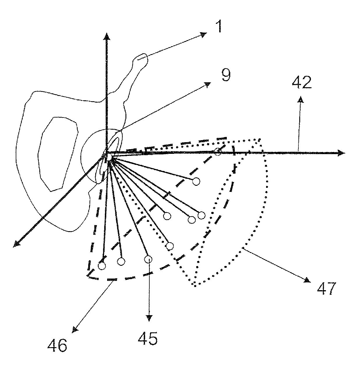
[0060]Referring to FIG. 3, a system has a central processing unit(s) 16, one or more output devices 14, and navigator technology 17. The navigator technology 17 may be passive localization or active positioning technologies. The system also has input devices 15 for example, keyboard, mouse, foot pedal, button probes, or virtual keyboards, specialized surgical instruments 18, and devices for localizing anatomies 19 using navigator technology 17. The central processing unit(s) 16 has the computational power to process localization data, perform complex mathematical calculations and, optionally, perform image processing. The central processing unit(s) 16 also has associated modules to carry out the various features and functions described herein, examples of such modules will be described later herein. The modules may be software in memory accessible to the central processing unit(s) 16. There is a means for communication between the central processing unit(s) 16, the other apparatus c...


PUM


Abstract

Systems and methods for determining placement of prosthetic components in joint including defining patient-specific frame of reference for joint, determining patient-specific postoperative range of motion of joint, evaluating patient-specific range of motion of joint, automatically planning placement of components balancing need for range of motion with prosthesis stability through bony coverage, and applying manual adjustments to the automatically planned placement of component by giving greater or lesser weight to need for range of motion or prosthesis stability through bony coverage. Apparatuses for defining center of prosthetic femoral head and axis of prosthetic femoral neck including primary cylinder, first alignment receptacle and second alignment receptacle, and a divot on exterior of primary cylinder, divot having normal parallel to longitudinal axis of second alignment receptacle and position of the divot being translated toward an opening of the first alignment receptacle on the primary cylinder. Methods for using apparatuses. Apparatus for mounting spatially tracked device to impactor for impacting prosthetic cup into reamed socket.

Description

CROSS-REFERENCE TO RELATED APPLICATIONS[0001]This application is a continuation of, and claims the benefit of, International Patent Application No. PCT / CA2007 / 001082, filed on Jun. 19, 2007, which claims the benefit of U.S. Provisional Patent Application Ser. No. 60 / 814,547, filed on Jun. 19, 2006. The entire disclosure of U.S. Provisional Patent Application Ser. No. 60 / 814,547 is hereby incorporated by reference.TECHNICAL FIELD[0002]The invention relates to joint placement generally, more specifically to placement of prosthetics for joint repair.BACKGROUND ART[0003]Joint repair, or arthroplasty, is a surgical intervention for repairing defects or injuries of a joint. The goal is to restore as much normal function as possible while at the same time reducing or eliminating pain and discomfort. Joint repairs allow patients to regain mobility, continue contributing to society and enjoy a greater quality of life.[0004]Arthroplasty attempts to correct how two bones interact with one anot...

Claims


Application Information

Patent Timeline
Patent Type & Authority Patents(United States)
IPC IPC(8): A61F2/32
CPCA61B19/52A61B19/5244A61F2/4657A61B19/50A61B2019/505A61B2019/5255A61F2/32A61F2/34A61F2/36A61F2002/3611A61F2002/3625A61F2002/4632A61B34/10A61B34/20A61B90/36A61B2034/105A61B2034/2055
Inventor RUDAN, JOHNELLIS, RANDY E.KUNZ, MANUELA
Owner IGO TECH
Learn More