Diagnostic panel of cancer antibodies and methods for use
a cancer antibody and diagnostic panel technology, applied in the field of epithelial cancer antibody panel, can solve the problems of poor overall compliance of patients undergoing procedures, and achieve the effect of determining the effectiveness of cancer therapy
- Summary
- Abstract
- Description
- Claims
- Application Information
AI Technical Summary
Benefits of technology
Problems solved by technology
Method used
Image
Examples
example 1
Antibody Immunity to a Panel of Oncogenic Proteins Predicts Presence of Colorectal Cancer and Stage of Disease
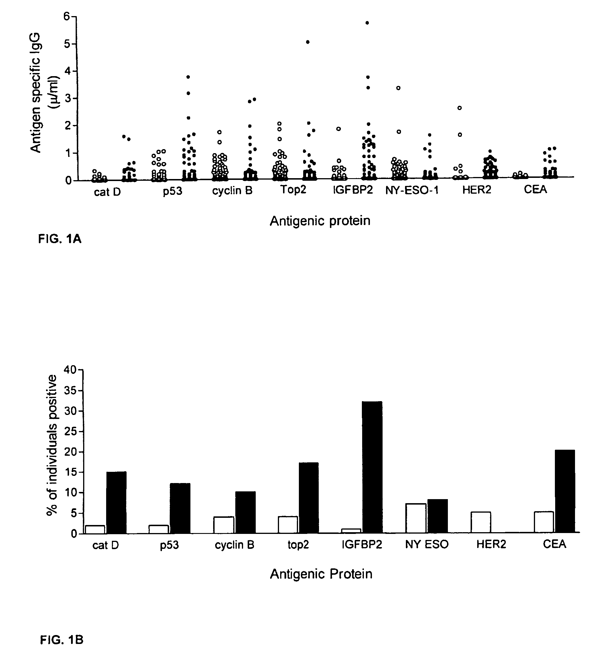
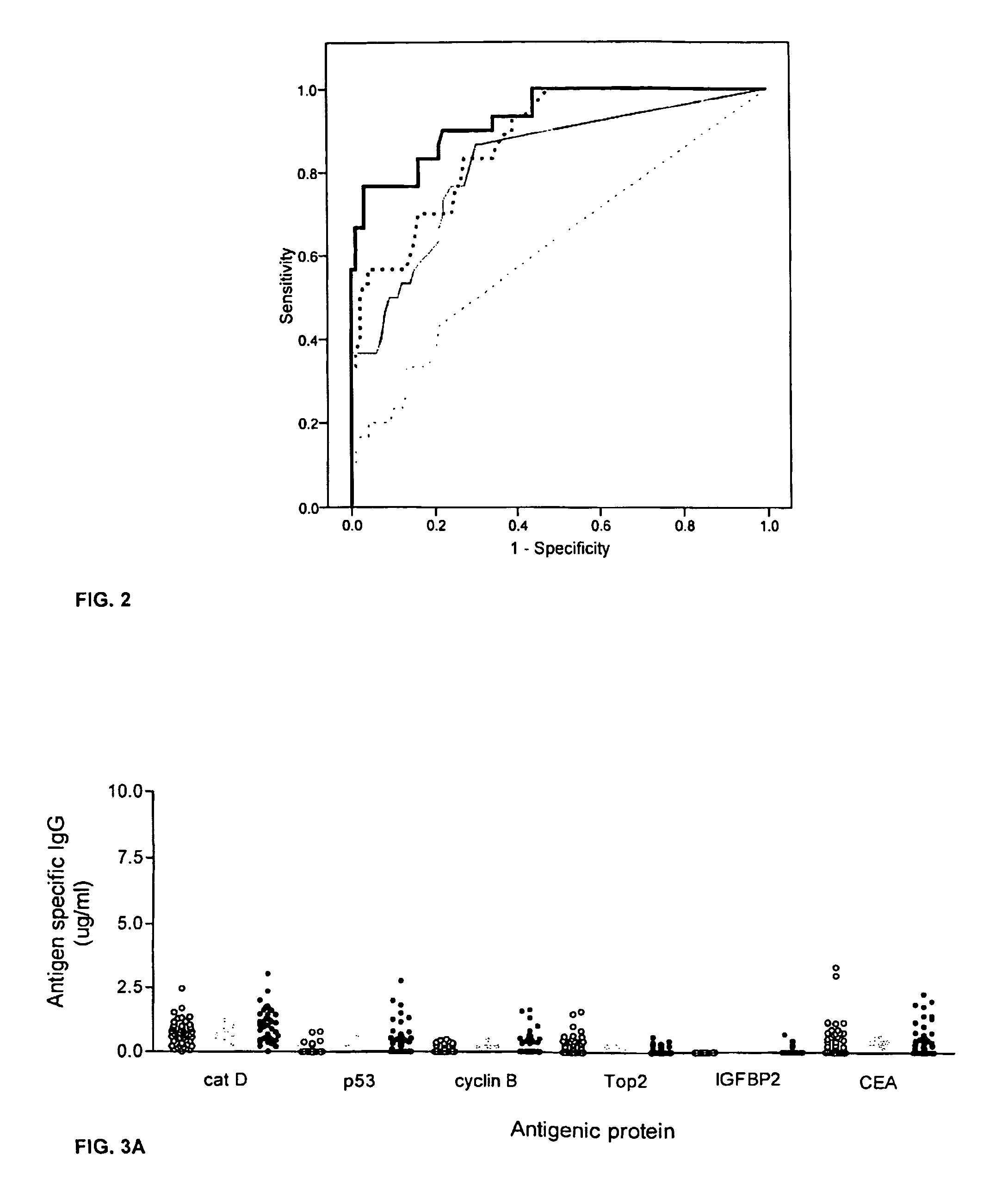
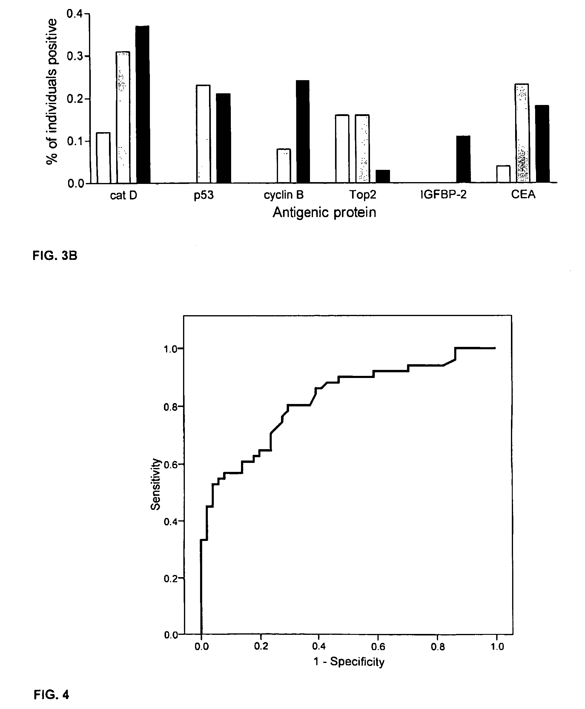
[0059]This example describes an assay of sera from patients with colorectal cancer and healthy controls that can identify antigens which indicate presence of disease. Antigens thus identified can be incorporated into a colorectal cancer specific screening panel. The example demonstrates that a panel of antigens identified by an exploratory sample set could be validated in an independent, blinded sample set for early detection of colorectal cancer.
[0060]The following abbreviations are used herein: HER2: HER-2 / neu; Cat D: cathepsin D; Top2: topoisomerase II α; CEA: carcinoembryonic antigen; ELISA: enzyme-linked immunosorbent assay; FCS: fetal calf serum; HRP: horseradish peroxidase; OD: optical density.
[0061]A preliminary set of 30 serum samples from colorectal cancer patients with late stage disease and 100 samples from normal donors were analyzed to compare antibody response...
example 2
Humoral Immunity Directed against Tumor-Associated Antigens as Biomarkers for the Early Diagnosis of Cancer
[0081]Many solid tumors are potentially curable if diagnosed at an early stage when the cancer can be completely surgically removed. Novel methods to aid in early diagnosis of cancer are sorely needed. A case in point is the need for early diagnosis in patients with breast cancer. Breast cancer is the most commonly diagnosed cancer in women.(1) Despite the availability of routine screening with mammography, about 40% of breast cancers, when first diagnosed, are not localized.(1) The development of new biomarkers that may help in the early detection of breast cancer will greatly facilitate the clinical management of the disease. Early detection by novel methods is critically important in younger premenopausal women whose mammograms may be compromised by increased breast density. The development of a serum-based assay that could indicate cancer exposure would be of great benefit....
example 3
Additional Data on using the Antibody Panel for Breast Cancer Diagnosis
[0170]Serum antibody responses to 8 tumor antigens were evaluated in 98 breast cancer patients (Tumor Vaccine Group, Seattle, Wash.; HER2 positive by FISH or IHC) and 98 age- and gender-matched volunteer control donors (Puget Sound Blood Bank, Seattle, Wash.). The female subjects ranged in age from 34-76 (cancer group) and 24-76 (control group), and each group had an average age of 52. The cancer patients had gone through surgery and chemotherapy and have stable disease. Samples were collected at their initial visit and prior to any vaccination treatment.
[0171]Tumor cell-based ELISA was used to measure antibody to HER2 / neu as described previously. In brief, the SKBR3 cell line was used as a source for HER2 / neu protein. 96-well microtiter plates were coated with 520C9, a monoclonal antibody specific to HER2 / neu before SKBR3 cell lysate (20 μg / ml) was added to the wells. Plates were washed again and serially dilute...
PUM
| Property | Measurement | Unit |
|---|---|---|
| equilibrium constant | aaaaa | aaaaa |
| survival time | aaaaa | aaaaa |
| concentration | aaaaa | aaaaa |
Abstract
Description
Claims
Application Information
 - R&D
- Intellectual Property
- Life Sciences
- Materials
- Tech Scout
- Unparalleled Data Quality
- Higher Quality Content
- 60% Fewer Hallucinations
Browse by: Latest US Patents, China's latest patents, Technical Efficacy Thesaurus, Application Domain, Technology Topic, Popular Technical Reports.
© 2025 PatSnap. All rights reserved.Legal|Privacy policy|Modern Slavery Act Transparency Statement|Sitemap|About US| Contact US: help@patsnap.com


