Mouse model for eye disease
a mouse model and eye disease technology, applied in the field of nonhuman animal eye disease model, can solve the problems of reduced visual acuity, loss of vision, retina impairment by elevated intraocular pressure,
- Summary
- Abstract
- Description
- Claims
- Application Information
AI Technical Summary
Benefits of technology
Problems solved by technology
Method used
Image
Examples
example 1
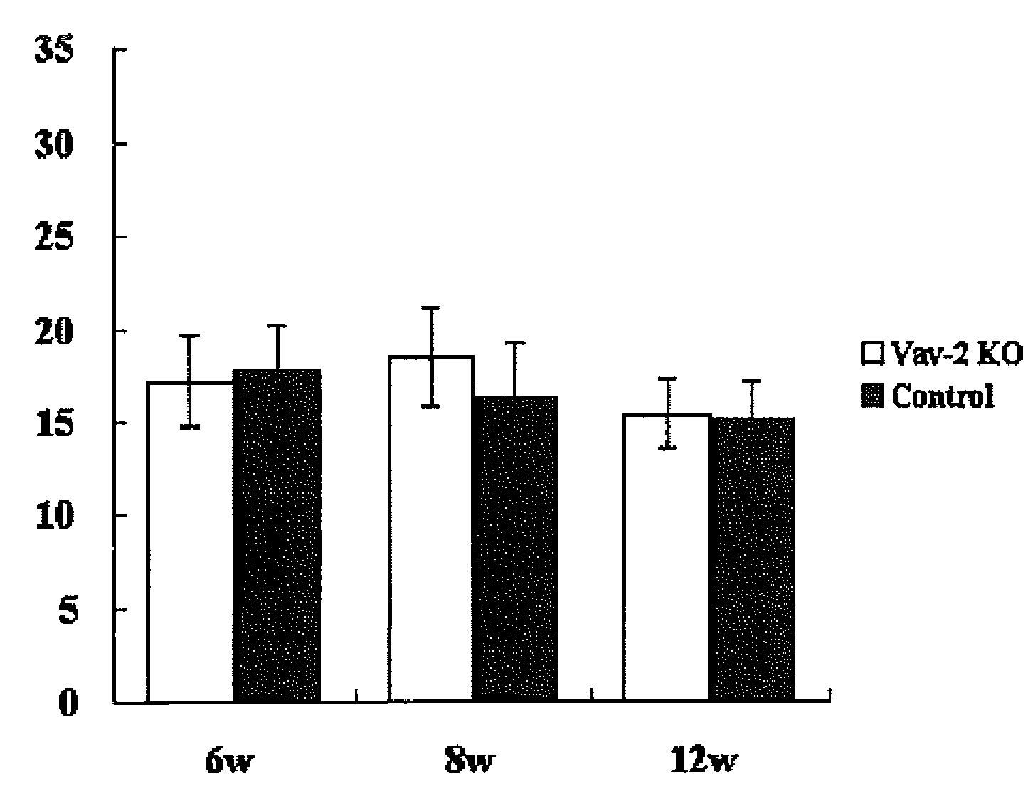
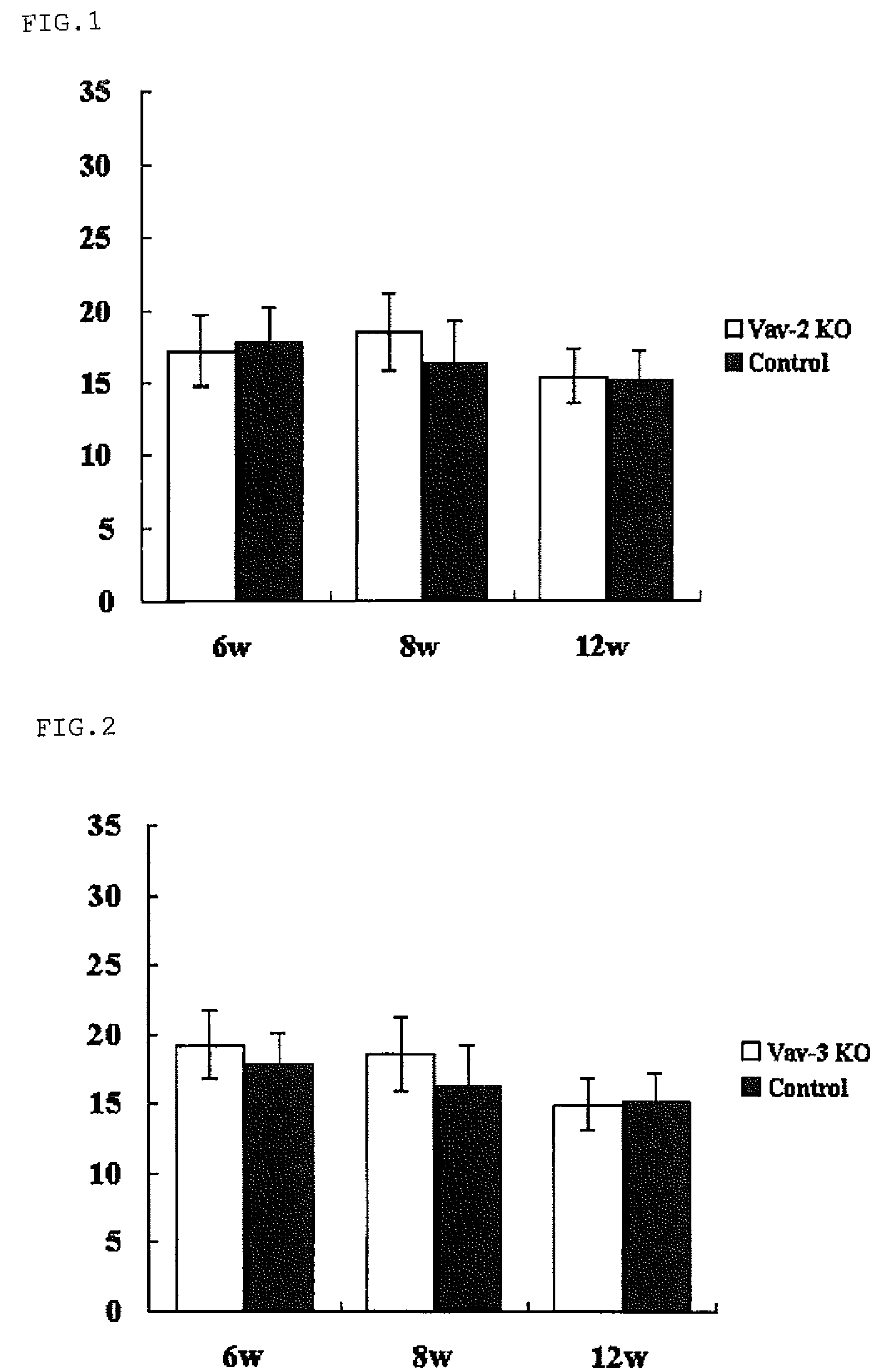
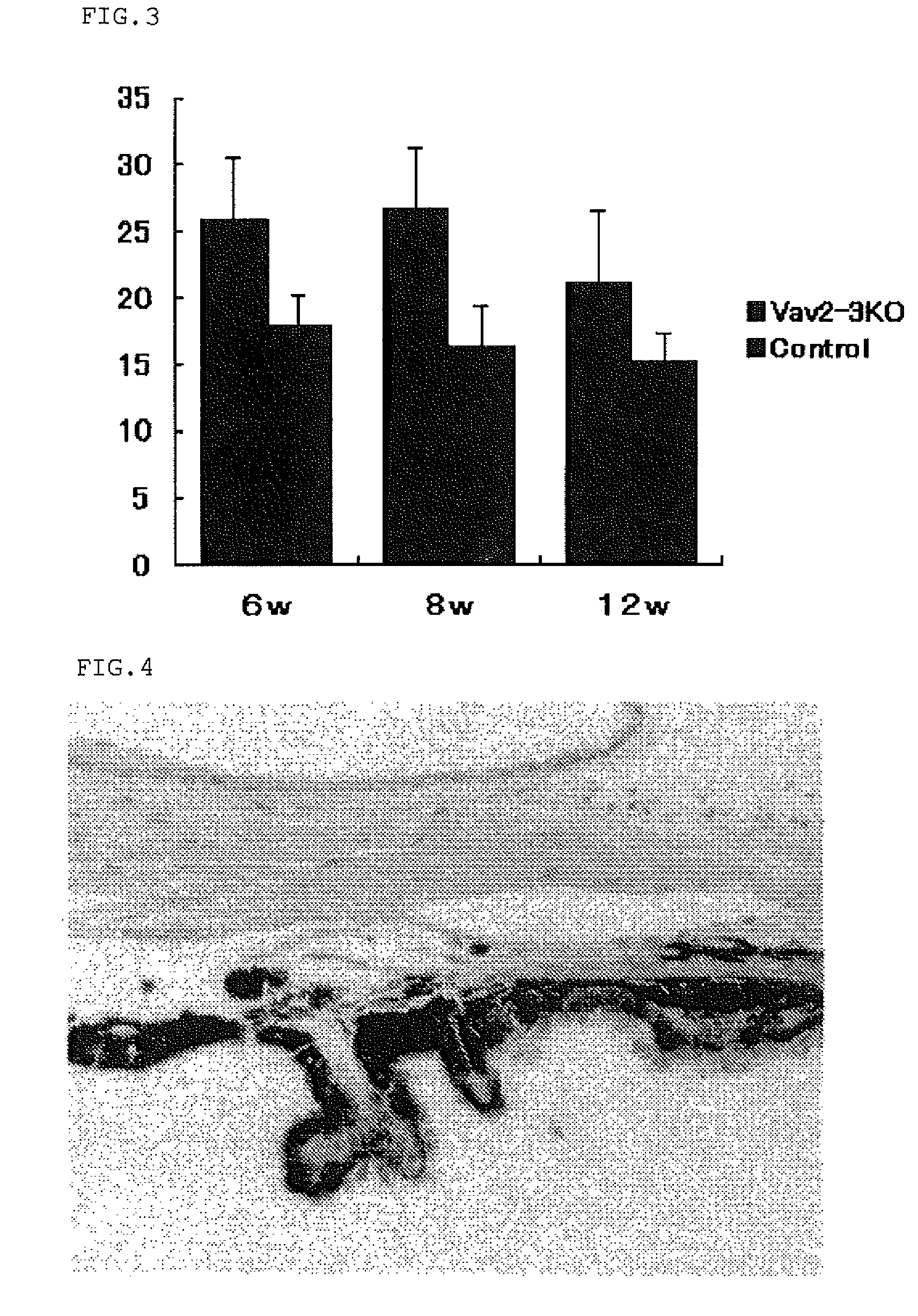
[0058]Vav2ko obtained from Dr. Swat W.'s laboratory at Washington University and Vav3ko obtained from Dr. Frederick W. Alt's laboratory at Harvard University (Massachusetts, USA) were separately subjected to back-crossing into C57BL / 6 mouse, and then Vav2ko and Vav3ko were bred with each other to obtain Vav2 / 3ko. Four animals for each of Vav2ko, Vav3ko and Vav2 / 3ko were raised under barrier free condition at SPF level designated by School of Medicine of Hokkaido University, according to the guidelines suggested by the Animal Committee. Further, using a Tonolab rebound tonometer (manufactured by Tiolat, Finland), intraocular pressure was measured every weak between 10 o'clock in the morning and noon under the condition recommended in the manual. The experiment was repeated four times using a different mouse every time, and overall sixteen animals were subjected to the experiment. The resulting data was analyzed based on two-tailed Student's t-test and standard deviation was obtained ...
example 2
[0061]From the control mouse (B6mouse) No. 22 (Table 1) and Vav2 / 3ko No. 18 (Table 2), both left and right eye balls were taken out, embedded for preservation, thinly sliced, and stained with hematoxylin-eosin (HE; Sigma) to give a pathological tissue specimen. The specimen was then examined under an optical microscope.
[0062]
TABLE 1GroupWild type (B6)Animal1Right eye ball ♂ 16DSameNumber2Left eye ball ♂ 16Dmouse3Right eye ball ♂ 16DSame4Left eye ball ♂ 16Dmouse5Right eye ball ♂ 4WSame6Left eye ball ♂ 4Wmouse7Right eye ball ♂ 4WSame8Left eye ball ♂ 4Wmouse9Right eye ball ♂ 4WSame10Left eye ball ♂ 4Wmouse11Right eye ball ♂ 4WSame12Left eye ball ♂ 4Wmouse13Right eye ball ♂ 9WSame14Left eye ball ♂ 9Wmouse15Right eye ball ♂ 9WSame16Left eye ball ♂ 9Wmouse17Right eye ball ♂ 11WSame18Left eye ball ♂11Wmouse19Right eye ball ♂ 21WSame20Left eye ball ♂ 21Wmouse21Right eye ball ♂ 21WSame22Left eye ball ♂ 21Wmouse
[0063]
TABLE 2GroupVav2-3ko1Right eye ball ♂ 20D #4Same2Left eye ball ♂ 20D #4mouse...
example 3
[0069]Vav2 / 3ko mouse of the present invention and wild type C57BL / 6 mouse (control) as a background were raised until Week 3, Week 10, Week 15 and Week 30 under the same condition as Example 1. After heavily anaesthetizing the mice with sodium pentobarbital solution, the eye balls were quickly removed and one of them was fixed with 2.5% glutaraldehyde solution (TAAB) for an electron microscope, which had been diluted with a deionized and neutral methanol solution containing 10% formalin, to examine the anterior chamber of the eye. The other eye ball was fixed for 12 hours by using Davidson's solution for observing retina. Fixed tissues were embedded in paraffin solution and cut into a 5 μm piece having cross-section view of an arrow by using a microtome. Then, the piece was de-paraffinized, dehydrated and stained with HE. Results of HE staining are shown in FIG. 13 and FIG. 14.
[0070]As shown in FIG. 13, from three-week old Vav2 / 3ko mouse, the optic nerve head appeared to be normal i...
PUM
| Property | Measurement | Unit |
|---|---|---|
| intraocular pressure | aaaaa | aaaaa |
| intraocular pressure | aaaaa | aaaaa |
| intraocular pressure | aaaaa | aaaaa |
Abstract
Description
Claims
Application Information
 - R&D
- Intellectual Property
- Life Sciences
- Materials
- Tech Scout
- Unparalleled Data Quality
- Higher Quality Content
- 60% Fewer Hallucinations
Browse by: Latest US Patents, China's latest patents, Technical Efficacy Thesaurus, Application Domain, Technology Topic, Popular Technical Reports.
© 2025 PatSnap. All rights reserved.Legal|Privacy policy|Modern Slavery Act Transparency Statement|Sitemap|About US| Contact US: help@patsnap.com


