Magnetic resonance method for quantification of transverse relaxation times

a magnetic resonance and transverse relaxation technology, applied in the field of magnetic resonance apparatus and methods for quantification of transverse relaxation times, can solve the problems of inconvenient data analysis, lack of diagnostic specificity, and the time-consuming of methods and apparatuses availabl

Active Publication Date: 2012-11-20
UNIV HOSPITAL OF BASEL
View PDF3 Cites 11 Cited by
  • Summary
  • Abstract
  • Description
  • Claims
  • Application Information

AI Technical Summary

Problems solved by technology

As a result, the contrast in an MR image is per se a mixture of all these effects and would, if used as such, lack diagnostic specificity.
It is, however, also evident from the term that in such an image only the contrast in the image is weighted by the specific tissue property but that it does not represent an actual measure of this specific tissue property.
Although quantification is in many ways superior to qualitative MRI, methods and apparatuses available require much more MRI scanner time and data analysis can be very time consuming.
Although the SE-based methods yield accurate estimates of T2 in combination with good anatomical information, they suffer from long acquisition times, making it clinically unattractive.

Method used


Image

  • Magnetic resonance method for quantification of transverse relaxation times
  • Magnetic resonance method for quantification of transverse relaxation times
  • Magnetic resonance method for quantification of transverse relaxation times

Examples


Embodiment Construction

[0031]For a more complete understanding of the present invention and advantages thereof, references is now made to the following description of various illustrative and non-limiting embodiments thereof, taken in conjunction with the accompanying drawings.

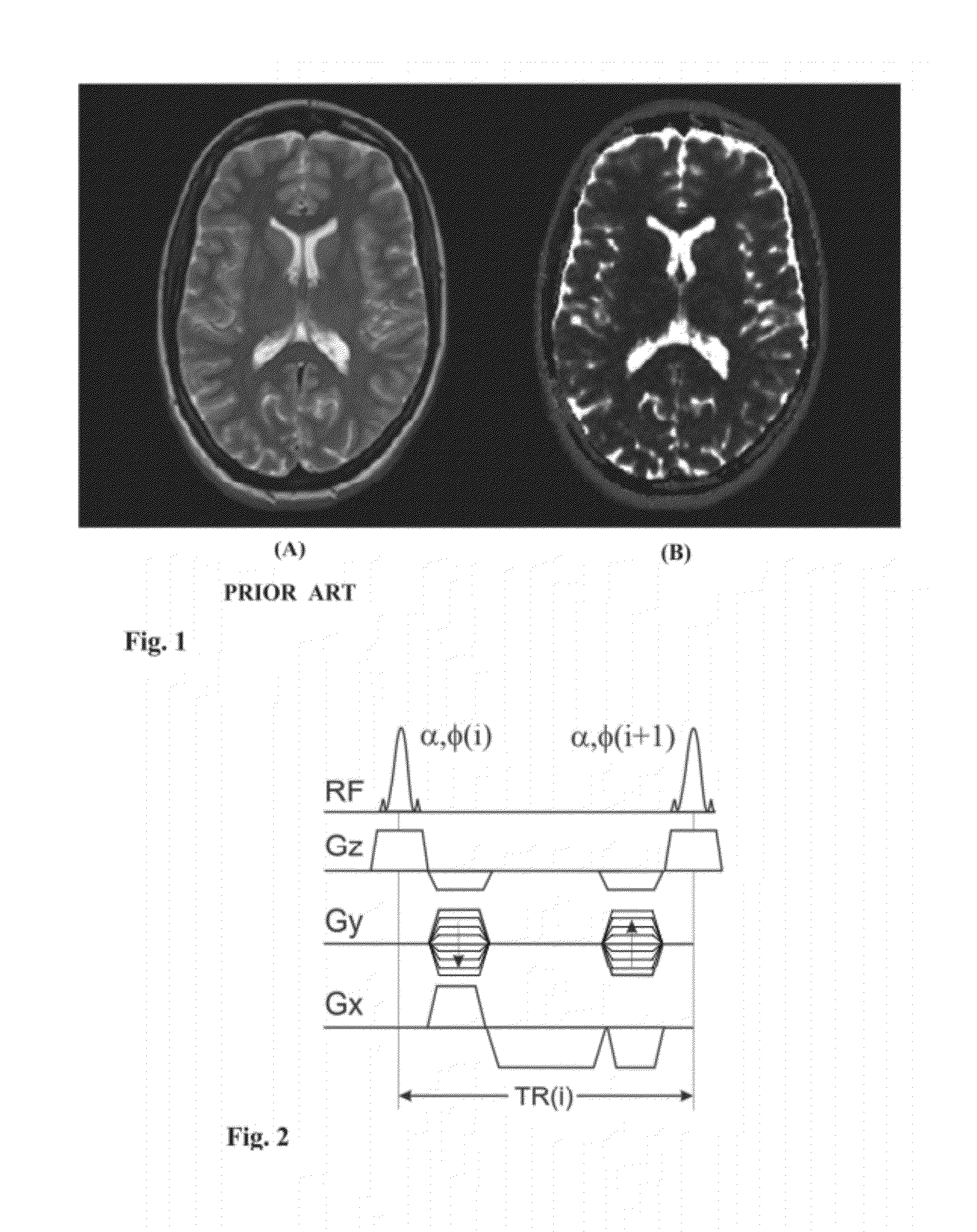
[0032]The difference between contrast-based and quantitative MRI is illustrated in FIG. 1, showing a T2-weighted image (A) and a quantitative T2 map (B) of the human brain. The axial image displayed in FIG. 1A was acquired using a SE sequence known in the art, whereas the image shown in FIG. 1B was acquired using one embodiment of the invention described herein. The contrast between pixels, such as between the cerebrospinal fluid (CSF) and gray matter (GM) or white matter (WM) in FIG. 1a is governed by the T2 relaxation times. This means, the longer the T2 value, the brighter the pixel. In contrast in FIG. 1B, a contrast similar to FIG. 1A is observed, but pixel intensities (PI) yield a direct quantitative measure of the T2 relaxati...


PUM


Abstract

Apparatus and methods for quantification of transverse relaxation times (T2) using steady-state free precession sequences (generally known as fast imaging sequences) and their sensitivity to a quadratic increase of the RF pulse phase, also known as RF spoiling. Using at least two image acquisitions with different partial RF spoiling increments, T2 can be assessed with high precision and with short acquisition times in the limit of large excitation angles being independent on the longitudinal relaxation time (T1) and magnetization transfer effects as compared to other SSFP based quantitative T2 methods. This invention is not restricted to any kind of target and may be applied in 3D as well as in 2D.

Description

FIELD OF THE INVENTION[0001]This invention relates to apparatus and methods for the quantification of transverse relaxation times (T2), for example, for biological tissues such as, but not limited to, brain including, e.g., grey and white matter, muscle and / or cartilage.BACKGROUND OF THE INVENTION[0002]Magnetic resonance imaging (MRI) refers to an imaging technique for visualization of primarily tissue structure and function. The strength of the magnetic resonance (MR) signal depends on the relative density of excited nuclei, on relaxation properties (longitudinal T1 and transversal T2 relaxation), on the signal upon excitation and other parameters such as, but not limited to, flow and chemical shifts. As a result, the contrast in an MR image is per se a mixture of all these effects and would, if used as such, lack diagnostic specificity. However, dedicated imaging sequences can emphasize one specific effect, such as the transverse relaxation time (T2), yielding a so called T2-weigh...

Claims


Application Information

Patent Timeline
Patent Type & Authority Patents(United States)
IPC IPC(8): G01V3/00
CPCG01R33/50G01R33/5613
Inventor BIERI, OLIVERSCHEFFLER, KLAUS
Owner UNIV HOSPITAL OF BASEL
Why Eureka
Try Eureka