Cartesian human morpho-informatic system

a morpho-informatics and cartesian technology, applied in the field of 3d cartesian coordinate systems, can solve the problems of large workload in medicinal diagnostics, inefficiency, and inability to accurately identify human morphology, and achieve efficient and accurate scanning protocols, reduce unnecessary radiation exposure, and reduce the number of scans a person.

Inactive Publication Date: 2012-12-11
UNIV OF SOUTH FLORIDA
View PDF14 Cites 2 Cited by
  • Summary
  • Abstract
  • Description
  • Claims
  • Application Information

AI Technical Summary

Benefits of technology

[0011]Use of a defined three dimensional coordinate system to align and register volumetric medical imaging data of the human body within three dimensional space makes possible the objective analysis of the morphometric organization of any individual human as well as the ability to compare and statistically define the morphological characteristics of populations of humans. This permits description of quantitative data of the morphometric features of the body of any individual human, relative to any set of three dimensional coordinates. Further, changes in any morphological feature over time that occur as a result of normal development, growth, aging, acute insult or progressive changes related to disease processes may be described using the three dimensional coordinate system. Moreover, patterns related to normal development, growth, aging, acute insult or disease processes may be documented, described, analyzed, and diagnosed using the present invention. Statistically-derived data sets of morphometric changes, patterns or changes in patterns of digital image data characteristics are also useful in the present invention.
[0017]Volumetric medical images are composed of arrays of rows and columns of voxels. Regardless of how that voxels are obtained (CT, MR, PET, etc) the final representation is organized in a 3D array. One of the features provided by the bounding box data approach is that the 3D grey scale voxel array patterns can be defined for normal conditions as well as for any and all variants of pathological conditions. With sufficient validated data sets of voxel grey scale array patterns, the scanning device computer may track the anatomical structures it is actively scanning and compare the active-scan structures to a validated database of voxel grey scan array patterns. Comparing the patient's array patterns with known patterns allows the computer to perform a real-time, first pass differential diagnosis of the image while the patient is still on the scan table. The results of the scan may be analyzed and, if the computer requires more information to make a decision, it has the opportunity to rescan the patient with an appropriate protocol the patient is still in the scanner.
[0020]A normative human morphology database may be developed for all relevant structures for a large population of normal healthy individuals to describe complete array of statistical descriptors of the morphological features. Medical imaging of millions of patients is performed each year. For each of these scans a radiologist provides a medical opinion as to whether the morphology is normal or abnormal. When abnormal, the pathology is described. Using normal and validated abnormal morphology, a database or series of databases may be developed to define the array patterns of normal anatomical structures and disease conditions for which medical imaging is utilized as a diagnostic tool. This database then can be used to provide a measure of limits between normal (healthy) and abnormal (diseased / pathological) morphological structure. The information is used to develop software for a first pass differential diagnosis and more efficient and accurate scanning protocols. This will ultimately reduce the number of scans a person may require, reduce unnecessary radiation exposure, and will result in faster, cheaper and more accurate imaging processes.
[0021]Data may be stored in any format known in the art, including Picker SPECT, GE MR SIGNA—including SIGNA 3, 5, and Horizon LX—Siemans Magnatom Vision, CTI, CTI ECAT 7, SMIS MRI, and ASI / Concorde MicroPET. The standard format for volumetric medical image data to be captured and stored is in the DICOM format. Images in DICOM format can be viewed, modeled and measured on a wide range of public domain and commercial software available today. Any and all volumetric medical image data oriented as described above to a defined human morphometric coordinate system can be data mined to provide precise and comparable measurements for any and all relationships of anatomical features. Software plug-ins for several software packages have been developed to permit efficient mining of data from the DICOM image sets oriented within the coordinate system. The plug-ins permit the point and click identification and storage of the 3D coordinate of specific anatomical features. Line distant length between two anatomical features can be determined. Any 2D area or 3D volume can be user defined by a point and click approach and the volume and 3D coordinate location recorded.
[0022]If and when volumetric image data of human morphology is placed in registration within a three dimensional coordinate system, a wide range of quantitative measurements can be made to better document the structural features of the body. When fully implemented computer based technology will be able to determine if all internal structures are normal, and if not what are the likely medical problems. Use of the coordinate system permits quantitative description and analysis of structural features includes but are not limited to: location, volume, orientation, length and diameter of all individual structure as well as the spatial relationships of any combination of morphological feature of the human body relative to a any three dimensional coordinate system. If a similar process is applied to a population of humans with image data all registered with the same three dimensional space relative to a consistent coordinate system, then detailed statistical analysis can be conducted to establish mathematical descriptors for all levels of normal human anatomy as well as each and every known pathological condition that has, as one of its features, an alteration in the patient's anatomical structure. The resulting human morphometric data sets can be used in the development of computer software for the automated analysis of medical images. The ability to quantitatively describe the structure of the human body will significantly increase the value of medical imaging in 21st century clinical medicine. This approach will ultimately provide faster, cheaper and more accurate initial stages of differential diagnosis of a wide range of medical conditions.

Problems solved by technology

Except for measurement protocols for skeletal morphology developed by physical anthropologists, there has been very little data developed to define human morphology.
Identification and extraction of quantitative anatomical data from three dimensional medical images consumes a vast amount of workflow in medicinal diagnostics.
The inefficiency is partially due to difficulties in generalizing the steps needed for successful image segmentation of medical imaging data.
Only recently has volumetric medical image data been possible to obtain, and few if any processes are in place to effectively clinically evaluate this volumetric data.
Computer technology currently plays very little role in the analysis of any medical images in regards to assisting the physician in making a differential diagnosis.
This approach is extremely inefficient, expensive and time consuming.
This approach also fails to utilize any technology resources to assist the physician with medical image analysis.
Much of the information contained in the volumetric image data is not taken into consideration because physicians currently do not have a recognized approach for utilizing the information and do not have reference normative data on which to base any level of diagnostic decision.
The rows and column array format of digital voxel data that is typical of most all volumetric medical images lends itself perfectly for the application of computer technology, but lack of a standard format and orientation for human anatomical image data has hindered the use of computer technology in any type of analysis of medical images.
Even current computerized, semi-automated techniques employed to analyze medical images require heavy user intervention, resulting in high variability in quantification.
Differences in the examining radiologist and the fatigue of the radiologist affect the segmentation input and impact the reliability of the data obtained.
The enumerated issues with traditional anatomical diagnosis affects the quality of the medical diagnosis and treatment provided to patients.

Method used


Image

  • Cartesian human morpho-informatic system
  • Cartesian human morpho-informatic system
  • Cartesian human morpho-informatic system

Examples


example

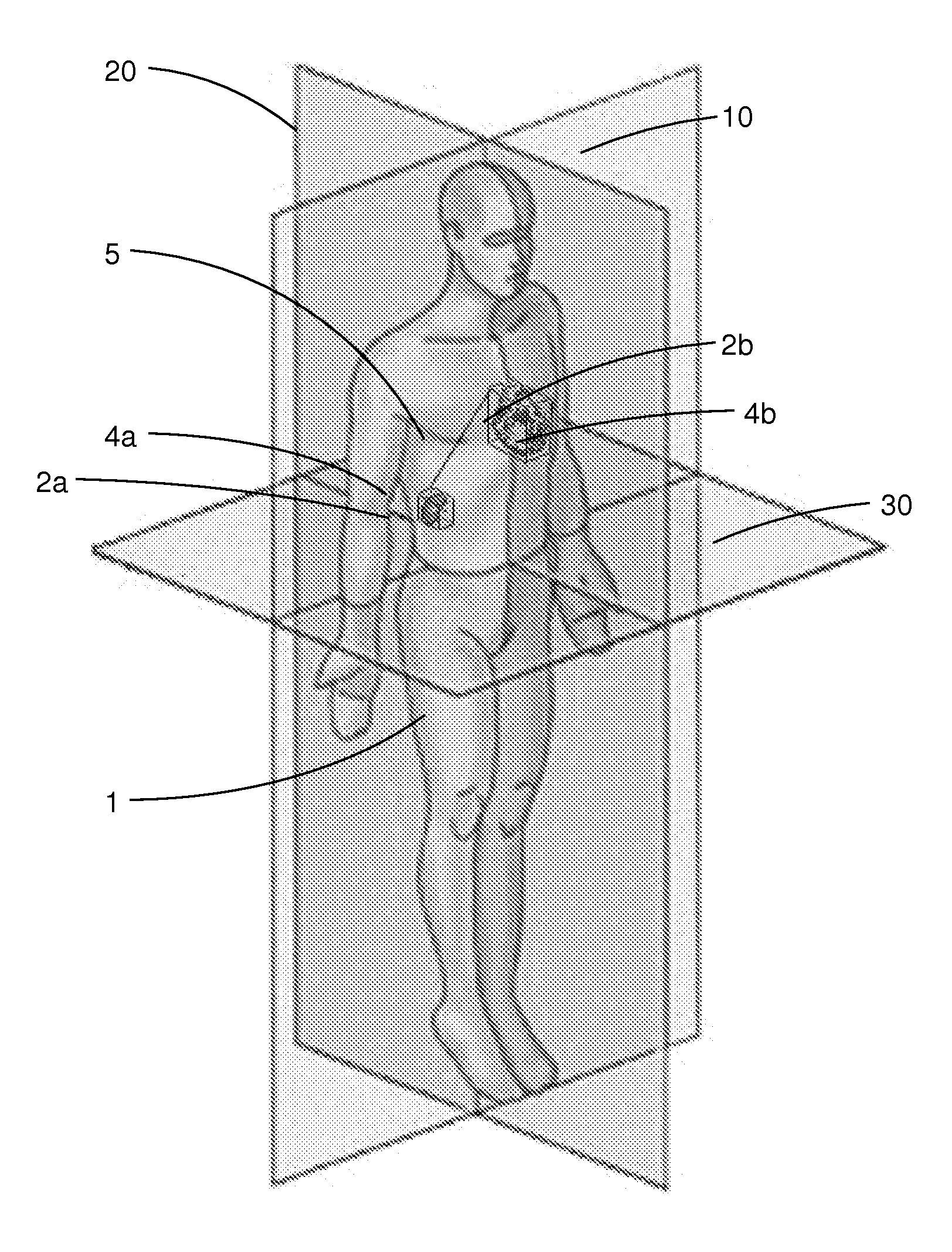
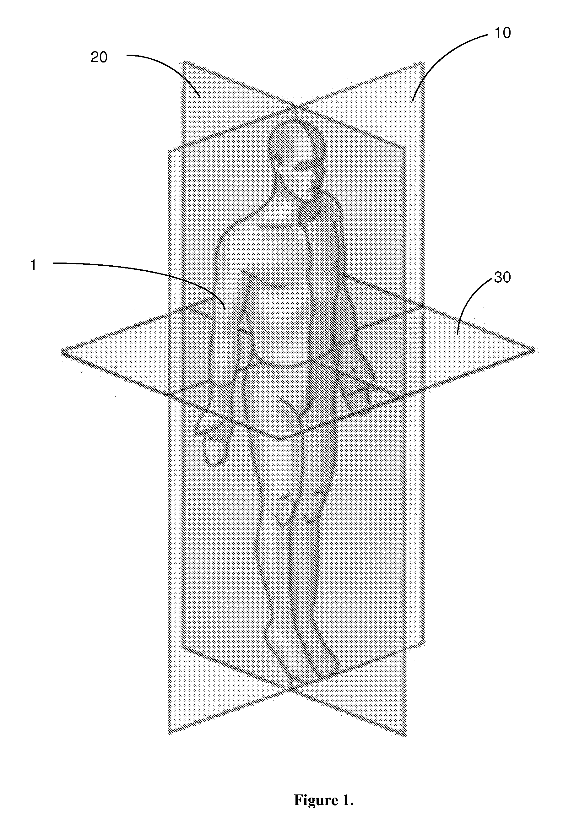
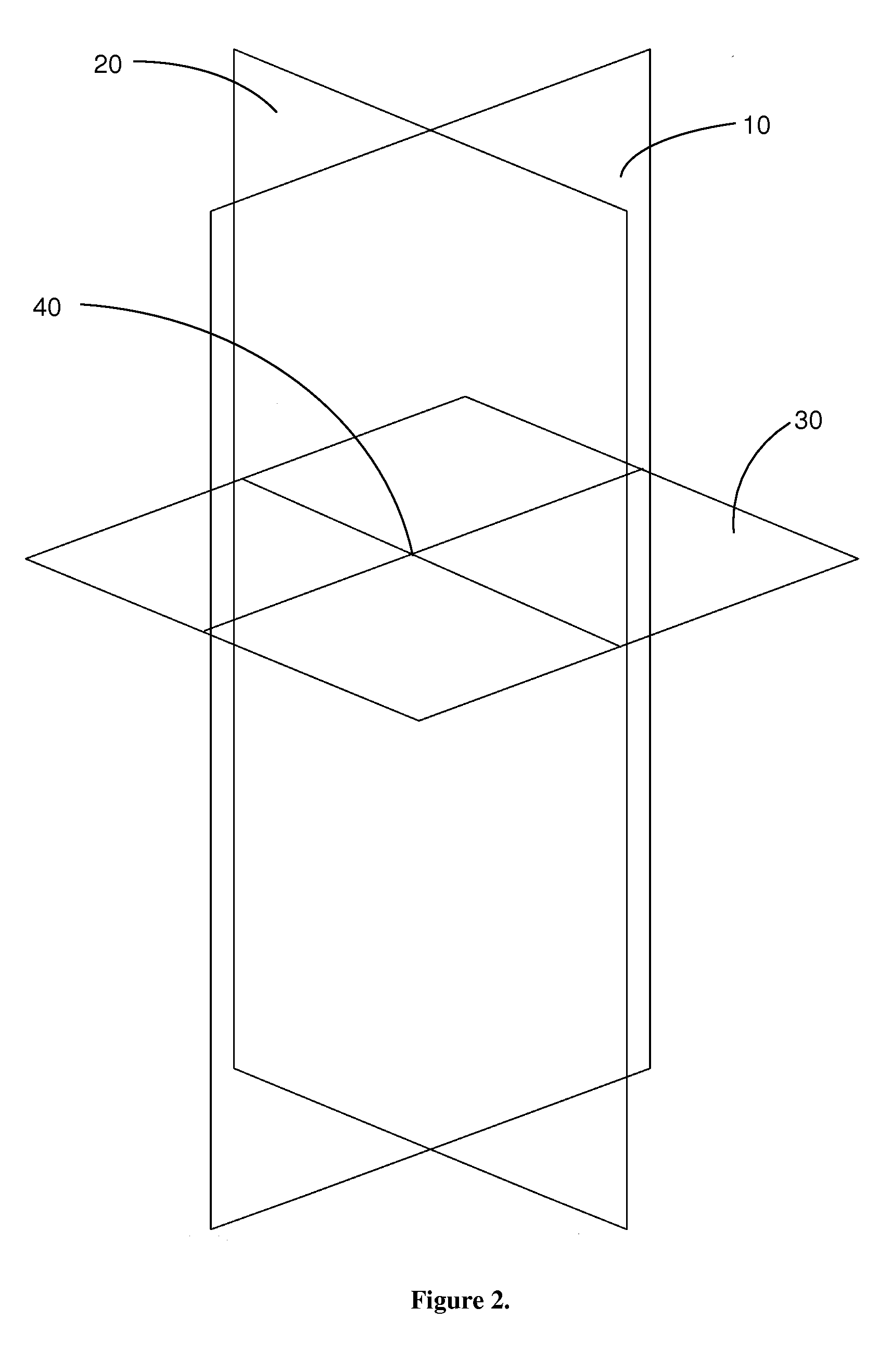
[0039]The present invention's ability to quantitatively describe the location of a structure in or on the human body is illustrated in the following example. Arbitrarily-chosen coordinates are utilized to define the location from intersection point 40 (0, 0, 0). As seen in FIG. 3, Sagittal plane 20 (X-axis) is defined from 50 (uppermost limit) to −50 (the lowermost limit); Transverse plane 30 (Y-axis) defined as 20 (right-most lateral limit) and −20 (the left-lateral-most limit); and Coronal plane 10 (Z-axis) defined as 10 (anterior-most limit) and −10 (the posterior-most limit). Body 1 is defined by body bounding box 3. Anatomical structure 2 is defined by structure bounding box 4. The points of the bounding box are determined on the coordinate system. In this example, the location of structure 2 would have a coordinate of (approximately) 10, 10, 10 (X, Y, Z). In this example, structure 2 is located approximately ⅕ of the distance upward on the coronal plane (X-axis) from the trans...


PUM


Abstract

The present invention is a three dimensional Cartesian coordinate system for the human body, having three perpendicular and intersecting planes. The present invention is based upon the use of the three cardinal planes, in the universally recognized orientations. The cardinal planes in accordance with the present invention are: Sagittal: midsagittal plane, Transverse: upper-most extent of the iliac crests, and Coronal: anterior-most aspect of the vertebral canal. The point at which these planes intersect defines the 0,0,0 location in the human body.

Description

CROSS REFERENCE TO RELATED APPLICATIONS[0001]This application is a continuation in part of co-pending U.S. patent application Ser. No. 11 / 428,926, filed Jul. 6, 2006. This application is also a non-provisional application of currently pending U.S. Provisional Patent Application 60 / 949,395, filed Jul. 12, 2007.FIELD OF INVENTION[0002]This invention relates to a 3D Cartesian coordinate system for use with human spatial morpho-informatics.BACKGROUND OF THE INVENTION[0003]Human anatomy has traditionally been a descriptive rather than an objective science. Except for measurement protocols for skeletal morphology developed by physical anthropologists, there has been very little data developed to define human morphology. For descriptive purposes classic anatomists have describe the human body as being placed in the “anatomical position”. When in anatomical position the body is standing erect with the arms at the sides with the palms of the hands facing forward. The feet are flat on the gro...

Claims


Application Information

Patent Timeline
Patent Type & Authority Patents(United States)
IPC IPC(8): G06K9/00
CPCG06T7/0012G06T7/0042G06T2207/30004G06T7/73
Inventor HILBELINK, DON R.
Owner UNIV OF SOUTH FLORIDA
Why Eureka
Try Eureka