Conjunctival tissue system

a tissue system and conjunctival technology, applied in the field of tissue systems, can solve the problems of limited long-term proliferative capacity, eye irritation, and lower regenerative potential after transplantation

Inactive Publication Date: 2012-12-25
RELIANCE LIFE SCI PVT
View PDF14 Cites 4 Cited by
  • Summary
  • Abstract
  • Description
  • Claims
  • Application Information

AI Technical Summary

Benefits of technology

[0019]The present disclosure describes a tissue system comprising conjunctival cells cultured on an appropriate support material, such as a biocompatible membrane or extracellular matrix, as well as methods for preparing such a conjunctival tissue system. In preferred embodiments, the conjunctival cells are cultured on amniotic membrane, such as human amniotic membrane (HAM), to generate the conjunctival tissue system. Preferably, the conjunctival tissue system disclosed herein is an equivalent of conjunctival tissues on the ocular surface. Surprisingly, the conjunctival tissue system disclosed herein, which may be prepared by culturing a conjunctival biopsy in a single medium, is sufficiently stable, stratified, and adherent that it can be used for clinical transplant. Preferably, the stable conjunctival tissue system is less likely to break apart after being introduced to the eye of the recipient, thereby increasing the clinical success of this method, as well as reducing the recovery time for the recipient.
[0024]Although the conjunctival tissue system disclosed herein may be transplanted to the appropriate site of the eye using methods well known in the art, the present disclosure also describes a suture-less method for delivering the tissue system to the eye of a subject. In the present disclosure, the terms “subject,”“patient,” and “recipient” are used interchangeably. In a preferred embodiment, the disclosure provides a suture-less delivery method for the conjunctival tissue system to the ocular surface of the eye of a subject. In certain embodiments, the tissue system uses a tissue adhesive or a biocompatible glue for adhering the tissue system of the present disclosure to the eye of the recipient. In some embodiments, the biocompatible glue comprises Amcrylate or fibrin sealant. In a preferred embodiment, the biocompatible glue is a sterile tissue adhesive selected from polylactic acid, polylactide glycolic acid, copolymers of polylactic acid and acrylates, and any combinations thereof. In other embodiments, the biocompatible glue comprises any adhesive which is biocompatible to the eye of the subject. The use of a biocompatible glue may be neat and less cumbersome, as well as safer than sutures, so that the cultured conjunctival transplant may be set on the injury with minimal disturbance to the recipient's eye, and with integration of at least some of the transplanted cells.

Problems solved by technology

Cholera toxin, which is a potential carcinogen, can cause eye irritation.
Terminally differentiated cells have limited long-term proliferative capacity, however, which results in a lower regenerative potential after transplantation.
While tissue equivalents developed may be transplanted by conventional methods, for example suturing the graft onto the eye, this can cause discomfort to the recipient.
Further, the act of suturing requires more precision then a suture-less method, since the site of the diseased or damaged eye cannot be clearly visualized.
In addition, such transplantations usually leave a scar at the site.

Method used


Image

  • Conjunctival tissue system
  • Conjunctival tissue system
  • Conjunctival tissue system

Examples


example 1

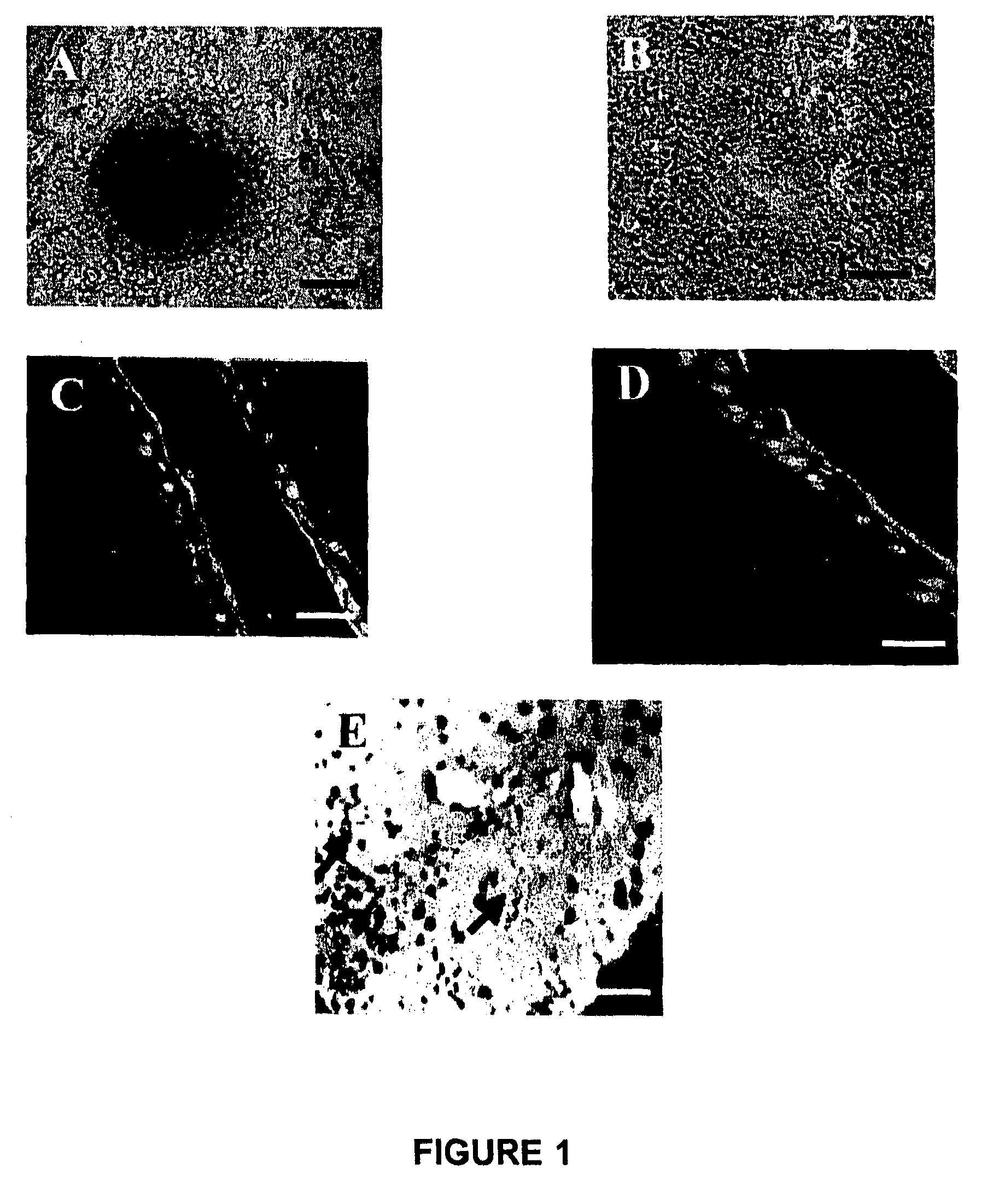
[0061]Generation of a Conjunctival Tissue System.

[0062]The process of culturing conjunctival cells from a tissue biopsy as disclosed in this Example is very simple and different from the existing millicell method. See Pellegrini et al., J. Cell Biol 145(4):769-782, 1999; Belyakov et al., Proc. Natl. Acad. Sci USA 102(40):14203-208, 2005. With the approval of an ethics committee, the collection of the conjunctival biopsies was done with prior informed consent of each patient and donor. The protocols followed were according to the tenets of the Declaration of Helsinki, and were approved by the Reliance Life Sciences (RLS) ethics committee. About 2 mm2 of human conjunctival tissue was surgically removed from the fornix during periocular surgery a the Zee Eye clinic, Bandra, Mumbai, India. The conjunctiva was collected carefully only from the periphery of the eyeball and on the inner surface of the eyelid. The excised tissue was then immediately placed in a transport medium consisting o...

example 2

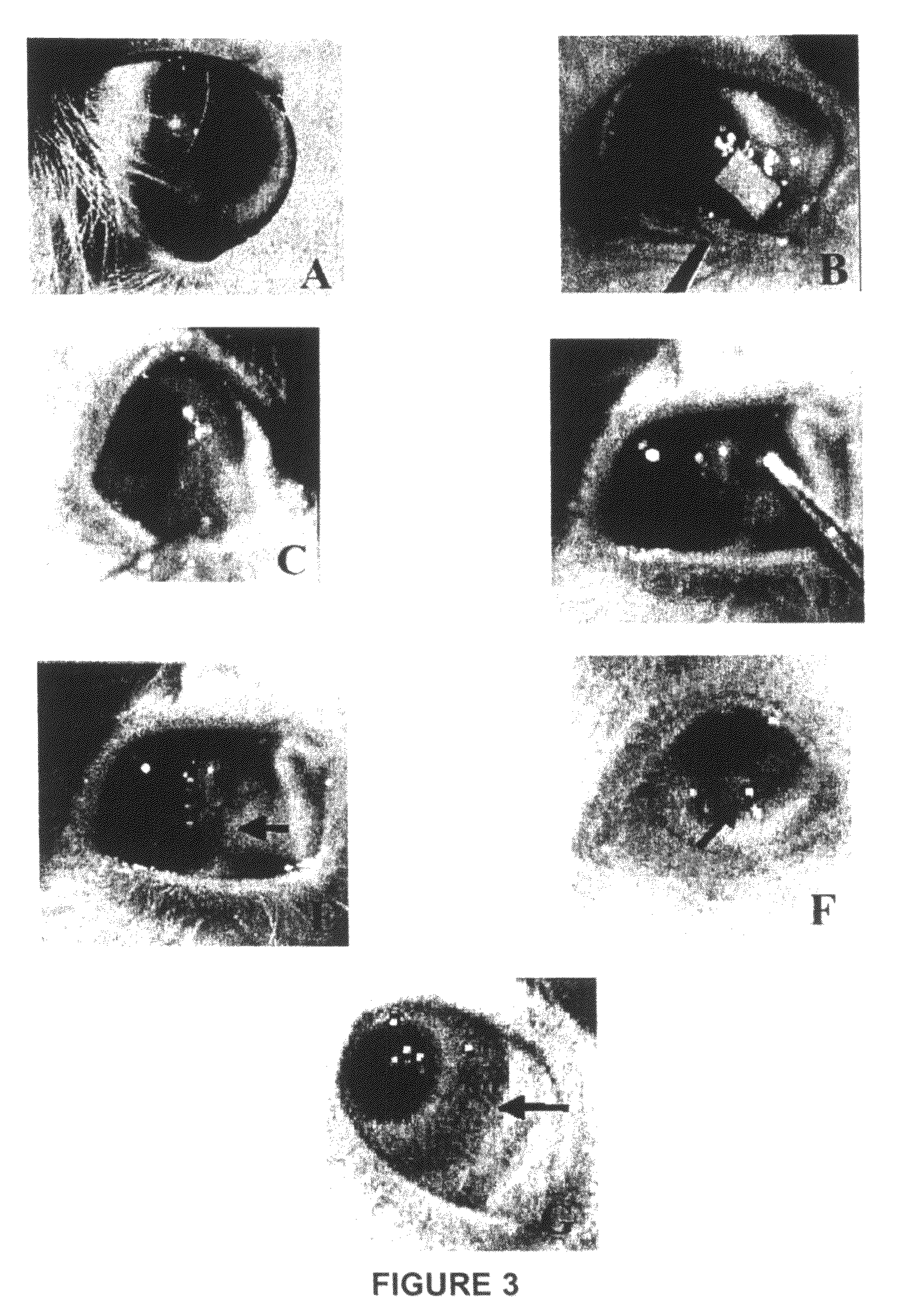
[0073]The present Example examines the ability of the conjunctival tissue system prepared using a plain culture scheme as described in Example 1 to treat the injured eye of a rabbit model of ocular conjunctival burn injury xeno-transplanted with the tissue system. The conjunctival tissue system was grafted using a tissue adhesive in the rabbit model. After transplantation, the survival and integration of the human cells were traced and evaluated using histopathology and immunostaining. The safety of the conjunctival tissue system in terms of abnormal vascularisation underneath, as well as polymorphonuclear leukocytes (PMN) infiltration, was checked in parallel with controls.

[0074]The rabbit model of chemical injury was prepared as follows. Prior approval of the internal review board (IRB), Institutional Animal Ethics Committee (IAEC), and Committee for the Purpose of Control and Supervision of Experiments on Animals (CPCSEA) was obtained for the animal protocols as described herein....


PUM

PropertyMeasurementUnit
sizeaaaaaaaaaa
sizeaaaaaaaaaa
concentrationaaaaaaaaaa

Abstract

The present disclosure describes a tissue system with conjunctival cells, including conjunctival stem cells. The conjunctival tissue system is derived from isolated tissue comprising conjunctival cells, and is suitable for restoring ocular surface impairments, particularly those that result from damaged or diseased conjunctiva. The tissue system is generated using a simple single medium culture scheme, and a support material, such as human amniotic membrane. The conjunctival tissue system generate is suitable for transplantation to treat the ocular surface of an eye of a subject that is damaged or diseased.

Description

CROSS-REFERENCES TO RELATED APPLICATIONS[0001]This application claims priority to Indian Application No. 267 / MUM / 2006, filed on Feb. 24, 2006, which is entirely incorporated by reference herein for all purposes.STATEMENT REGARDING FEDERALLY SPONSORED RESEARCH OR DEVELOPMENT[0002]Not applicable.INCORPORATION-BY-REFERENCE[0003]The Sequence Listing file “11710623SeqListing_ST25.txt” is hereby incorporated by reference into the present specification in its entirety.REFERENCE TO A “MICROFICHE APPENDIX”[0004]Not applicable.BACKGROUND OF THE INVENTION[0005]1. Field of the Invention[0006]The present disclosure relates to tissue systems, methods of producing tissue systems and methods of treatment using tissue systems comprising conjunctival cells for restoring damaged or diseased ocular surfaces. The tissue system of the present disclosure comprises mammalian conjunctival cells, preferably progenitor conjunctival cells derived from the fornix region of the conjunctiva. The present disclosur...

Claims


Application Information

Patent Timeline
Patent Type & Authority Patents(United States)
IPC IPC(8): C12N5/00C12N5/02C12N5/071
CPCA61L27/3804A61L27/3839A61L27/3895C12N5/0621A61L2430/16C12N2500/25C12N2501/11C12N2501/115C12N5/0602C12N5/00A61L27/60
Inventor DEVI, KASHYAP SUBHADRAREDDY, KISHOREVASANIA, VIRAF
Owner RELIANCE LIFE SCI PVT
Why Eureka
Try Eureka