Method and system for anatomic landmark detection using constrained marginal space learning and geometric inference

a technology of geometric inference and marginal space learning, applied in image enhancement, tomography, instruments, etc., can solve the problems of detecting and segmenting human anatomical structures in 3d medical image volumes, which are typically more difficul

Active Publication Date: 2013-01-29
SIEMENS HEALTHCARE GMBH
View PDF5 Cites 4 Cited by
  • Summary
  • Abstract
  • Description
  • Claims
  • Application Information

AI Technical Summary

Benefits of technology

[0006]The present invention provides efficient detection of 3D anatomical landmarks in medical images using constrained marginal space learning (MSL) and geometric inference. Embodiments of the present invention can be used to constrain the search space for landmark detection by exploiting geometric constraints among object pose parameters (i.e., position, orientation, and scale) of multiple 3D anatomical landmarks.

Problems solved by technology

Detecting and segmenting human anatomic structures in 3D medical image volumes (e.g., CT, MRI, etc.) is a challenging problem, which is typically more difficult than detecting anatomic structures in 2D images.

Method used


Image

  • Method and system for anatomic landmark detection using constrained marginal space learning and geometric inference
  • Method and system for anatomic landmark detection using constrained marginal space learning and geometric inference
  • Method and system for anatomic landmark detection using constrained marginal space learning and geometric inference

Examples


Embodiment Construction

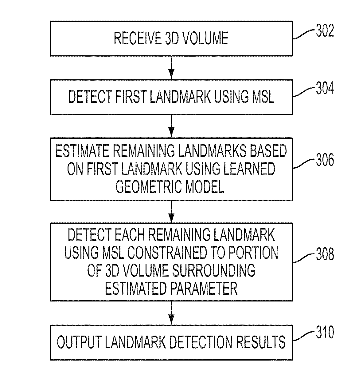
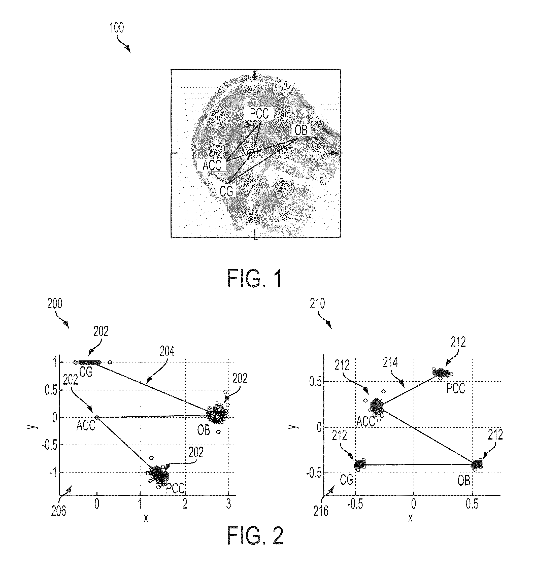
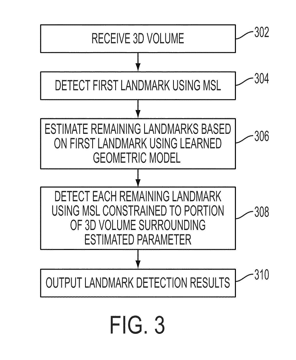
[0016]The present invention is directed to a method for detecting anatomical landmarks in medical images, such as computed tomography (CT), magnetic resonance imaging (MRI), ultrasound, etc. Embodiments of the present invention are described herein to give a visual understanding of the anatomical landmark detection method. A digital image is often composed of digital representations of one or more objects (or shapes). The digital representation of an object is often described herein in terms of identifying and manipulating the objects. Such manipulations are virtual manipulations accomplished in the memory or other circuitry / hardware of a computer system. Accordingly, is to be understood that embodiments of the present invention may be performed within a computer system using data stored within the computer system.

[0017]According to an embodiment of the present invention, an anatomical structure is detected in a 3D medical image using constrained marginal space learning (MSL). MSL i...


PUM


Abstract

A method and apparatus for detecting multiple anatomical landmarks in a 3D volume. A first anatomical landmark is detected in a 3D volume using marginal space learning (MSL). Locations of remaining anatomical landmarks are estimated in the 3D volume based on the detected first anatomical landmark using a learned geometric model relating the anatomical landmarks. Each of the remaining anatomical landmarks is then detected using MSL in a portion of the 3D volume constrained based on the estimated location of each remaining landmark. This method can be used to detect the anatomical landmarks of the crista galli (CG), tip of the occipital bone (OB), anterior of the corpus callosum (ACC), and posterior of the corpus callosum (PCC) in a brain magnetic resonance imaging (MRI) volume.

Description

[0001]This application claims the benefit of U.S. Provisional Application No. 61 / 113,705, filed Nov. 12, 2008, the disclosure of which is herein incorporated by reference.BACKGROUND OF THE INVENTION[0002]The present invention relates to 3D object detection in images, and more particularly, to automated detection of 3D anatomical structures in medical images using marginal space learning.[0003]Efficiently localizing anatomical structures (e.g., heart, liver, kidney, etc.) in medical images is often a prerequisite for further diagnostic image processing procedures, such as segmentation, measuring, and classification. Detecting and segmenting human anatomic structures in 3D medical image volumes (e.g., CT, MRI, etc.) is a challenging problem, which is typically more difficult than detecting anatomic structures in 2D images.[0004]Previously, marginal space learning (MSL) has been proposed for efficient and automatic 3D object localization based on learning of discriminative classifiers....

Claims


Application Information

Patent Timeline
Patent Type & Authority Patents(United States)
IPC IPC(8): A61B6/03G06V10/25
CPCG06K9/3233G06K9/6857G06T7/0046G06T7/0048A61B2019/505G06K2209/051G06T2207/10081G06T2207/10088G06T2207/10132G06T2207/30016A61B2034/105G06T7/75G06T7/77G06V10/25G06V30/2504G06V2201/031
Inventor SCHWING, ALEXANDERZHENG, YEFENGHARDER, MARTINCOMANICIU, DORIN
Owner SIEMENS HEALTHCARE GMBH
Why Eureka
Try Eureka