Apparatus and method for processing biological information

a biological information and apparatus technology, applied in the field of apparatus and method for processing biological information, can solve the problem of difficult to accurately estimate the light intensity (z), and achieve the effect of accurate estimation, high accuracy estimation and high accuracy estimation

Inactive Publication Date: 2013-01-29
CANON KK
View PDF3 Cites 10 Cited by
  • Summary
  • Abstract
  • Description
  • Claims
  • Application Information

AI Technical Summary

Benefits of technology

[0014]The present invention provides a biological information processing apparatus and method capable of accurately estimating a light intensity in a local region of interest with high accuracy, thus allowing for high accuracy estimation of an absorption coefficient based on an acoustic signal of the PAT.
[0017]According to the biological information processing apparatus and method of the present invention, a light intensity in a local region of interest is estimated accurately using the AOT technique, so that an absorption coefficient can be estimated with high accuracy using an acoustic signal of PAT. Thus, a distribution of absorption coefficients in biological tissue can be imaged with high resolution.

Problems solved by technology

If the specimen is heterogeneous optically, however, it is difficult to accurately estimate the light intensity Φ(z).

Method used


Image

  • Apparatus and method for processing biological information
  • Apparatus and method for processing biological information
  • Apparatus and method for processing biological information

Examples


second embodiment

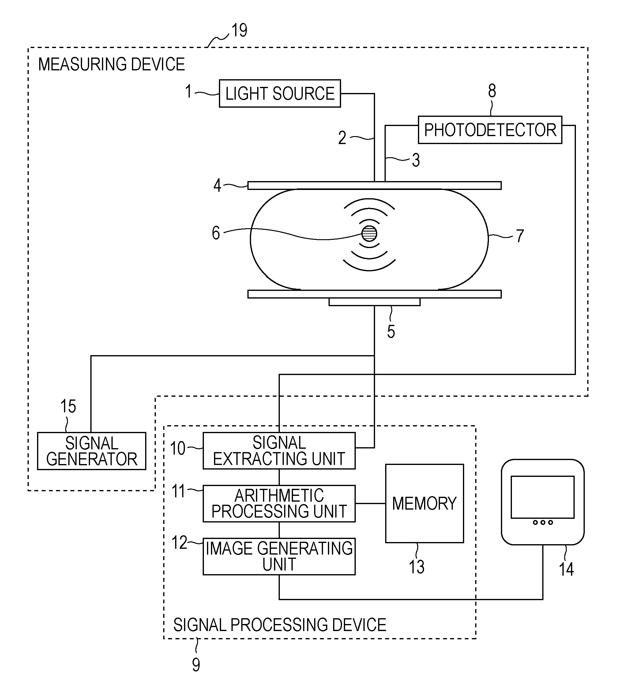
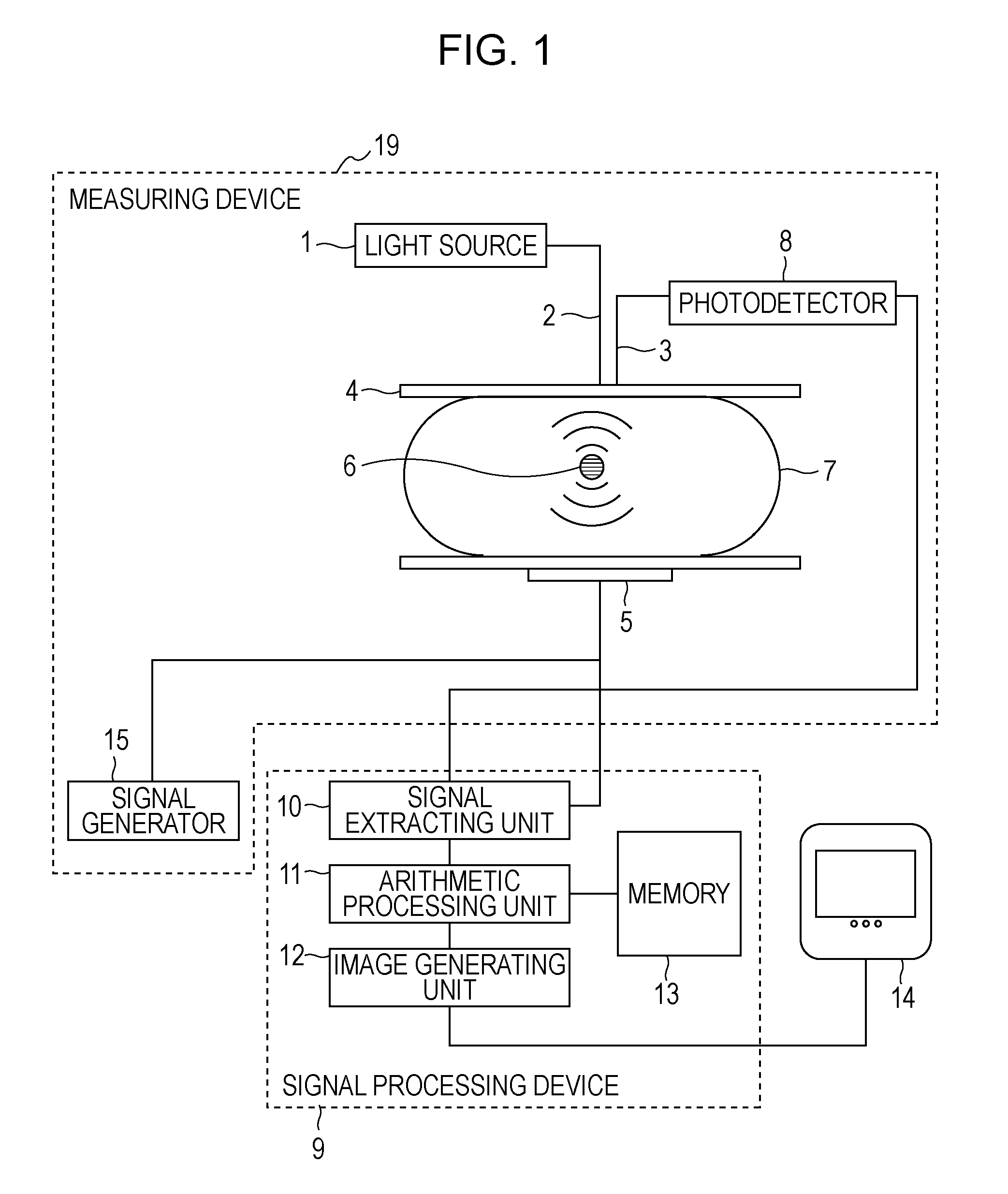
[0088]A biological information method according to a second embodiment of the present invention will now be described. The configuration of an apparatus in this embodiment is the same as that of the apparatus in the first embodiment. FIG. 9 illustrates a flowchart of a measuring process in the present embodiment. In step S200, the PAT measurement is performed while the surface of one of the specimen fixing plates 4 is two-dimensionally scanned by the incidence optical fiber 2, thus obtaining PAT measurement values in all of sections in the specimen 7. At that time, it is preferable that measurement be performed at a plurality of wavelengths to obtain spectral information.

[0089]In step S201, image reconstruction is performed on the basis of signals obtained in step S201 using a well-known method disclosed in, for example, Non-patent Document 1, thus obtaining a distribution of sound pressures inside the specimen 7. At that time, the attenuation of an ultrasonic wave upon propagating ...

third embodiment

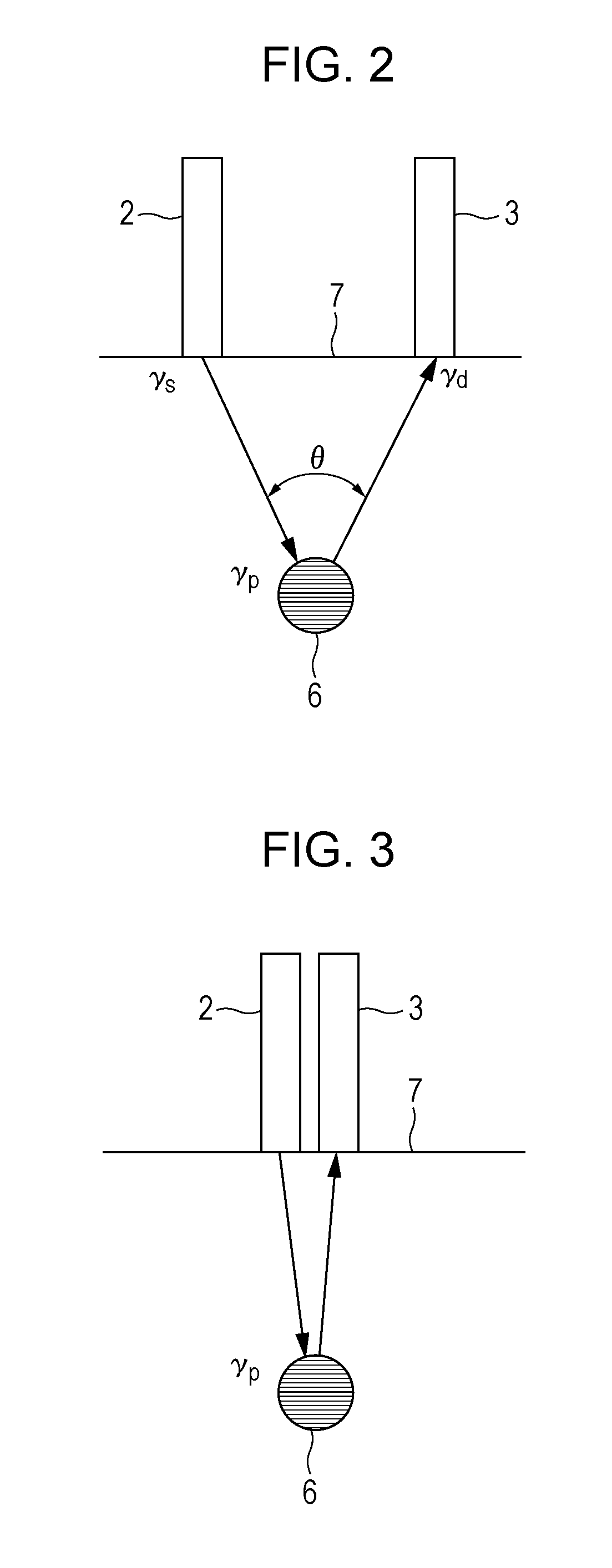
[0097]A biological information processing method according to a third embodiment of the present invention will now be described. The configuration of an apparatus in the present embodiment is the same as that in the first embodiment. In the first embodiment, the incidence optical fiber 2 and the detection optical fiber 3 are arranged so that their positions are regarded as substantially the same position relative to the probe region 6. In the present embodiment, the optical fibers are arranged at any distance therebetween. Typically, it is preferable to arrange the optical fibers at a distance of several centimeters. In the present embodiment, a method of calculating a light intensity in a local region using the AOT is different from that in the first embodiment.

[0098]Referring to FIG. 10, modulated-light measurement regions 6a in each of which an AOT signal along is acquired are arranged below the surface of the specimen 7 such that the respective regions 6a are located relative to...

fourth embodiment

[0111]A biological information processing method according to a fourth embodiment of the present invention will now be described. The configuration of an apparatus in the present embodiment is the same as that in the first embodiment. Prior to the AOT measurement or the PAT measurement, a pulsed ultrasonic wave is transmitted from the ultrasonic transducer 5 and an ultrasonic echo, serving as a reflected wave, is received by the ultrasonic transducer 5. The measurement is performed while the direction in which the pulsed ultrasonic wave is transmitted is changed relative to the specimen 7, thus obtaining structural data regarding the inside of the specimen 7. The data is stored into the memory 13.

[0112]In the arithmetic processing unit 11, the structural data regarding the inside of the specimen 7 obtained by the ultrasonic echo measurement is read out from the memory 13, the specimen 7 is segmented into sections using structural features, and structural information regarding the se...


PUM

PropertyMeasurementUnit
coherence lengthaaaaaaaaaa
wavelength bandaaaaaaaaaa
light intensityaaaaaaaaaa

Abstract

A biological information processing apparatus includes the following elements. A light source irradiates biological tissue with light. A transducer functions as an ultrasonic wave transmitting unit that applies an ultrasonic wave to a local region in the biological tissue. A photodetector detects modulated light obtained by modulating the light from the light source with the ultrasonic wave in the local region. The transducer also functions as an acoustic wave detecting unit that detects an acoustic wave emitted from the local region at a time when the local region absorbs the light from the light source. An absorption characteristic in the local region is calculated using an acoustic signal, serving as an output of the acoustic wave detecting unit, on the basis of a light intensity in the local region calculated based on a modulation signal, serving as an output of the photodetector.

Description

BACKGROUND OF THE INVENTION[0001]1. Field of the Invention[0002]The present invention relates to an apparatus and method for processing biological information.[0003]2. Description of the Related Art[0004]Research on optical imaging apparatuses for irradiating biological tissue with light emitted from a light source such as a laser, and imaging biological information obtained on the basis of incident light is being advanced actively in the medical field.[0005]One of optical imaging techniques is photoacoustic tomography (PAT) called photoacoustic imaging. The photoacoustic imaging is a method of obtaining a distribution of optical characteristic values in biological tissue using the properties of an ultrasonic wave that is scattered in vivo less than light (refer to U.S. Pat. No. 5,840,023 and M, Xu, L. V. Wang, “photoacoustic imaging in biomedicine”, Review of scientific instruments, 77, 041101, 2006 (hereinafter, referred to as “Non-patent Document 1”).[0006]According to the method...

Claims


Application Information

Patent Timeline
Patent Type & Authority Patents(United States)
IPC IPC(8): G06F19/00A61B8/08A61B10/00G01N21/00G01N29/00G01N29/44
CPCA61B5/0073A61B5/0095A61B5/0097G01N21/1702G01N29/0672G01N29/2418A61B5/0059G01N2291/02475
Inventor MASUMURA, TAKAHIRO
Owner CANON KK
Why Eureka
Try Eureka