Method to restore cartilaginous phenotype of chondrocytes after cultured and expanded in vitro

a cartilaginous phenotype and chondrocyte technology, applied in the field of restoring the cartilaginous phenotype of chondrocytes after culture and expansion in vitro, can solve the problems of limited source of normal tissues or cells, restricting the development and clinical application of cell-based therapies, and serious physical debilitation, so as to achieve the effect of restoring the “dedifferentiated” chondrocyte and reducing experimental variability

Inactive Publication Date: 2013-04-30
TAIPEI MEDICAL UNIV
View PDF3 Cites 1 Cited by
  • Summary
  • Abstract
  • Description
  • Claims
  • Application Information

AI Technical Summary

Problems solved by technology

Damage of cartilage caused by trauma or disease, e.g., rheumatoid and osteoarthritis, can lead to serious physical debilitation.
However, the limited source for sufficient amount of normal tissues or cells restricts the development and clinical application of these cell-based therapies.
Loss of chondrocyte phenotype during serial expansion in vitro poses a key limitation to the commercialization and / or clinical use of orthobiologic approaches to articular cartilage repair.
Accordingly, the numbers of expanded chondrocytes which are still retaining the ability to restore native chondrocyte phenotype can only be achieved at about 23.8 folds of expansion of the original cell number, which are insufficient for commercial and / or clinical use.
Furthermore, the procedure for three-dimensional culture in alginate bead is complex and may take at least two weeks to induce restoration of cartilaginous phenotype.
All the above disadvantages make the three-dimensional methods ineffective.
Yaeger P. C. et al. demonstrated that synergistic action of TGF-β1 and IGF-I induces expression of type II collagen and aggrecan genes in adult human articular chondrocytes.30 However, addition of cytokines, hyaluronic acid or growth factors for restoration of chondrocyte phenotype in chondrocyte cell culture is cost ineffective.
Accordingly, the numbers of redifferentiated chondrocytes can only achieve about 23.8 folds of the original number, which is insufficient for commercial and / or clinical use.

Method used


Image

  • Method to restore cartilaginous phenotype of chondrocytes after cultured and expanded in vitro
  • Method to restore cartilaginous phenotype of chondrocytes after cultured and expanded in vitro
  • Method to restore cartilaginous phenotype of chondrocytes after cultured and expanded in vitro

Examples


example 1

Chondrocyte Morphology & Responses to Various Combinations of Growth Factors

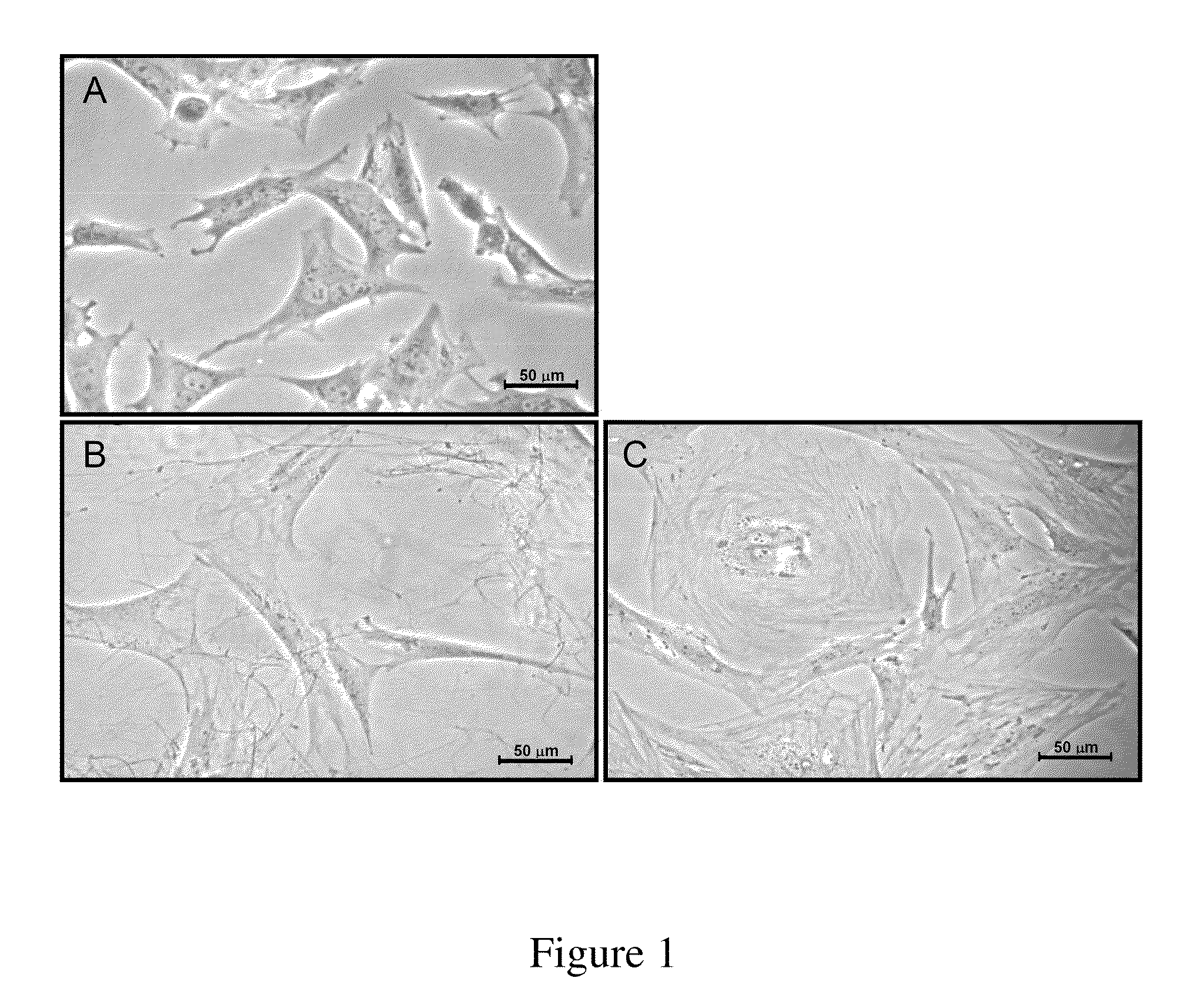
[0053]FIG. 1A shows that P1 chondrocytes appeared polygonal in shape and small in size. In the presence of type II collagen (COL II), stratification was formed in COL II-treated groups, probably through complex cell-matrix interaction. Noteworthy, P9 rabbit chondrocytes cultured in media containing exogenous COL II appeared predominantly polygonal or fibroblast-like (FIG. 1B). However, P9 cells without COL II-supplement became greatly enlarged in size, flattened down on substratum and fully extended, considered as ‘rag-like’ in general appearance (FIG. 1C).

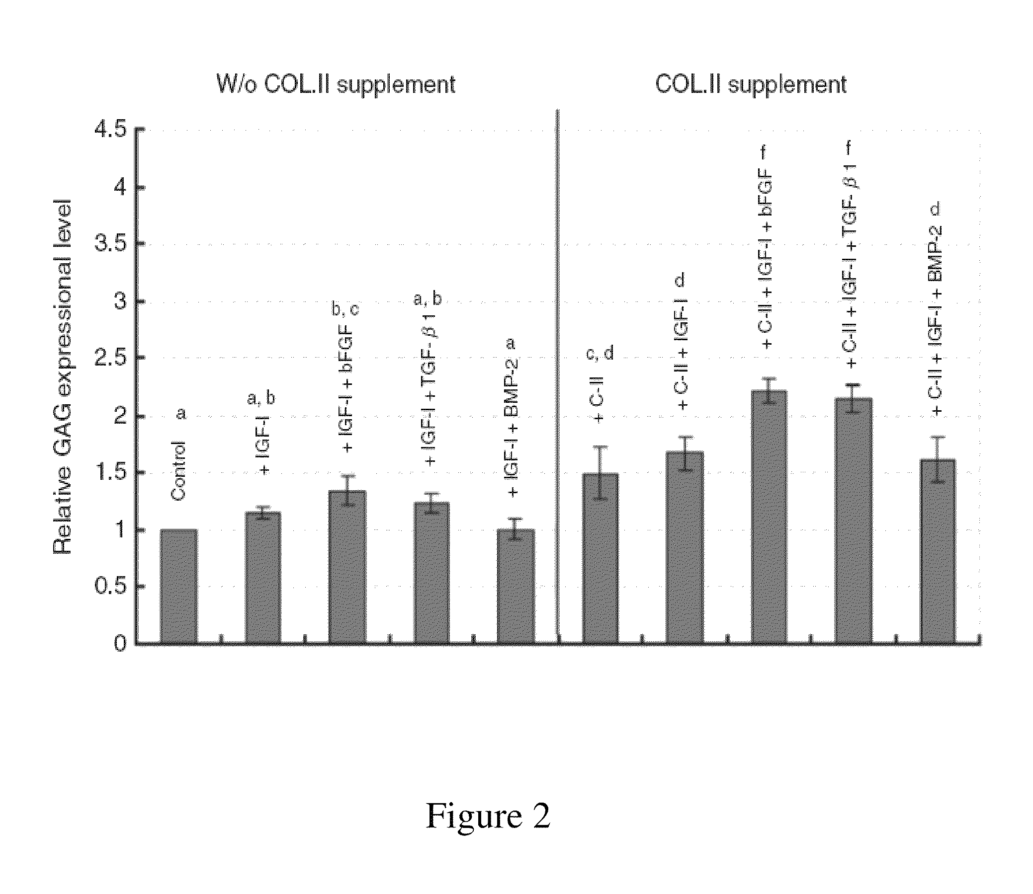
[0054]The effects of combination with cartilaginous growth factors on the induction of GAG expression in rabbit chondrocytes were screened in this study. As this was a preliminary survey, P4 chondrocytes were used. The results were summarized in FIG. 2. In the absence of type II collagen (w / o COL II supplement group), only the treatment of cells with IGF-I...

example 2

Effects of Type II Collagen on Chondrocyte Proliferation

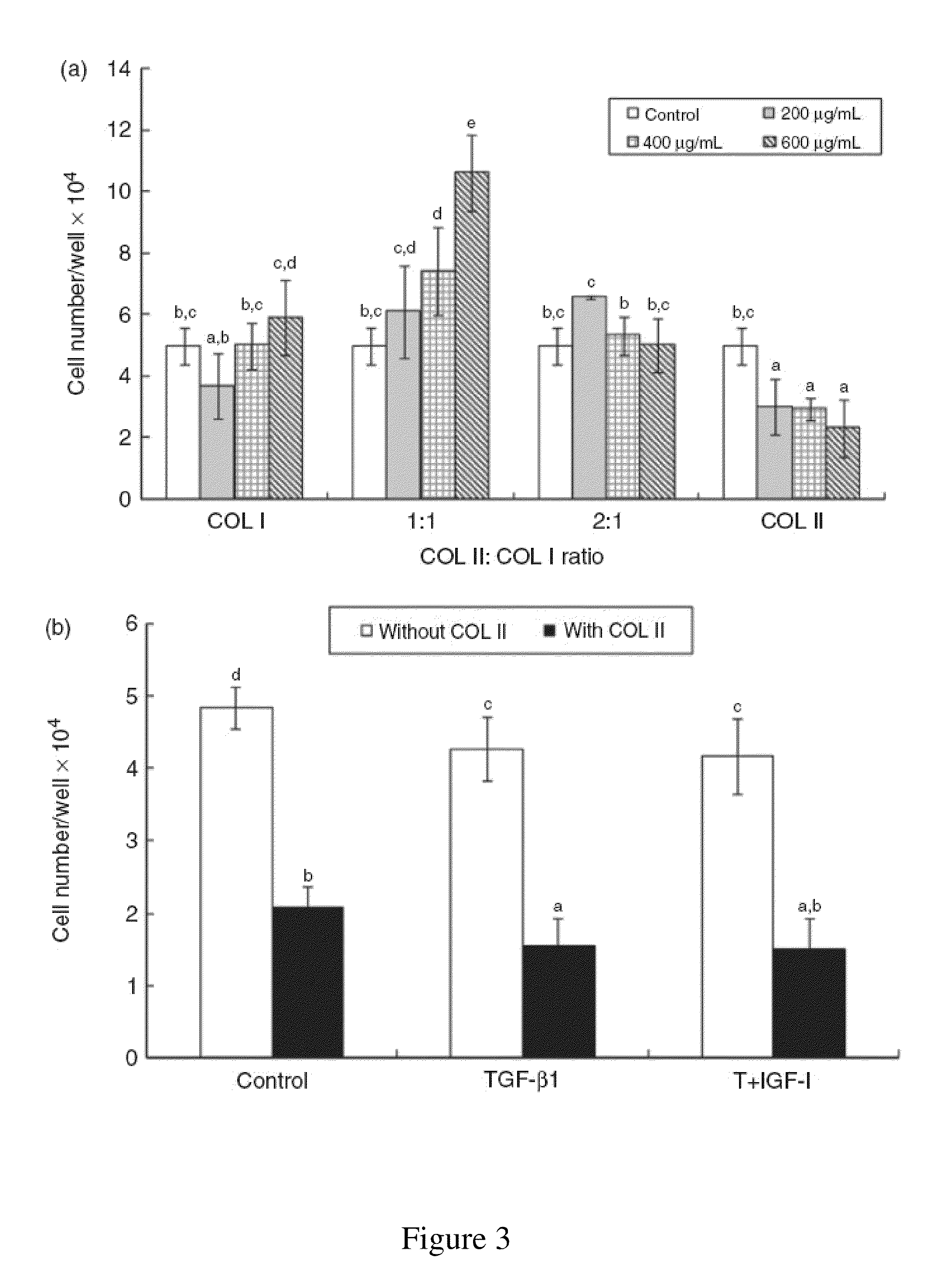
[0056]Among P9 chondrocytes treated with different compositions of ECM components, type I collagen (COL I) alone exhibited little effect on cell proliferation rate except at 600 μg / ml. The effect of combining COL II with COL I at 1:1 ratio on cell proliferation was stimulatory at all three matrix concentrations studied and appeared dose-dependent. At the concentration of 600 μg / ml, the P9 chondrocyte proliferation rate was up to 2.2 folds. In COL II combined with COL I at a ratio of 2:1 group, no significant effect on total cell number were observed when high concentrations of collagen were added. On the contrary, in COL II alone group, COL II greatly inhibited cell proliferation rate at all added concentrations. The final cell number was reduced to 60% and 46%, respectively, of that in the control group when the exogenous COL II concentrations rose from 200 to 600 μg / ml (FIG. 3A).

[0057]On the other hand, the proliferation rate...

example 3

Effects of Type II Collagen on Chondrocytes Glycosaminoglycan Level

[0058]In the group of COL II alone or COL II:COL I at a ratio of 2:1, total GAG levels of P9 chondrocytes were greatly increased up to several folds (3.7 to 4.4 or 2.2 to 3.8 folds, respectively) at all 3 collagen concentrations ranged from 200 to 600 μg / ml (FIG. 4). Nevertheless, in the presence of COL I alone, the total GAG levels of P9 chondrocytes increased only about 1.8 to 2.6 folds with a reverse dose effect. Various concentrations of COL II:COL I at 1:1 ratio also stimulated total GAG levels of chondrocytes to around 1.8 to 2.3 folds (FIG. 4).

[0059]As shown in FIG. 5, both TGF-β1 and TGF-β1 plus IGF-I affected total GAG levels of P9 chondrocytes in the absence of COL II. On the other hand, in the presence of COL II, TGF-β1 alone slightly but significantly reduced GAG level while IGF-I plus TGF-β1 greatly increased GAG level from that of the TGF-β1 group or COL II alone group. The data clearly demonstrated tha...


PUM

PropertyMeasurementUnit
concentrationaaaaaaaaaa

Abstract

The present invention provides a method for restoring native chondrocyte phenotype and functions of, and / or increasing type II collagen as well as aggrecan mRNA expression levels and GAG accumulation level in dedifferentiated chondrocytes which have been subcultured and expanded in vitro, which comprising culturing the said dedifferentiated chondrocytes in vitro with a medium comprising type II collagen, or its biologically active peptide fragment(s) or analogs with or without growth factor(s), wherein the type II collagen or its biologically active peptide fragment(s) or analogs are effective to restore chondrocyte phenotype and functions of, and / or to increase type II collagen and aggrecan expression levels and GAG accumulation level in the said dedifferentiated chondrocytes.

Description

FIELD OF THE INVENTION[0001]The present invention relates to a method of restoring chondrocyte phenotype in dedifferentiated chondrocytes. Particularly, the chondrocytes produced from the invention are suitable for cartilage repair.BACKGROUND OF THE INVENTION[0002]Articular cartilage is composed of chondrocytes encased within the complex extracellular matrix produced by these cells. The unique biochemical composition of this matrix provides for the smooth, nearly frictionless motion of articulating surfaces of the knee joint. With age, tensile properties of human articular cartilage change as a result of biochemical changes. After the third decade of life, the tensile strength of articular cartilage decreases markedly. Damage of cartilage caused by trauma or disease, e.g., rheumatoid and osteoarthritis, can lead to serious physical debilitation. The repair of defective cartilages has been improved recently by transplantation of autogenous or allogenous chondrocytes, or through the u...

Claims


Application Information

Patent Timeline
Patent Type & Authority Patents(United States)
IPC IPC(8): C12N5/00C12N5/02
CPCC12N5/10C12N5/0655C12N2501/105A61K35/32C12N2501/15C12N2501/115A61K38/39
Inventor TSAI, YU-HUICHIU, LI-HSUANWONG, CHIN-CHEANLAI, WEN-FUCHEN, SHIH-CHING
Owner TAIPEI MEDICAL UNIV
Why Eureka
Try Eureka