

Drug delivery to human tissues by single chain variable region antibody fragments cloned by phage display
a technology of antibody fragments and human tissues, applied in the direction of antibody medical ingredients, drug compositions, peptides, etc., can solve the problem of suprabasilar blistering extension to the epidermis
- Summary
- Abstract
- Description
- Claims
- Application Information
AI Technical Summary
Benefits of technology
Problems solved by technology
Method used
Image

Examples
experimental examples
[0206]The invention is further described in detail by reference to the following experimental examples. These examples are provided for purposes of illustration only, and are not intended to be limiting unless otherwise specified. Thus, the invention should in no way be construed as being limited to the following examples, but rather, should be construed to encompass any and all variations which become evident as a result of the teaching provided herein.
Materials and Methods
Phage display Library construction.
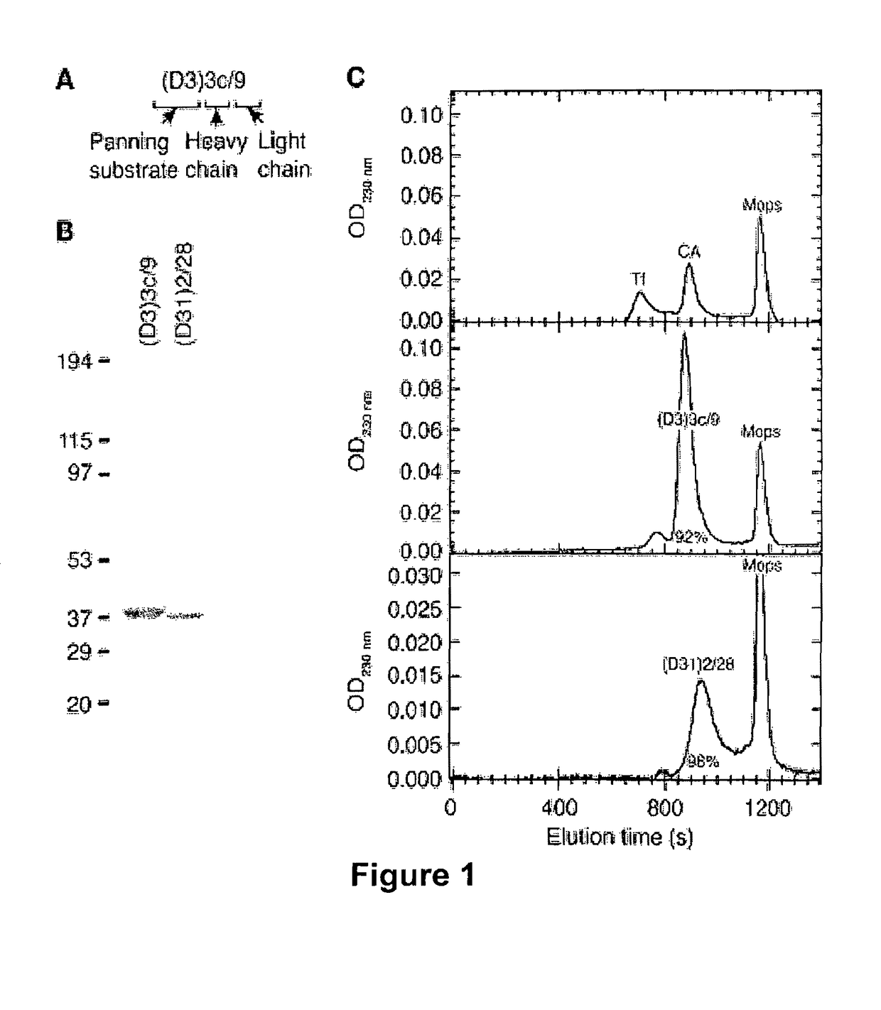
[0207]All human and animal studies were approved by the Institutional Review Board of the University of Pennsylvania. Using previously described methods (protocol 9.2, Barbas, C. F. I., Burton, D. R., Scott, J. K., and Silverman, G. J. 2001. Phage display: a laboratory manual. Cold Spring Harbor Laboratory Press. Cold Spring Harbor, N.Y., USA.), separate IgGκ and IgGλ, phage libraries were constructed using the phagemid vector pComb3X (Scripps Research Institute) from 4×10⁷ mono...
example 1
Isolation of Human Anti-Dsg3 and Anti-Dsg1 mAbs from a Phage Display Library Constructed from Lymphocytes from a Mucocutaneous PV Patient
[0217]RT-PCR was used to amplify mRNA for the Ig variable-region heavy chain (VH) and variable-region light chain (VL) fragments from peripheral blood lymphocytes isolated from a patient with active acute mucocutaneous PV. The resulting cDNA was then cloned into a phagemid vector, which facilitated the creation of an antibody phage display library, comprising approximately 4×10⁸ independent transformants. Phage particles, each displaying a particular VH and VL pair on their surface (folded into a single monovalent antigen-binding site) with the corresponding cDNA encoding the pair within the particle, were selected by panning against the extracellular domain of desmoglein substrate immobilized on ELISA plates. Four rounds of panning were conducted with scFvs selected against only Dsg3 (D3), only Dsg1 (D1), or both Dsg3 and Dsg1 (D31). Phage clones ...
example 2
The Repertoire of Human PV scFv mAbs Shows Various Patterns of Desmoglein-Specific Binding as Determined by Indirect Immunofluorescence and ELISA
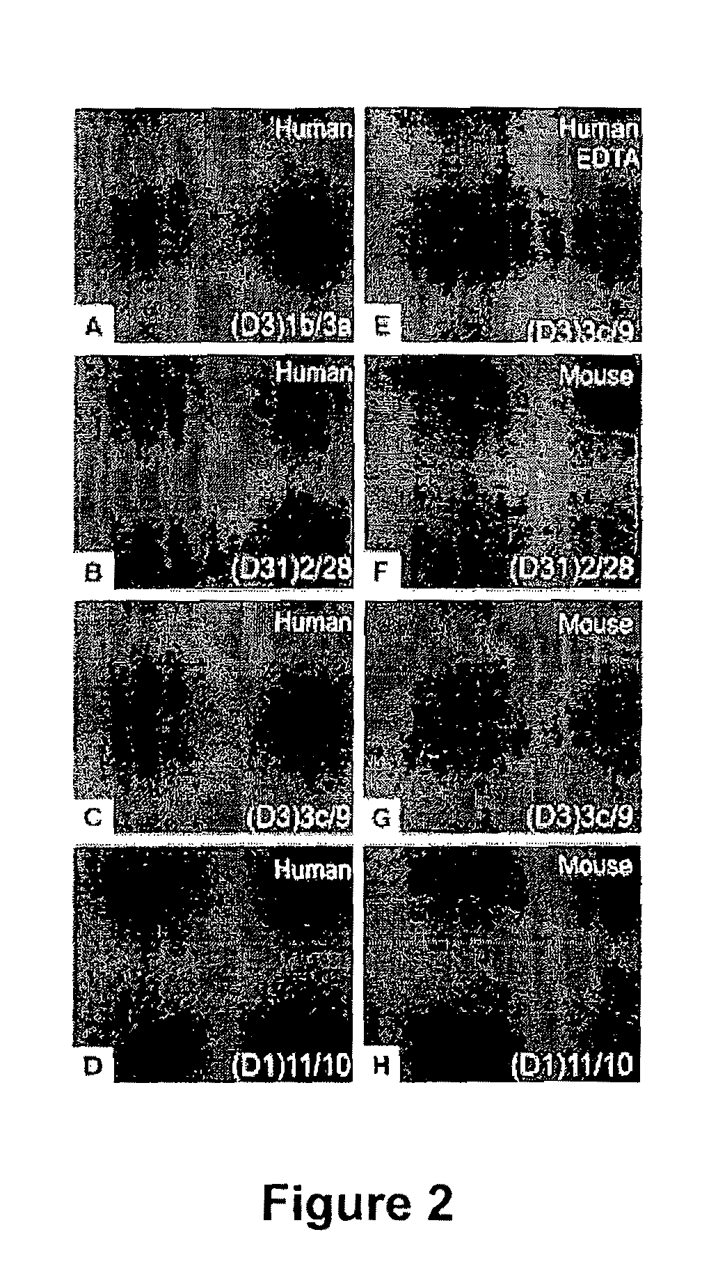
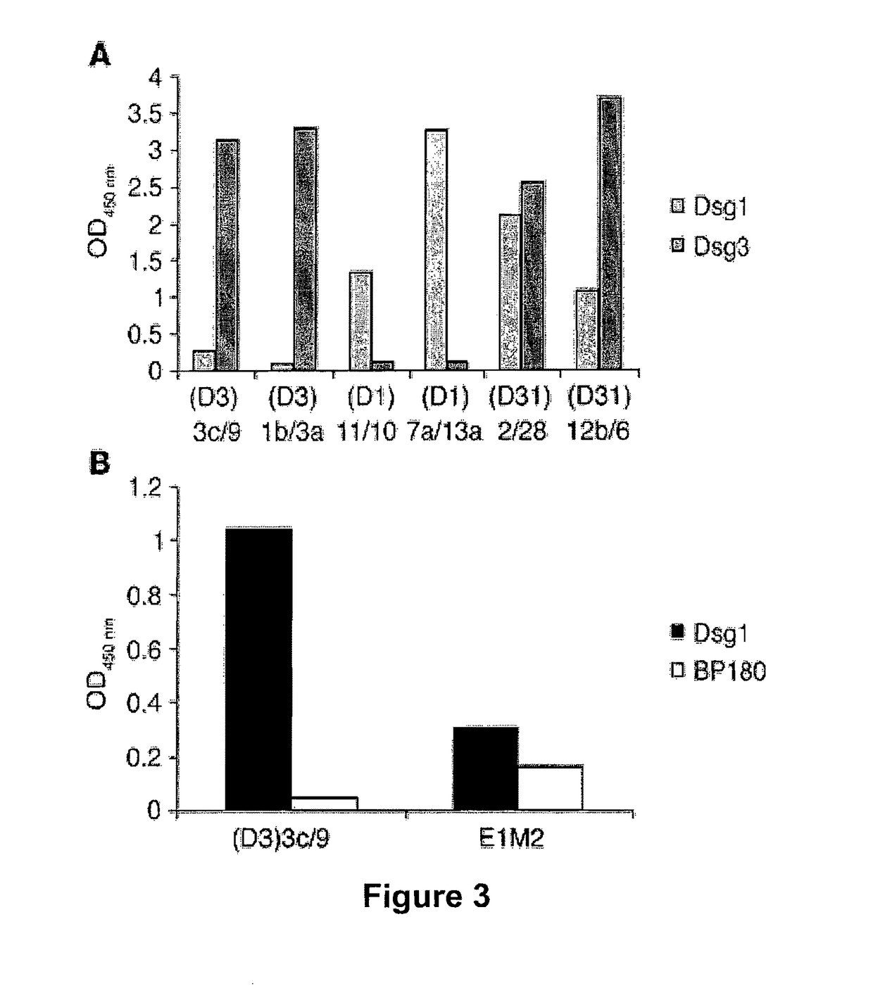
[0218]ScFvs were tested by indirect immunofluorescence (IIF) on normal human skin using anti-HA antibodies for detection in order to evaluate the ability of recombinant mAbs to bind native desmogleins in human tissue. In general, scFvs derived from phage libraries panned on Dsg3 showed the expected binding to the keratinocyte cell surface in the basal and immediate suprabasal layers, where Dsg3 is expressed (FIG. 2A), and those derived from libraries panned on Dsg3 and Dsg1 bound throughout the epidermis (FIG. 2B). However, 2 clones panned only on Dsg3, (D3)3c / 9 and (D3)3a / 9, bound throughout the epidermis (FIG. 2C and data not shown). As discussed below, by ELISA these scFv mAbs showed not only binding to Dsg3 but also slight binding to Dsg1. Interestingly, only 1 of the scFv mAbs derived from a phage library selected on Dsg1 stained human...
PUM
| Property | Measurement | Unit |
|---|---|---|
| time | aaaaa | aaaaa |
| time | aaaaa | aaaaa |
| pH | aaaaa | aaaaa |
Abstract
Description
Claims
Application Information