Method and magnetic resonance tomography apparatus for triggered acquisition of magnetic resonance data

a technology of magnetic resonance data and tomography equipment, which is applied in the direction of diagnostic recording/measuring, instruments, and using reradiation, can solve the problems of shortening the wait time, and achieve the effect of reliable and disruption-free data acquisition

Active Publication Date: 2013-08-27
SIEMENS HEALTHCARE GMBH
View PDF12 Cites 5 Cited by
  • Summary
  • Abstract
  • Description
  • Claims
  • Application Information

AI Technical Summary

Benefits of technology

[0013]An object of the present invention is to provide a method for triggering a magnetic resonance measurement that allows a reliable and disruption-free data acquisition even given variable heart frequency of the patient.
[0015]Instead of waiting for the next detection of the reference point (usually the R-spike), this is detected independently of the partial measurement. If the heart beat now accelerates, the time span of the RR-interval accordingly shortens. In spite of this shortening, however, no RR interval is skipped since the reference point is detected independently of the implementation of a partial measurement. If the reference point still occurs during a partial measurement, the beginning of the next partial measurement is accordingly delayed, meaning that the wait time is shortened.
[0018]The method according to the invention produces a more robust data acquisition given an irregular heart beat. Since the detection of the R-spike ensues independently of the end of a partial measurement, RR-intervals (and therefore no cardiac cycles) are no longer skipped. In methods whose magnetization preparation achieves a certain steady state, this steady state is accordingly maintained, while the magnetization decays into an undefined state upon omitting a cardiac cycle. The measurement times of the implemented measurements can therefore furthermore be shortened, and in particular measurements during breath-hold times are implemented optimally.
[0030]The time span can advantageously be determined depending on the ending of the data acquisition of the image data set acquired during the detection of the reference point. This means that the partial measurement is not interrupted immediately, but rather that at least the currently measured k-space line is completely acquired, and only after this is the partial measurement terminated. The data acquisition of a k-space line shifts in the range from 1-2 ms and is therefore markedly shorter than the additional steps necessary for data acquisition, for example preparation by an inversion pulse. Accordingly, the ending of the actual data acquisition of the currently acquired k-space line is normally less time-critical, which is why the acquisition of these data is still conducted.
[0031]The time span can be determined depending on the complete data acquisition of a specific number of image data sets. This guarantees that a minimum number of image data sets—and therefore of slice images—exists that the user considers to be necessary for the diagnostic finding. If the heart beat accelerates, for example, such that only a single slice is acquired per RR-interval, this could lead to a significant limitation of the significance of the acquired data. It is therefore reasonable to give the user the possibility to allow a minimum number of image data sets or, respectively, slices to be acquired in order to acquire the data set that the user requires.
[0033]Even when conducting perfusion measurements, the determination of the number of image data sets to be acquired advantageously ensues depending on the time interval of at least two of the last detected reference points. As already described, this procedure allows an adaptive adaptation to the accelerated heart beat. A threshold that cannot be fallen below can be particularly advantageously provided for the number of image data sets. The user can thus start with an arbitrarily high number of image data sets to be acquired; the partial measurements or the number of image data sets to be acquired are / is automatically adapted to the provided time span between the detected reference points (thus the RR-interval). A required minimum number of image data sets or slices are maintained. Complicated adaptations before the actual measurement thus are not necessary, so the residence time of the patient in the magnetic resonance tomography apparatus is shortened. If one considers that the actual measurement or, respectively, the measurement time often accounts for half of the time during which the patient is located within the magnetic resonance tomography apparatus, this type of EKG triggering entails a doubled savings potential with regard to the residence time of the patient. Moreover, setting up the measurement—thus the adjustment of the acquisition parameters—takes place faster. Additionally, the duration of the measurement as such can also be reduced to a minimum by the utilization of every occurring RR-interval. The total residence duration of the patient in the magnetic resonance tomography apparatus can thus be significantly shortened. Moreover, operating errors can be avoided.

Problems solved by technology

If the reference point still occurs during a partial measurement, the beginning of the next partial measurement is accordingly delayed, meaning that the wait time is shortened.

Method used


Image

  • Method and magnetic resonance tomography apparatus for triggered acquisition of magnetic resonance data
  • Method and magnetic resonance tomography apparatus for triggered acquisition of magnetic resonance data
  • Method and magnetic resonance tomography apparatus for triggered acquisition of magnetic resonance data

Examples


Embodiment Construction

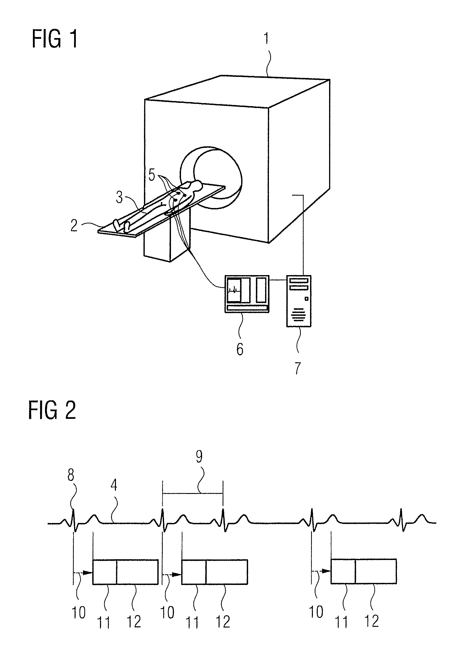
[0046]FIG. 1 shows a magnetic resonance system 1 having a bore in which a patient bed 2 can be inserted. To acquire magnetic resonance data from a patient 3, the patient 3 is supported on the patient bed 2 and electrodes 5 are located on the body of the patient 3 to detect an EKG signal 4. The signals detected by the electrodes 5 are relayed to the EKG device 6. The EKG device 6 communicates with the control device 7 of the magnetic resonance system 1, wherein the EKG device 6 and the control device 6 can naturally be spatially separated devices or can also be arranged in a single housing.

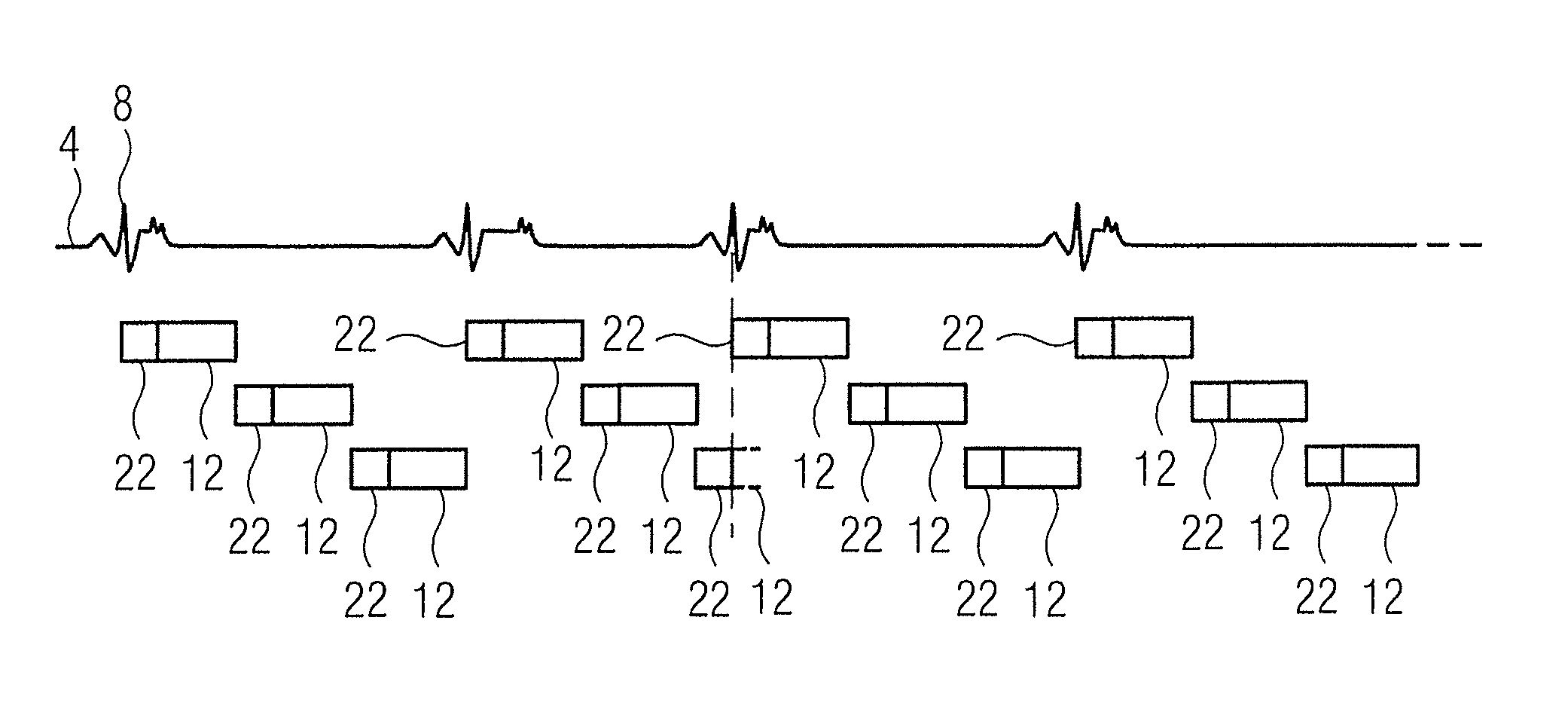
[0047]FIG. 2 shows a known method from the prior art for EKG-triggered implementation of a measurement. The R-spike 8 of the EKG signal 4 is used as a reference point of the cardiac phase. The time between the occurrence of two R-spikes 8 is designated as an RR-interval 9. The control device 7 of the magnetic resonance system 1 polls the trigger signal in the EKG device 6. If this occurs, a partial...


PUM


Abstract

In a method and magnetic resonance tomography apparatus for triggered implementation of a measurement (composed of partial measurements) in the magnetic resonance tomography apparatus, at least one image data set is determined from the data acquired within the scope of the partial measurements, and for triggering a reference point of the movement phase of the movement is used. The image data set is acquired in segments; the reference point is detected by a control device independent of a partial measurement, and the partial measurement following the detected reference point is conducted depending on the independently detected reference point. The wait time that specifies the interval from the end of the partial measurement to the beginning of the next partial measurement is adapted depending on the point in time of detection.

Description

BACKGROUND OF THE INVENTION[0001]1. Field of the Invention[0002]The present invention concerns a method for triggered implementation of a measurement composed of partial measurements in a magnetic resonance tomography apparatus, of the type wherein at least one image data set is determined from the data acquired within the scope of the partial measurements, and wherein a reference point of the movement phase of the movement is used for triggering.[0003]2. Description of the Prior Art[0004]For data acquisition of magnetic resonance tomography data, it is known to acquire the data in an EKG-triggered manner in order to minimize movement artifacts from the post-processed image data. The EKG signal (wherein EKG is the generally known abbreviation for electrocardiogram) is a measure of the electrical activity of the heart muscle fibers. The contraction of the heart muscle is produced by an electrical excitation, which is detected with the EKG apparatus. The EKG signal is a periodic signa...

Claims


Application Information

Patent Timeline
Patent Type & Authority Patents(United States)
IPC IPC(8): G01V3/00A61B5/352
CPCA61B5/055A61B5/7285G01R33/56308G01R33/56366G01R33/5673A61B5/0456A61B5/721G01R33/5601A61B5/352
Inventor LANDSCHUETZ, WILFRIEDSCHMIDT, MICHAELASPEIER, PETER
Owner SIEMENS HEALTHCARE GMBH
Why Eureka
Try Eureka