
Image analysis method and system

a technology of image analysis and system, applied in the field of image analysis, can solve the problems of false indication of metabolic information for a particular organ, and affecting the accuracy of data analysis

Inactive Publication Date: 2013-09-03
GE HEALTHCARE LTD
View PDF6 Cites 11 Cited by
  • Summary
  • Abstract
  • Description
  • Claims
  • Application Information

AI Technical Summary

Benefits of technology

This approach effectively separates tissues with different kinetic behaviors, enhances signal extraction, and reveals clinically significant features that were previously obscured, improving the accuracy of image analysis and diagnosis.

Problems solved by technology

Although many tracers are currently used in PET studies to good effect, where various different tracers are used, e.g. for comparative studies of the same anatomical region, their differing biochemical properties can give rise to false indications of metabolic information for a particular organ.
Hence, when using various tracers, data analysis is more difficult and clinicians' attention might not be accurately drawn to the most clinically important organs, or regions of organs, since, for example, tissues or vessels surrounding them may show a higher tracer uptake than those more clinically significant areas.

Method used


Image

  • Image analysis method and system
  • Image analysis method and system
  • Image analysis method and system

Examples


Embodiment Construction

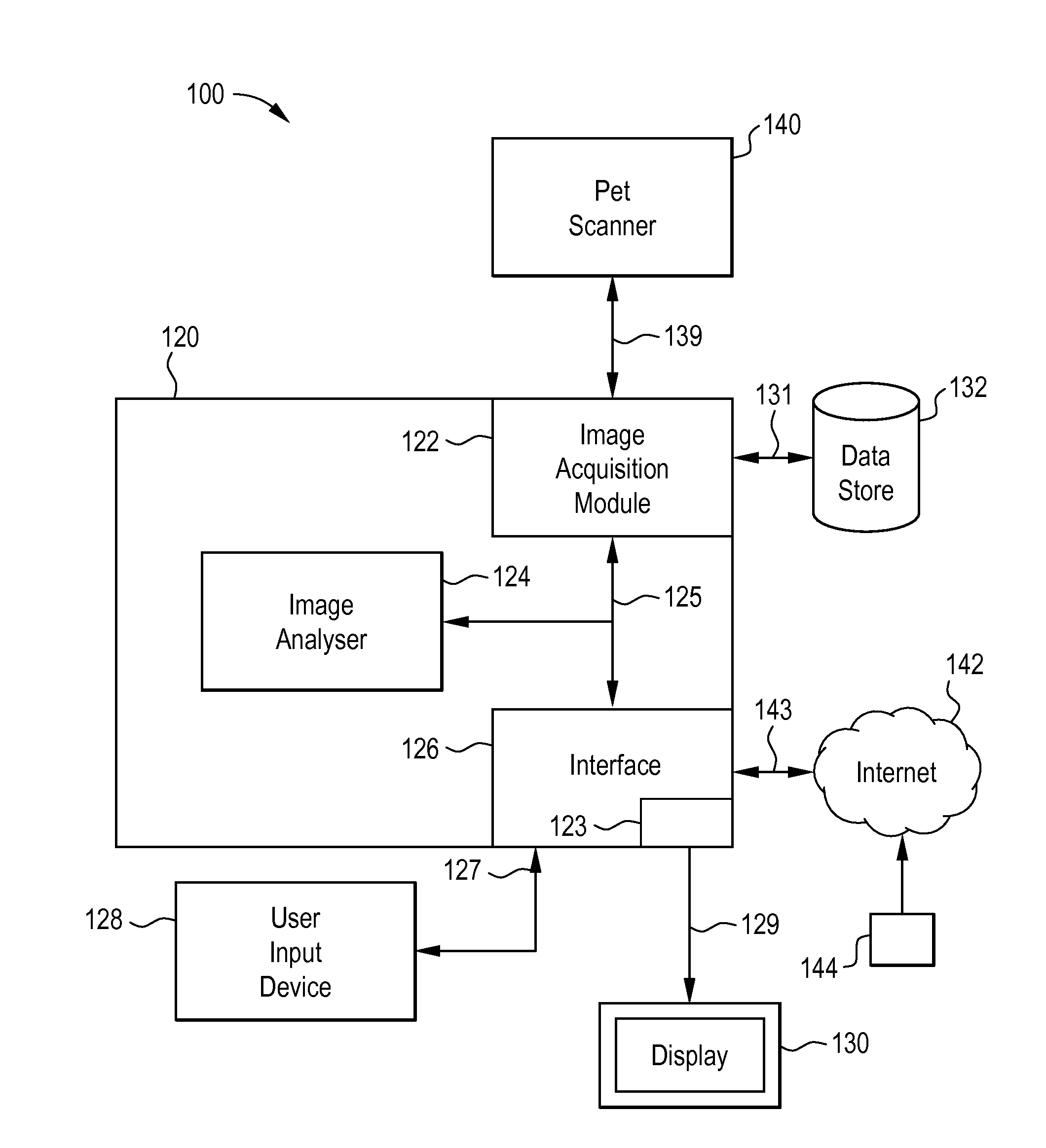
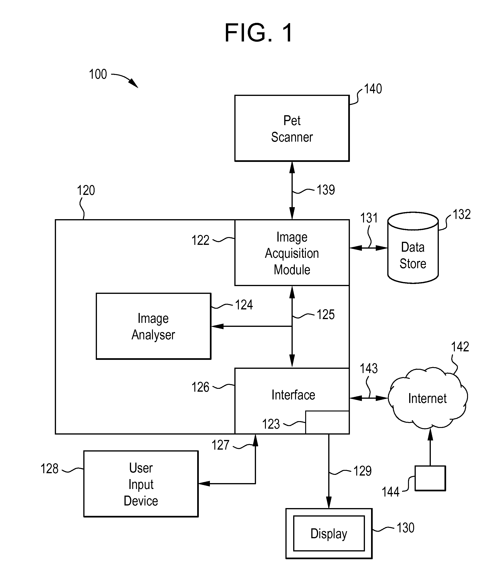
[0027]FIG. 1 shows a system 100 for aiding clinical diagnosis of a subject according to an embodiment of the present invention. The system 100 includes a data processing apparatus 120 that is configured to provide various interfaces 123,126, an image acquisition module 122 and an image analyser 124. The interfaces 123,126, image acquisition module 122 and image analyser 124 can be logically coupled together by way of a data bus 125 under the control of a central processing unit (not shown).

[0028]The data processing apparatus 120 provides a first general purpose interface 126 for interfacing the data processing apparatus 120 to external components. In this embodiment the external components include: an input data link 127 coupled to at least one user input device 128 (e.g. a mouse / keyboard / etc.), a network data link 143 coupled to the Internet 142, and a display data link 129 coupled to a display 130. Additionally, the general purpose interface 126 also provides a GUI 123 through whi...


PUM


Abstract

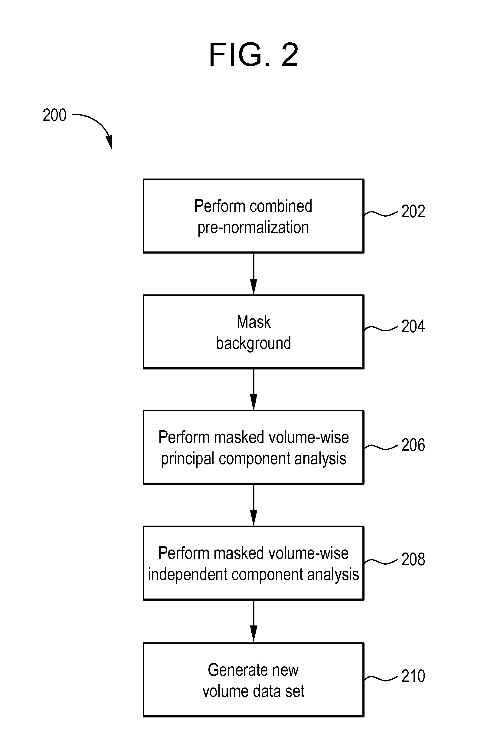
The invention relates to a system and method for enhancing image data obtained from a positron emission tomography (PET) scan. In various embodiments, the method comprises transforming an original image data set to provide a first modified image data set by performing a masked volume-wise principal component analysis (MVW-PCA) on the original image data set. The first modified image data set is then transformed to provide a second modified image data set by performing a masked volume-wise independent component analysis (MVW-ICA) on the first modified image data set, the second modified image data set thereby comprising enhanced image data.

Description

[0001]This application is a filing under 35 U.S.C. 371 of international application number PCT / US2010 / 034477, filed May 12, 2010, which claims priority to U.S. application No. 61 / 177,374 filed May 12, 2009, the entire disclosure of which is hereby incorporated by reference.FIELD OF THE INVENTION[0002]The present invention generally relates to image analysis. In particular, the present invention relates to methods and apparatus for enhancing image data obtained from a positron emission tomography (PET) scan, including computer tomography (CT) images from combined PET / CT studies.BACKGROUND OF THE INVENTION[0003]Various medical imaging techniques exist to aid clinicians in the diagnosis of pathological conditions caused, for example, by anatomic or functional manifestations of a disease. Many such techniques produce one or more image frames that can be used to highlight to the clinician various instantaneous or temporal variations in anatomical and / or functional properties of a patient...

Claims


Application Information

Patent Timeline
Patent Type & Authority Patents(United States)
IPC IPC(8): G06K9/00G01T1/164
CPCA61B6/037G06T5/003A61B6/03G06T2207/10084G06T2207/10104G06T2207/20016G06T2207/30016G06T5/73
Inventor RAZIFAR, PASHALANGSTROM, BENGT
Owner GE HEALTHCARE LTD
Learn More