Medical image data alignment apparatus, method and program

a technology for aligning apparatus and medical images, applied in image enhancement, tomography, instruments, etc., can solve the problems of low resolution of spect image, long time to perform alignment with sufficient accuracy and reproducibility, and increase the number of false positives of ct image alone, etc., to achieve high correlation in image signal values, high accuracy, and sufficient reproducibility

Active Publication Date: 2013-10-15
CANON MEDICAL SYST COPRPORATION
View PDF5 Cites 10 Cited by
  • Summary
  • Abstract
  • Description
  • Claims
  • Application Information

AI Technical Summary

Benefits of technology

[0021]With the foregoing in view, it is an object of the present invention to provide a medical image data alignment apparatus, method and program which can align different kinds of image data on a tissue to be objected, between a medical image obtained by a first kind image diagnostic apparatus which can generate an image of form information on a tissue to be objected and a medical image obtained by a second kind image diagnostic apparatus which can generate an image of function information on the tissue to be objected, with sufficient accuracy and reproducibility, in a short time.
[0042]According to the medical image data alignment apparatus, method and program of the present invention, alignment of the specified first medical image data and the specified second medical image data is performed in steps via the atlas second medical image data, whereby the following functional effects are exhibited.
[0045]The specified first medical image data holds form information of the tissue to be objected, so it is easy to add the first coordinate information to the specified first medical image data, at high accuracy with sufficient reproducibility. The specified second medical image data and the atlas second medical image data, which are the same kind of data, have high correlation in the image signal values, and the alignment of these image data based on this correlation can be performed in a short time and at high accuracy. The atlas second medical image data and the specified first medical image data, on the other hand, are different kinds of image data from each other, but these image data are aligned based on the first coordinate information added to the first medical image data and the second coordinate information added to the atlas second medical image data, therefore this alignment can be performed in a short time at high accuracy as well. The final alignment of the specified first medical image data and the specified second medical image data can be performed by arithmetic processing using the determined first transformation matrix and the second transformation matrix, therefore this alignment can also be performed in a short time at high accuracy.
[0046]Hence according to the medical image data alignment apparatus, method and program of the present invention, different kinds of image data on the tissue to be objected, between the medical image obtained by the first kind image diagnostic apparatus which can generate an image of the form information of the tissue to be objected, and the medical image obtained by the second kind image diagnostic apparatus which can generate an image of the function information of the tissue to be objected, can be aligned in a short time with sufficient accuracy and reproducibility.

Problems solved by technology

However a stricture of coronary artery does not always cause immediate ischemia, so diagnosis by CT image alone increases the number of false positives.
However the information to be generated into an image is different and therefore correlation is low between a CT image and a SPECT image, and the resolution of the SPECT image is low, so even if the physician has extensive experience in image diagnosis, it takes a long time to perform alignment with sufficient accuracy and reproducibility.
The methods according to the Non-patent Documents 2 and 3 mentioned above perform stable alignment by extracting the left ventricle portion, of which correlation with the SPECT image is relatively high, from the CT image or MRI image, and using this portion for alignment, but alignment accuracy greatly depends on the result of segmenting the left ventricle, so a problem is that the alignment accuracy falls considerably unless a good segmentation result is obtained.
H10-137231 mentioned above, on the other hand, must extract some anatomical form information from the SPECT image of which resolution is low, so this method is effective if an area of which form information can be extracted relatively easily, like the case of a skull, is imaged, but the problem is that it takes an enormous amount of time to extract the form information from the SPECT image if such an appropriate area does not exist.

Method used


Image

  • Medical image data alignment apparatus, method and program
  • Medical image data alignment apparatus, method and program
  • Medical image data alignment apparatus, method and program

Examples


Embodiment Construction

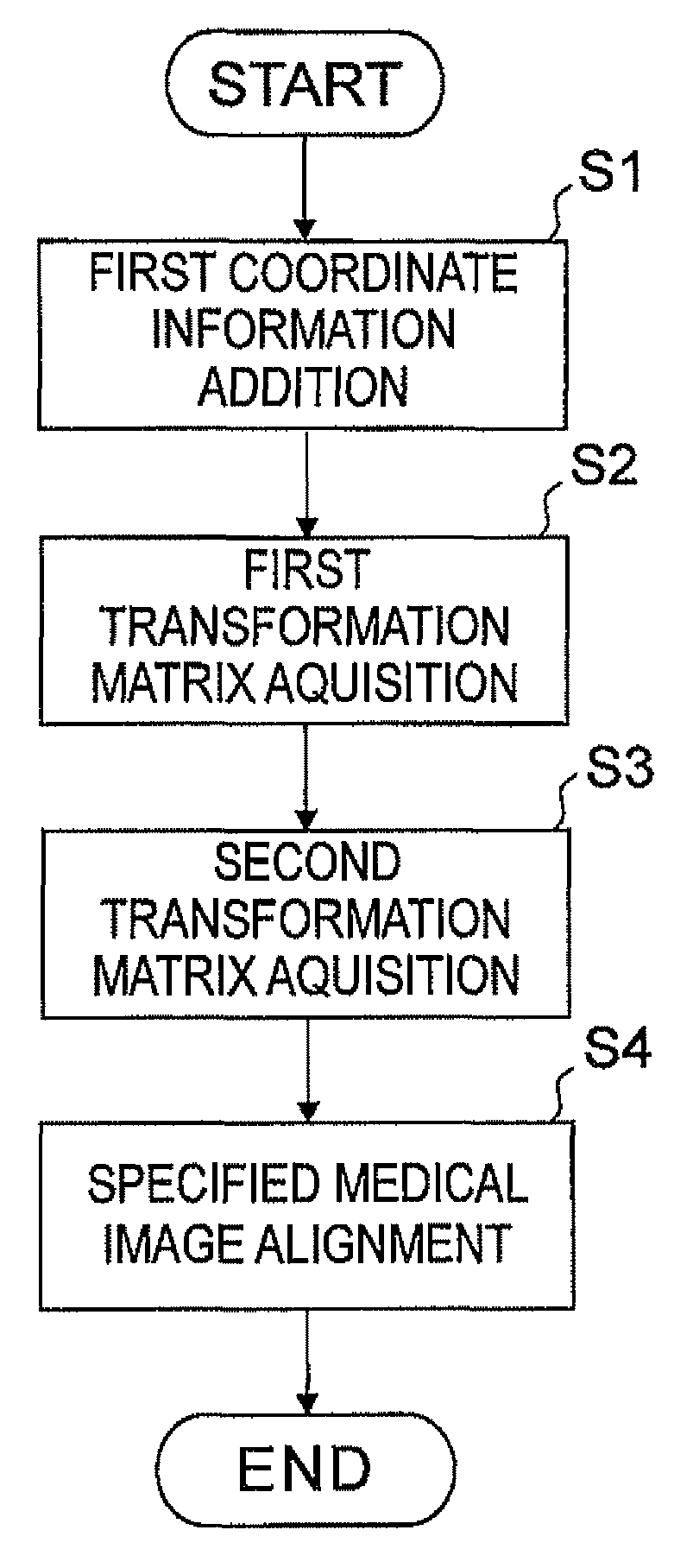
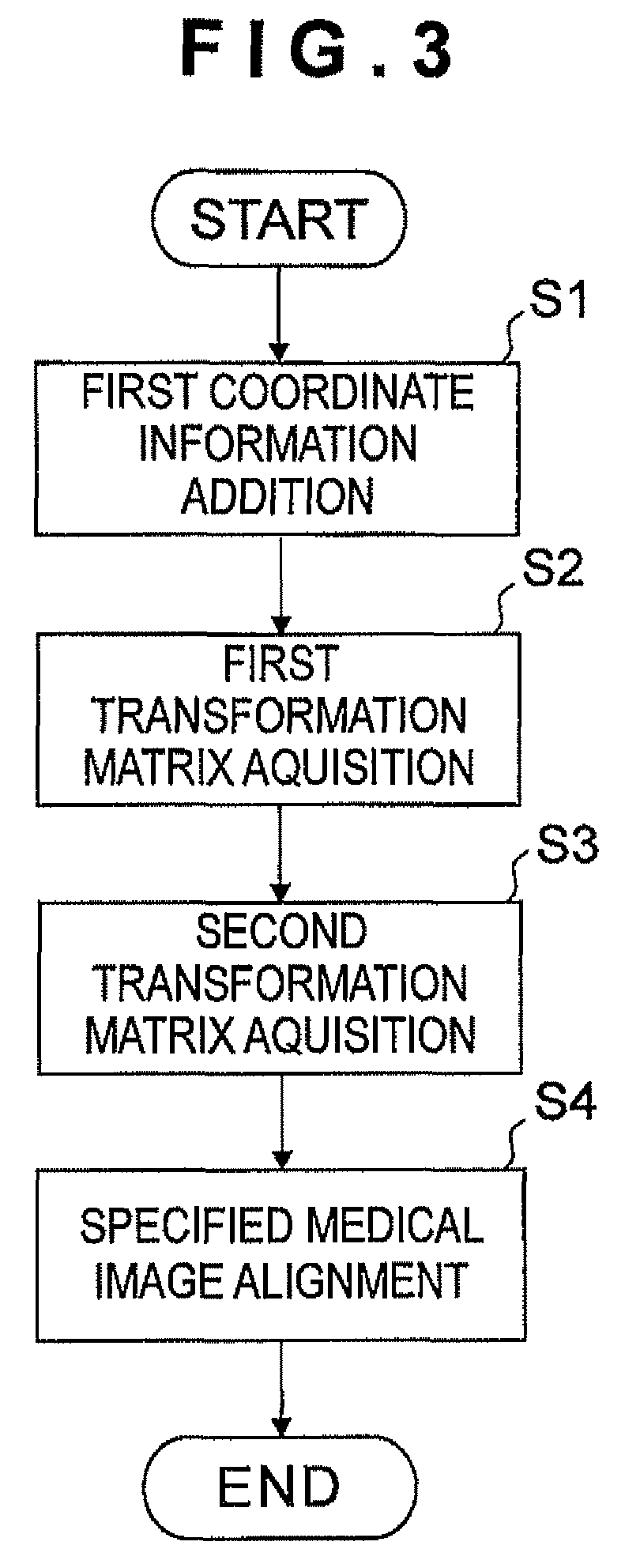
[0055]Embodiments of the present invention will now be described in detail with reference to the above mentioned drawings. First, a medical image data alignment apparatus according to an embodiment of the present invention will be described with reference to FIG. 1 and FIG. 2.

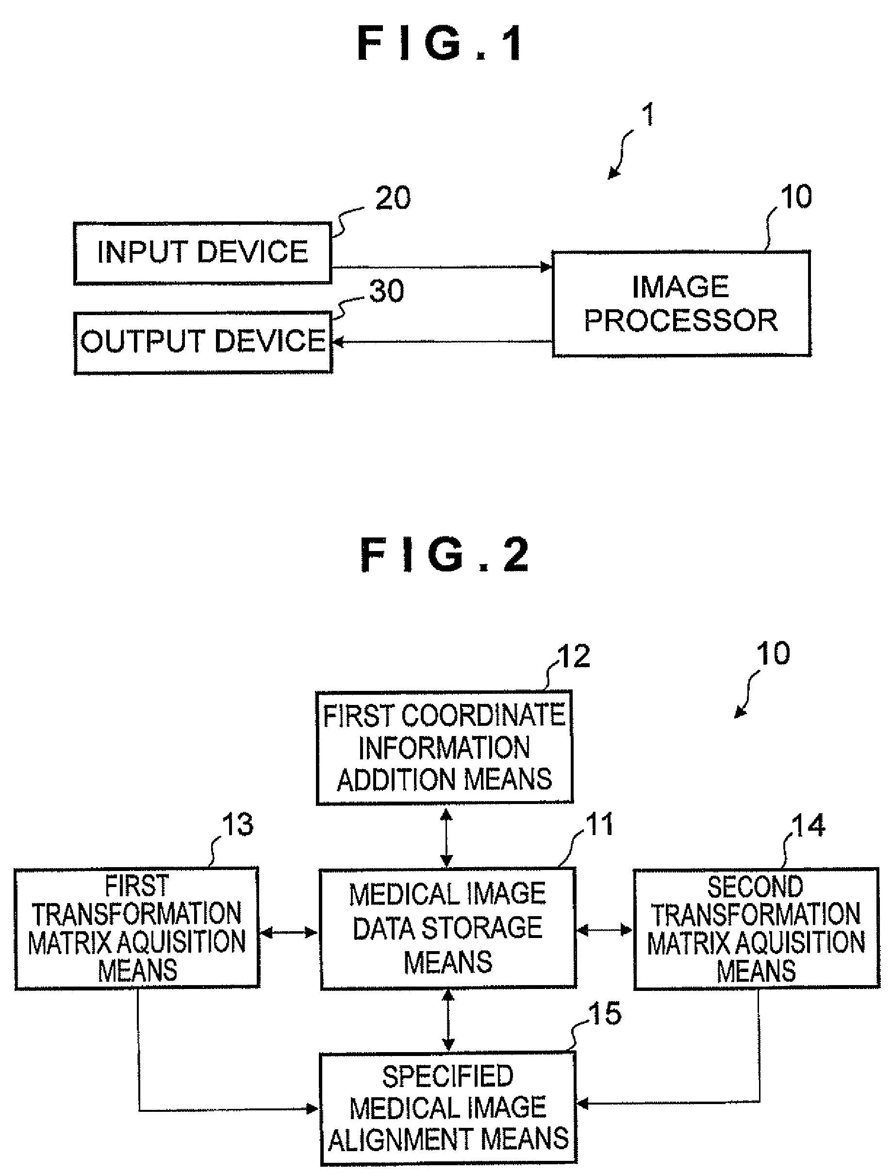
[0056]The medical image data alignment apparatus 1 shown in FIG. 1 aligns specified first medical image data holding form information of a tissue to be objected of a specified subject (specified patient) obtained by a first kind image diagnostic apparatus, and specified second medical image data holding function information of the tissue to be objected of the specified subject, obtained by a second kind image diagnostic apparatus, and has an image processor 10 which is a computer or the like, an input device 20 such as a mouse and keyboard, and an output device 30 which is an image display device, for example.

[0057]In the present embodiment, a case when the first kind image diagnostic apparatus is an X-ray CT a...


PUM


Abstract

An object of the present invention is to provide a medical image data alignment apparatus, method and program capable of aligning different kinds of image data on a tissue to be objected at high precision and in a short time. According to the present invention, a patient SPECT and a patient CT are aligned via atlas data. The atlas data is a standard of SPECT image data, and is created based on SPECT image data of a plurality of patients. The patient SPECT and the atlas data are aligned based on the correlation of image signal values thereof, and a first transformation matrix T1 is determined. The atlas data and the patient CT are aligned based on the coordinate information added to these data in advance, and a second transformation matrix T2 is determined. The patient SPECT and the patient CT are aligned using the first transformation matrix T1 and the second transformation matrix T2.

Description

RELATED APPLICATIONS[0001]This application claims the priority of Japanese Patent Application No. 2009-218831 filed on Sep. 24, 2009, which is incorporated herein by reference.BACKGROUND OF THE INVENTION[0002]1. Field of the Invention[0003]The present invention relates to a technology for aligning, among a plurality of medical images, image data on a tissue to be objected to be an object of diagnosis and observation, including such organs as a heart, liver, pancreas, kidney, spleen, brain and lung, or such biotissue as a tumor (coordinating, among the images, each image point corresponding to each portion of a tissue to be objected in each image), and more particularly to a medical image data alignment apparatus, method and program for aligning different kinds of image data on a tissue to be objected, between a medical image obtained by an image diagnostic apparatus (hereafter called “first kind image diagnostic apparatus”) capable of generating an image of anatomical form informati...

Claims


Application Information

Patent Timeline
Patent Type & Authority Patents(United States)
IPC IPC(8): G06K9/00
CPCG06T7/0026A61B6/032A61B6/037A61B6/463G06T2207/10081G06T2207/10088G06T2207/10108G06T2207/30004G06T7/32
Inventor AZEMOTO, SHOGOGOKO, YASUAKIHOASHI, MASAKATSU
Owner CANON MEDICAL SYST COPRPORATION
Why Eureka
Try Eureka