Medical image processing apparatus, method, and program
a technology of image processing and image enhancement, applied in image analysis, image enhancement, instruments, etc., can solve the problems of not directly detecting an abnormal pattern itself, not being able to detect a pattern present in an area not contrast enhanced, and ignoring abnormal patterns
- Summary
- Abstract
- Description
- Claims
- Application Information
AI Technical Summary
Benefits of technology
Problems solved by technology
Method used
Image
Examples
first embodiment
[0063]Hereinafter, the present invention will be described with reference to the accompanying drawings.
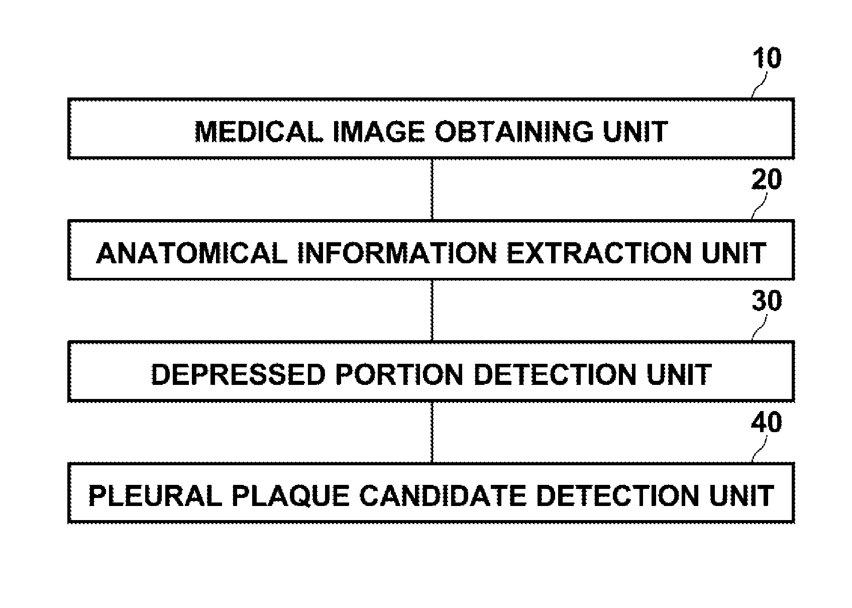
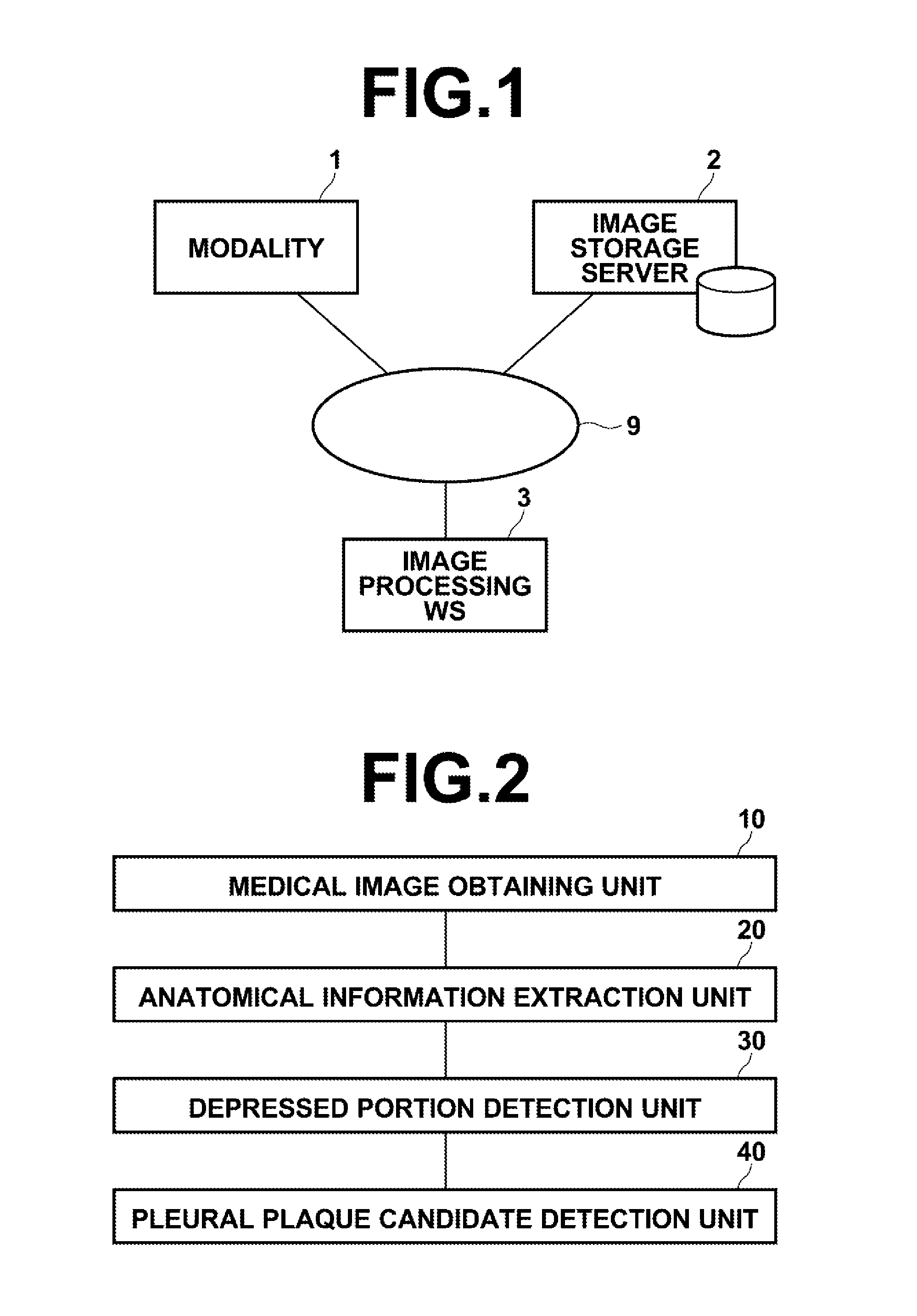
[0064]FIG. 1 is a hardware configuration diagram of a medical image processing apparatus, illustrating an overview thereof. As shown in FIG. 1, the system includes modality 1, image storage server 2, and image processing workstation 3 communicatably linked via network 9.
[0065]Modality 1 is equipment for obtaining a medical image V representing a test body, and more specifically it is CT equipment, MRI equipment, PET, ultrasonic diagnostic equipment, or the like.
[0066]Image storage server 2 is a computer for storing / managing a medical image V obtained by modality 1 and a medical image V generated in workstation 3 by image processing, and includes an external large capacity recording medium and database management software (for example, ORDB (object relational database) management software).
[0067]In the present embodiment, a software program that provides a function to detect a pleur...
second embodiment
[0112]Hereinafter, the present invention will be described.
[0113]As described above, the pleural plaque is irregular white plate like thickening developed in the parietal pleura, which characteristically appears in morphology, size, and peripheral or inner density in a diagnostic image. The second embodiment focuses attention on the morphology and density of a pleural plaque and performs determination of a pleural plaque candidate using a normal medical image, in addition to the detection of a first pleural plaque candidate based on the depressed portion described in the first embodiment. Note that the hardware configuration is identical to that of the first embodiment.
[0114]A configuration related to a medical image processing function according to the second embodiment of the present invention will now be described.
[0115]FIG. 5 is a block diagram illustrating a portion of workstation 3 related to the medical image processing function according to the second embodiment of the prese...
third embodiment
[0134]Hereinafter, a third embodiment will be described.
[0135]As described above, the pleural plaque, in general, is irregular white plate like thickening developed in the parietal pleura. There may be a case in which such thickening outside of a lung is developed in patches and in such a case a pleural plaque may be detected as an uneven portion of the parietal pleura.
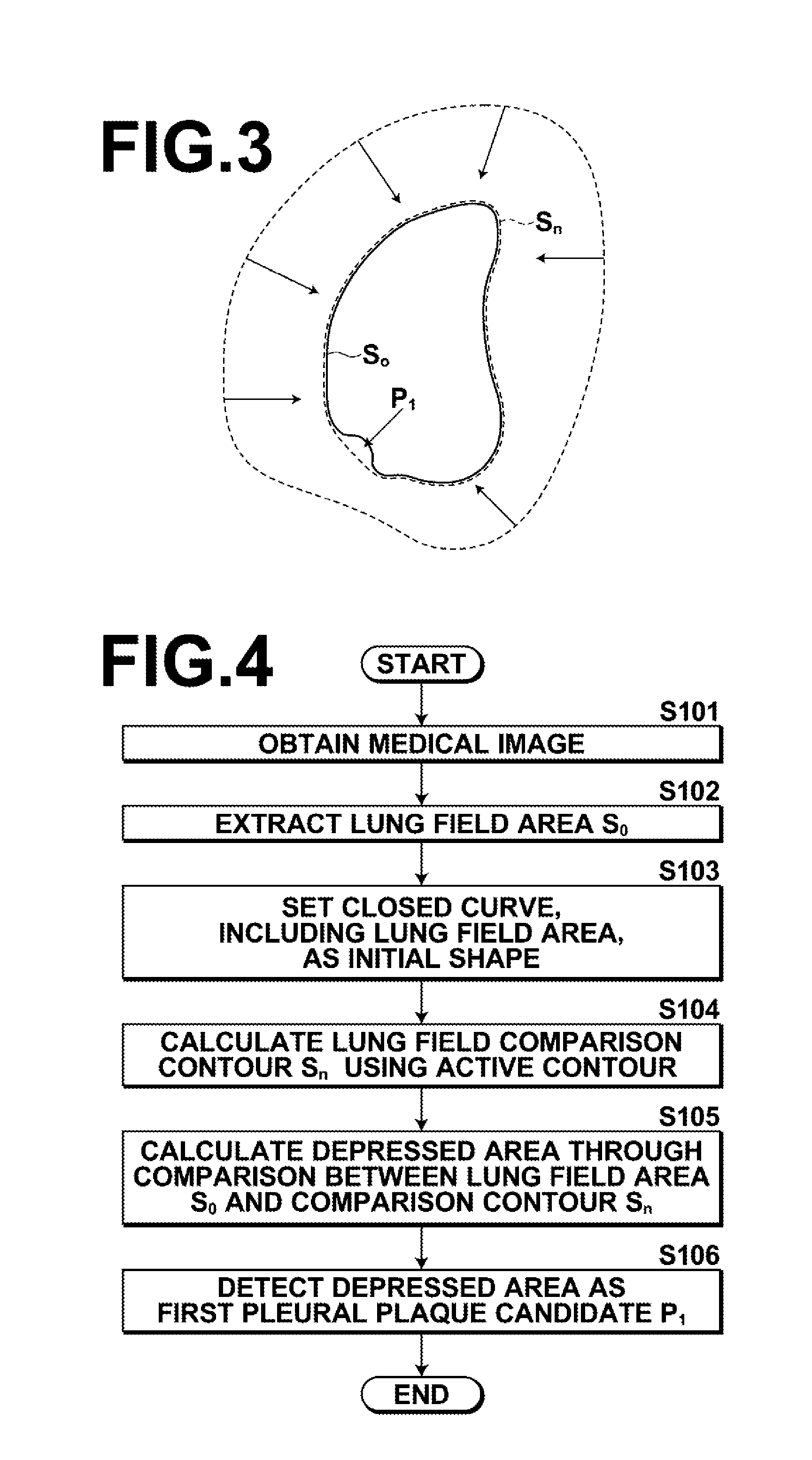
[0136]Consequently, in the third embodiment, a portion of a detected lung field contour S0 having an unevenness value greater than a threshold value is detected as a third pleural plaque candidate, thereby providing diagnostic assistance.
[0137]In the first embodiment, a first pleural plaque candidate P1 is detected based on a depressed portion, while in the third embodiment, a pleural plaque candidate is detected by taking into account an uneven portion of a lung field contour in addition to a depressed portion. Note that the hardware configuration is identical to that of the first embodiment. Further, it is possible ...
PUM
 Abstract
Description
Claims
Application Information
 - R&D
- Intellectual Property
- Life Sciences
- Materials
- Tech Scout
- Unparalleled Data Quality
- Higher Quality Content
- 60% Fewer Hallucinations
Browse by: Latest US Patents, China's latest patents, Technical Efficacy Thesaurus, Application Domain, Technology Topic, Popular Technical Reports.
© 2025 PatSnap. All rights reserved.Legal|Privacy policy|Modern Slavery Act Transparency Statement|Sitemap|About US| Contact US: help@patsnap.com


