Medical devices with encapsulated visibility particles

a technology of visibility particles and medical devices, applied in the field of medical devices, can solve the problems of affecting the flexibility and other mechanical properties of segments, aging degradation, and inability to withstand aging, and achieve the effects of reducing hydrolysis and/or oxidation, and enhancing stability

Active Publication Date: 2013-11-26
MEDTRONIC INC
View PDF20 Cites 1 Cited by
  • Summary
  • Abstract
  • Description
  • Claims
  • Application Information

AI Technical Summary

Benefits of technology

[0013]In another embodiment, the present disclosure provides a method of increasing the shelf life of at least a portion of a medical device. In certain embodiments, the method includes: providing a base polymeric material (i.e., a first polymeric material); and distributing coated tungsten and / or tungsten carbide particles within the base polymeric material, wherein the coated particles include an additional polymeric coating material (i.e., a second polymeric material) that is more hydrophobic than the base polymeric material. In other embodiment, the method includes providing a base polymeric material (i.e., a first polymeric material); and distributing coated tungsten and / or tungsten carbide particles within the base polymeric material, wherein the coated particles include a coating material that is a substantial barrier to the transmission of water and / or water vapor.

Problems solved by technology

Although it is known that polyether block amides can be loaded with tungsten and / or tungsten carbide to provide visibility using both fluoroscopy and ultrasonic imaging, these compositions can, in some instances, suffer from degradation during aging.
The degradation can adversely impact the flexibility and other mechanical properties of the segments in which the visibility materials are provided.

Method used


Image

  • Medical devices with encapsulated visibility particles
  • Medical devices with encapsulated visibility particles

Examples


Embodiment Construction

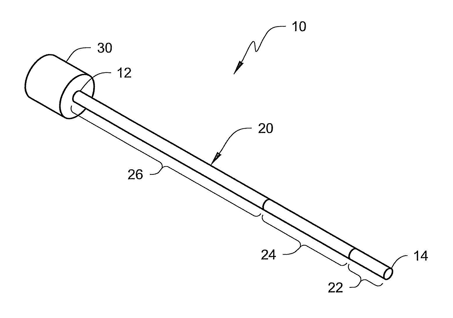
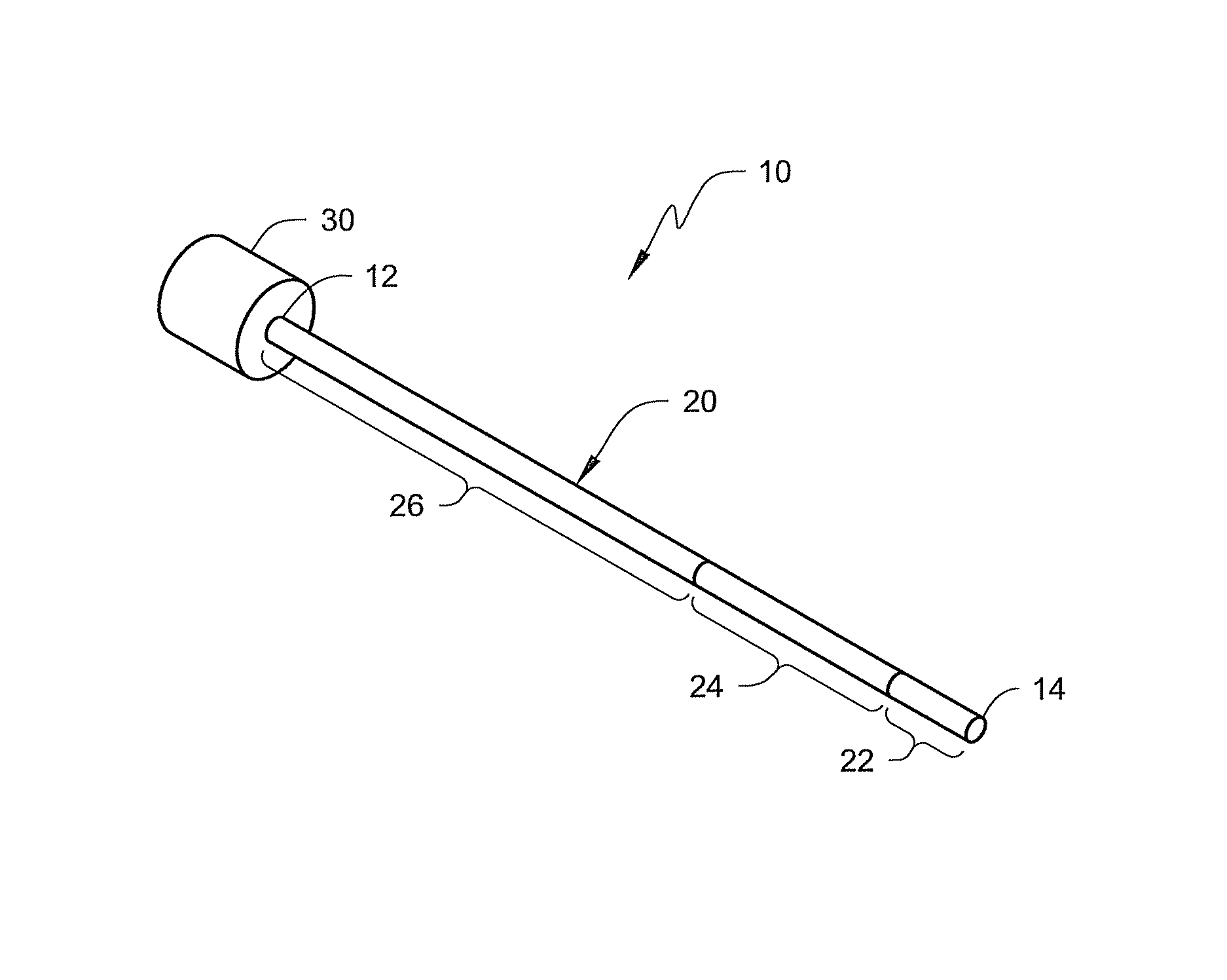
[0007]The present disclosure is directed to medical devices (such as, e.g., catheters, guide catheters, etc.) that include one or more selected segments that are constructed using visibility materials compounded with one or more polymeric materials that make the selected segments visible using both fluoroscopy and ultrasonic imaging. The visibility material may take the form of tungsten and / or tungsten carbide particles dispersed within a polymeric material.

[0008]The use of visibility material as described herein can potentially promote enhanced visibility of a medical device (such as a guide catheter) using both fluoroscopic and ultrasonic imaging techniques. This feature provides a physician with flexible imaging options. The medical device can be used by physicians who prefer fluoroscopic imaging, for example, as well as those who prefer ultrasound. In each case, the physician may select the same guide catheter without regard to the desired imaging modality.

[0009]In some embodime...


PUM

PropertyMeasurementUnit
thicknessaaaaaaaaaa
particle sizeaaaaaaaaaa
diameteraaaaaaaaaa

Abstract

The preparation and use of medical devices having radiopaque and echogenic materials including coated tungsten and / or tungsten carbide particles are disclosed herein.

Description

[0001]This application claims the benefit of U.S. Provisional Application Ser. No. 61 / 117,198, filed Nov. 23, 2008, which is herein incorporated by reference in its entirety.TECHNICAL FIELD[0002]The present disclosure relates generally to medical devices and, more particularly, to medical devices that include selected segments that are visible using both fluoroscopy and ultrasonic imaging.BACKGROUND[0003]It may be desirable to monitor the position of medical devices such as, e.g., catheters, while they are within a patient's body. For example, it may be useful to monitor the position of guide catheters which are used to place catheters, electrode leads and the like in desired locations within the body of a patient. A guide catheter typically includes an elongated sheath that is inserted into a blood vessel or another portion of the body. A catheter or lead is introduced through an inner channel defined by the sheath.[0004]To enable precise positioning of a medical device, one or mor...

Claims


Application Information

Patent Timeline
Patent Type & Authority Patents(United States)
IPC IPC(8): A61M25/098B05D7/00
CPCA61L29/06A61L29/085A61L29/106A61L29/18A61M25/0108C08L77/12A61M25/0662A61M2025/0681
Inventor EBERT, MICHAEL J.WALDHAUSER, STEVEN L.SCHNEIDER, MARK D.SHIPE, GREGORY P.
Owner MEDTRONIC INC
Why Eureka
Try Eureka