Method and apparatus for continuous guidance of endoscopy

a technology of endoscopy and guidance apparatus, which is applied in the direction of static indicating devices, instruments, applications, etc., can solve the problems of navigation errors, difficulty in identifying airways in ct slices, and exacerbated difficulties

Active Publication Date: 2014-03-18
PENN STATE RES FOUND
View PDF81 Cites 158 Cited by
  • Summary
  • Abstract
  • Description
  • Claims
  • Application Information

AI Technical Summary

Benefits of technology

[0012]This invention is directed to continuous guidance of endoscopy during a live procedure. A data-set based on 3D image data is pre-computed including reference information representative of a predefined route through a body organ to a final destination. A plurality of live real endoscopic (RE) images are displayed as an operator maneuvers an endoscope within the body organ. Information is presented corresponding to an initial reference location along the predefined route enabling the operator to move the endoscope toward the reference location. A registration and tracking algorithm is invoked that registers the data-set to one or more of the RE images and continuously maintains the registration as the endoscope is locally maneuvered. Information corresponding to another reference location along the predefined route is presented, enabling the endoscope operator to move the endoscope close to this new reference location, and these steps are repeated until the endoscope is within the vicinity of the final destination. Additional information related to the final destination is then presented enabling the endoscope operator to decide on a final maneuver for the procedure.

Problems solved by technology

It has been shown, however, that the path is often chosen incorrectly using this approach and that physicians have difficulty in identifying airways in CT slices as early as the second generation.12,13
Central to this procedure is the task of navigating the endoscope tip to the previously-defined ROIs, relying on both the live endoscopic video feed as well as the analysis of the 3D image performed in Phase I. This task is difficult for several reasons: 1) the ROIs can lie beyond the walls of the hollow organ and are not visible in the endoscopic video; 2) the endoscopic video is markedly different from the 3D radiologic images in which these ROIs are defined; and 3) endoscopic views from different locations within the organ can be visually indistinguishable.
In bronchoscopy, these difficulties are exacerbated by the complexity of the tracheobronchial airway tree in which the bronchoscope operates.
These effects result in navigation errors and are known to contribute to large variations in skill level between different physicians.14,15 Such errors may also result in missed diagnoses, necessitating invasive and potentially fatal follow-up procedures.3,4 Previously, image-based and electromagnetic (E / M) guidance techniques have been proposed to aid physicians both in navigation to ROIs and in improving the biopsy accuracy of ROIs.
E / M guidance techniques generate an E / M field around the patient, and locate the bronchoscope using an E / M sensor inserted through the instrument channel of the bronchoscope.16-21 These techniques require special hardware that adds to the cost of each procedure and limits the type of bronchoscope that can be used.
Because the E / M probe occupies the bronchoscope's instrument channel, these techniques are not suitable for the smaller channels present on small-diameter endoscopes designed for pediatric or peripheral lung procedures.
In addition, E / M registration can only locate the bronchoscope relative to an external field.
However, it has previously been computationally intensive, limiting its use to single-frame or buffered-video applications, and requiring the physician to wait several seconds for each registration result.

Method used


Image

  • Method and apparatus for continuous guidance of endoscopy
  • Method and apparatus for continuous guidance of endoscopy
  • Method and apparatus for continuous guidance of endoscopy

Examples


Embodiment Construction

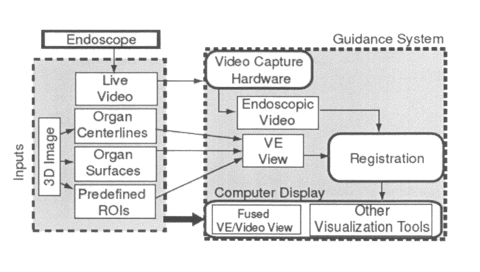
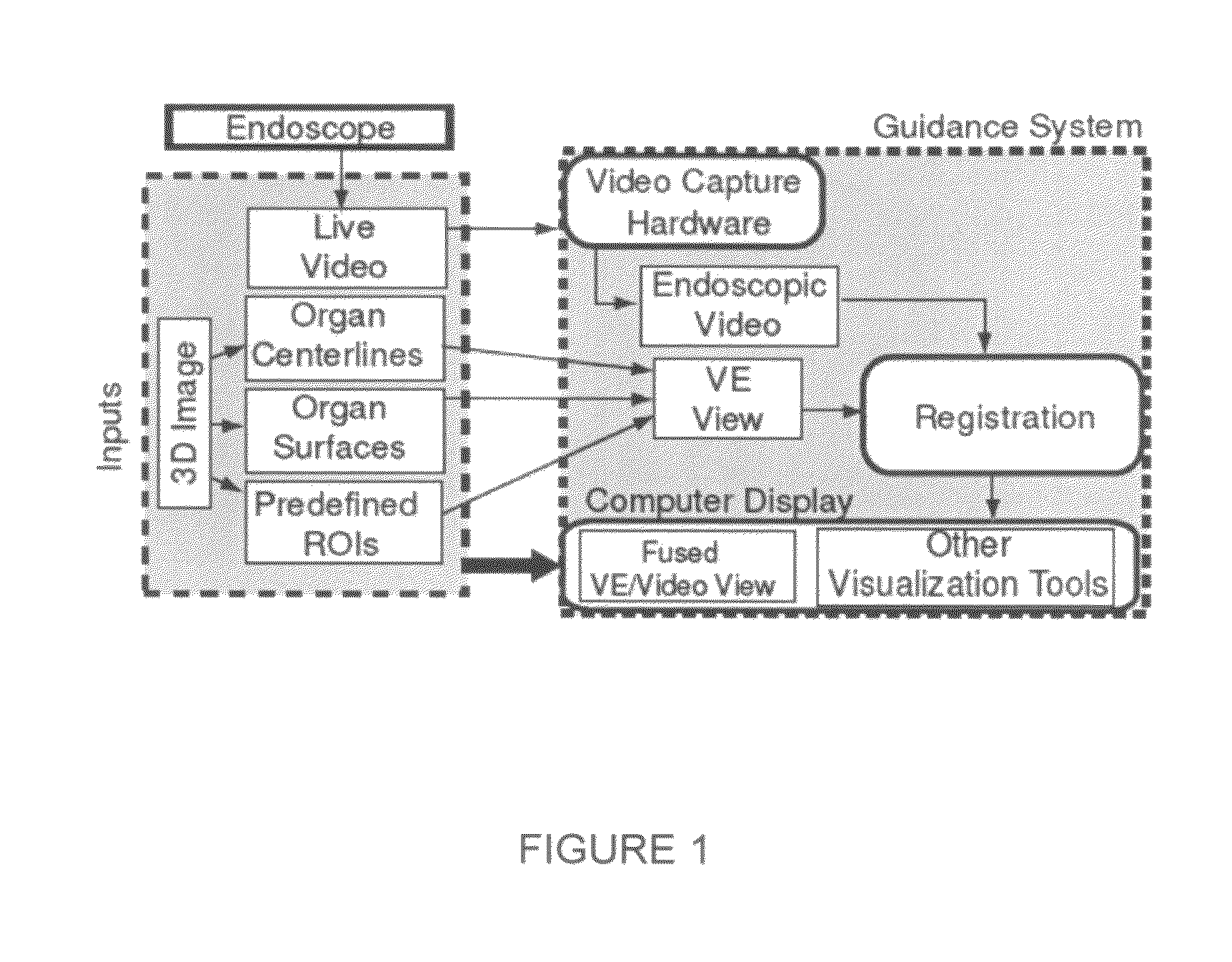
[0022]This invention resides in a system-level approach to guidance of endoscopy, including a complete paradigm for real-time image-based guidance providing a physician with continuously-updated navigational and guidance information.

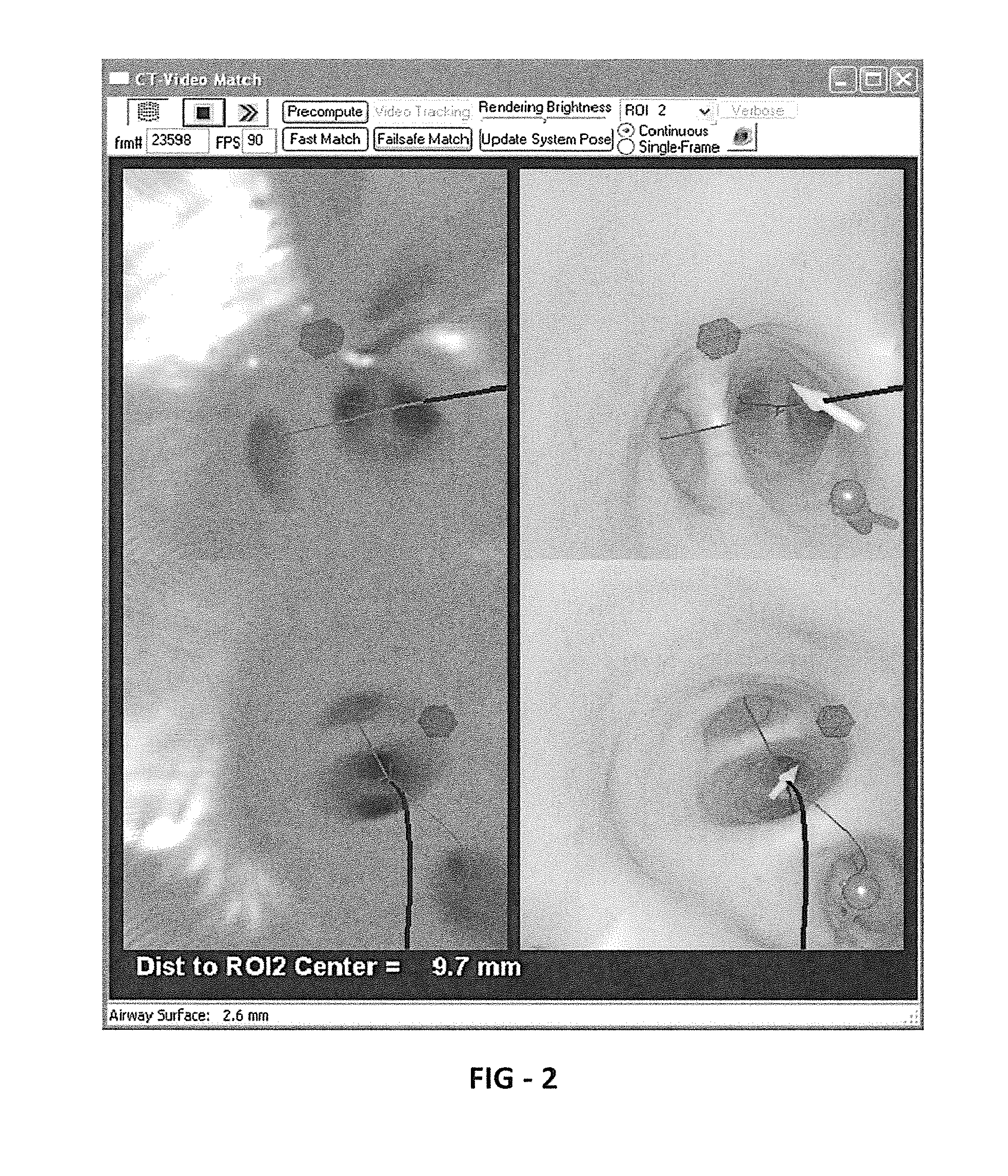
[0023]At least three novel embodiments for guidance of endoscopy are disclosed. Additional elements such as global surface rendering, local cross-sectional views, and pertinent distances provide additional utility to the physician. Phantom results were generated using bronchoscopy performed on a rapid prototype model of a human tracheobronchial airway tree. The system has also been tested in ongoing live human tests. Ten such tests have been performed thus far and focus on bronchoscopic intervention of pulmonary patients using 3D chest CT.

[0024]This disclosure presents generally applicable methods, but focuses on the chest and bronchoscopy. In this domain, Phase I centers around acquisition and analysis of an MDCT image, where the ROIs may be lymph nodes...


PUM


Abstract

Methods and apparatus provide continuous guidance of endoscopy during a live procedure. A data-set based on 3D image data is pre-computed including reference information representative of a predefined route through a body organ to a final destination. A plurality of live real endoscopic (RE) images are displayed as an operator maneuvers an endoscope within the body organ. A registration and tracking algorithm registers the data-set to one or more of the RE images and continuously maintains the registration as the endoscope is locally maneuvered. Additional information related to the final destination is then presented enabling the endoscope operator to decide on a final maneuver for the procedure. The reference information may include 3D organ surfaces, 3D routes through an organ system, or 3D regions of interest (ROIs), as well as a virtual endoscopic (VE) image generated from the precomputed data-set. The preferred method includes the step of superimposing one or both of the 3D routes and ROIs on one or both of the RE and VE images. The 3D organ surfaces and routes may correspond to the surfaces and paths of a tracheobronchial airway tree extracted, for example, from 3D MDCT images of the chest.

Description

REFERENCE TO RELATED APPLICATION[0001]This application claims priority from U.S. Provisional Patent Application Ser. No. 60 / 887,462, filed Jan. 31, 2007, the entire content of which is incorporated herein by reference.GOVERNMENT SPONSORSHIP[0002]This invention was made with government support under Grant No. CA074325, awarded by The National Institutes of Health. The Government has certain rights in the invention.BACKGROUND OF THE INVENTION[0003]Flexible endoscopy is a class of minimally-invasive procedures wherein a flexible device is maneuvered through the body's existing hollow organs (e.g., tracheobronchial airway tree, colon, sinuses) in order to perform diagnosis or deliver treatment.1,2 Endoscopy reduces and often eliminates the need for incisions, thereby reducing patient risk and recovery time. Flexible endoscopy is therefore preferable to more invasive surgeries, which typically require an incision large enough for the physician to directly view and manipulate the target o...

Claims


Application Information

Patent Timeline
Patent Type & Authority Patents(United States)
IPC IPC(8): G09G5/00G06T15/10A61B1/04A61B1/00
CPCA61B19/5244A61B19/56A61B1/0005A61B19/52A61B2019/505A61B1/00009A61B2019/5291A61B2019/507A61B19/5212A61B90/36A61B34/20A61B34/25A61B90/361A61B2034/105A61B2034/107A61B2090/365A61B1/000094
Inventor HIGGINS, WILLIAM E.MERRITT, SCOTT A.RAI, LAVGIBBS, JASON D.YU, KUN-CHANG
Owner PENN STATE RES FOUND
Why Eureka
Try Eureka