Closed loop monitoring of automated molecular pathology system

a technology of molecular pathology and monitoring system, which is applied in the field of automatic methods and devices for iterative staining of biological samples, can solve the problems of limited shelf life, high cost of reagents used in the staining process, and time-consuming manual techniques that are susceptible to errors

Active Publication Date: 2014-03-18
LEICA MICROSYSTEMS CMS GMBH
View PDF12 Cites 3 Cited by
  • Summary
  • Abstract
  • Description
  • Claims
  • Application Information

AI Technical Summary

Problems solved by technology

The staining process is typically performed using time-consuming manual techniques that are susceptible to error.
The reagents used in the staining process are often expensive and have limited shelf life thereby requiring special handling techniques

Method used


Image

  • Closed loop monitoring of automated molecular pathology system
  • Closed loop monitoring of automated molecular pathology system

Examples


Embodiment Construction

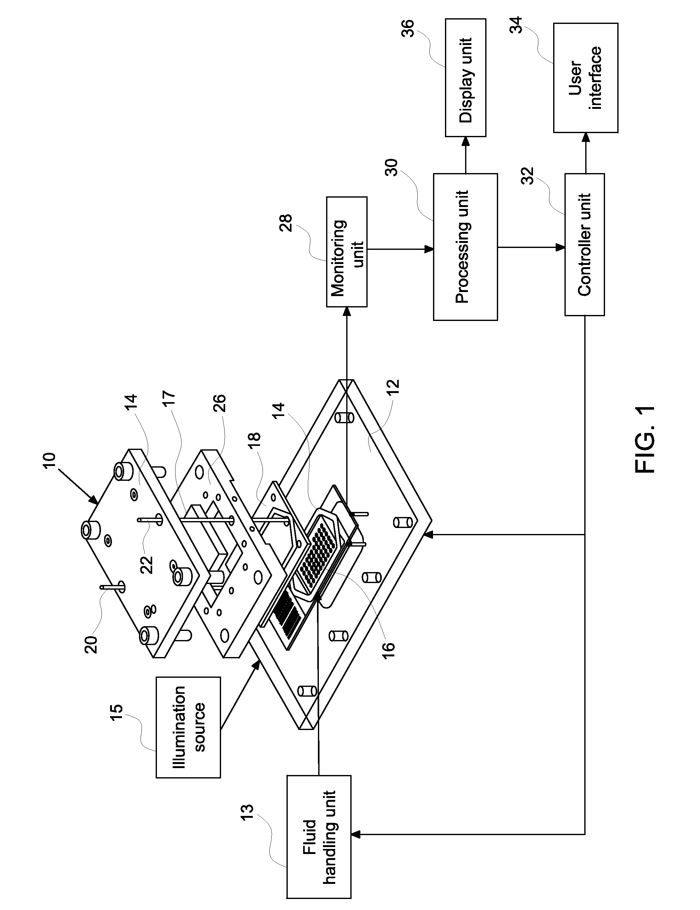
[0010]Embodiments relate to methods and systems for closed loop monitoring of an automated molecular pathology system for molecular imaging. The methods and systems enable optimized operation in molecular imaging. In certain embodiments, the automated molecular pathology system may operate with minimal operator intervention by reducing or eliminating the need to transfer samples (e.g., tissue samples on a slide within the flow cell). The systems and methods may reduce or eliminate the need to displace samples between the staining component and the imaging component. Closed loop monitoring minimizes both reagent volume and reagent dwell time within the system thereby saving on expensive reagents, such as fluorescence labeled antibodies, and minimizing reagent decomposition or side reactions.

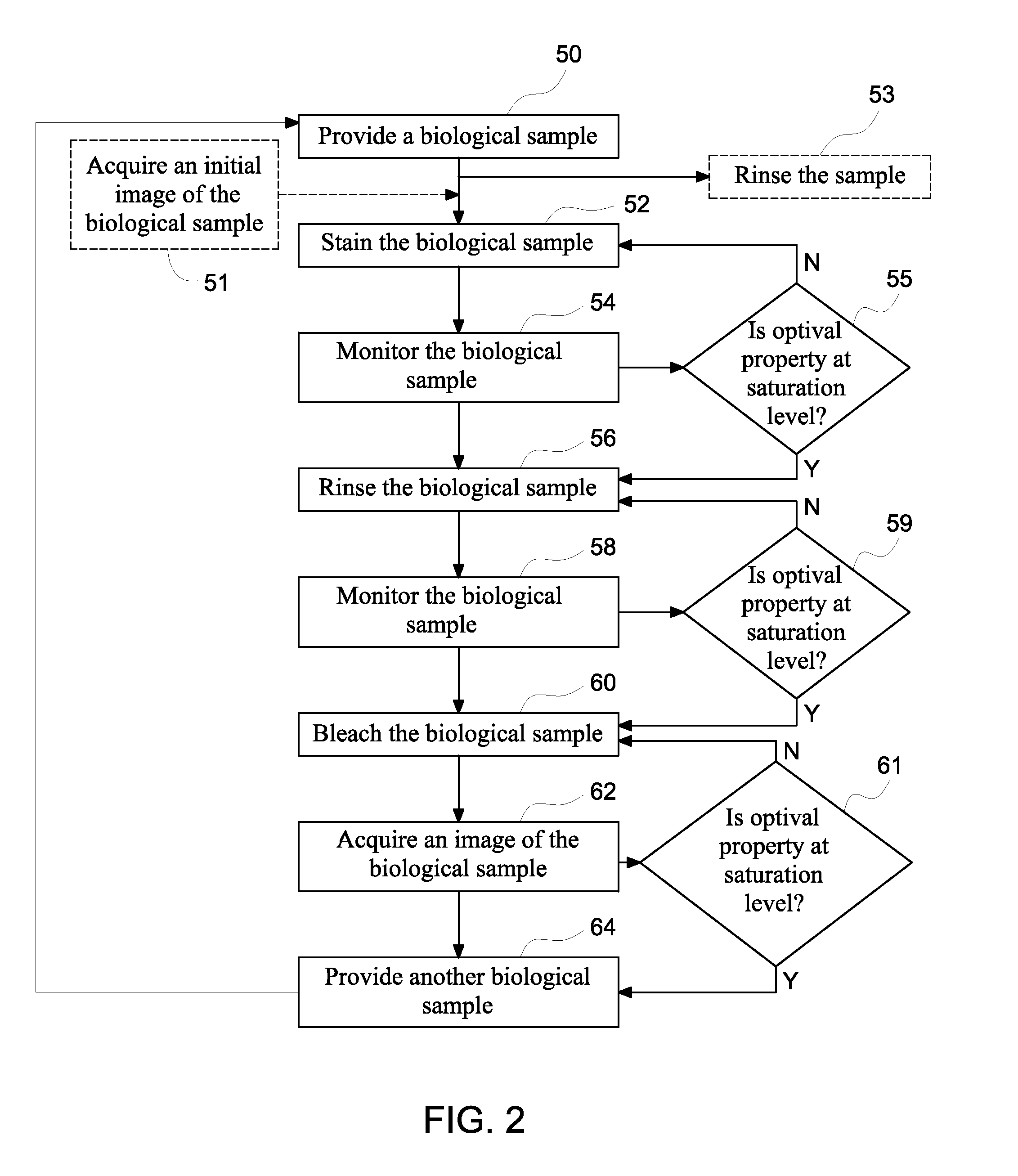
[0011]In certain embodiments, a closed loop automated method for staining a biological sample comprises providing a biological sample, staining at least a portion of the biological sample, monitor...


PUM

PropertyMeasurementUnit
thicknessaaaaaaaaaa
thicknessaaaaaaaaaa
thicknessaaaaaaaaaa

Abstract

A closed loop automated method for staining of a biological sample is provided. The method comprises providing a biological sample, staining at least a portion of the biological sample by flowing in a reagent, monitoring one or more optical characteristics of the biological sample, and calculating a figure of merit based on at least one of the optical characteristics. An automated device for iterative staining of a biological sample is also provided.

Description

BACKGROUND[0001]The invention relates generally to automated methods and devices for iterative staining of biological samples for imaging applications.[0002]Biological samples are used for analytic and diagnostic purposes, such as diagnosing diseased tissue at a molecular level. A biological sample, such as tissue micro arrays (TMA), on which tissue samples are fixed are typically stained with a morphological stain or biomarker. The stained TMA are then analyzed manually with a microscope, or an image may be taken of the TMA for subsequent analysis or comparison. After the first stain is applied and imaged, one or more serial or successive stains or biomarkers may be applied and the TMA may be analyzed again. The two or more serial images may then be compared. A single staining cycle may comprise the steps of flowing a stain (antibody) over the tissue, incubating the stain for an appropriate time, rinsing away the stain to reduce background fluorescence, imaging the slide, and bleac...

Claims


Application Information

Patent Timeline
Patent Type & Authority Patents(United States)
IPC IPC(8): G01N33/53G01N35/02G01N33/48B05D3/00G01N1/30G01N1/31
CPCG01N2001/368G01N1/312G01N1/31G01N33/48Y10T436/115831
Inventor CORWIN, ALEX DAVIDFILKINS, ROBERT JOHNXIE, JUNSEVINSKY, CHRISTOPHER JAMESSHAIKH, KASHAN ALI
Owner LEICA MICROSYSTEMS CMS GMBH
Why Eureka
Try Eureka