Medical image and vessel characteristic data processing system

a data processing system and medical image technology, applied in image analysis, image enhancement, instruments, etc., can solve the problems of time-consuming and burdensome tasks, and achieve the effect of enhancing the structure of the vessel

Active Publication Date: 2014-05-20
SIEMENS HEALTHCARE GMBH
View PDF22 Cites 9 Cited by
  • Summary
  • Abstract
  • Description
  • Claims
  • Application Information

AI Technical Summary

Problems solved by technology

A user is responsible for identifying the vessel phases of each image frame which is a burdensome and time consuming task.

Method used


Image

  • Medical image and vessel characteristic data processing system
  • Medical image and vessel characteristic data processing system
  • Medical image and vessel characteristic data processing system

Examples


Embodiment Construction

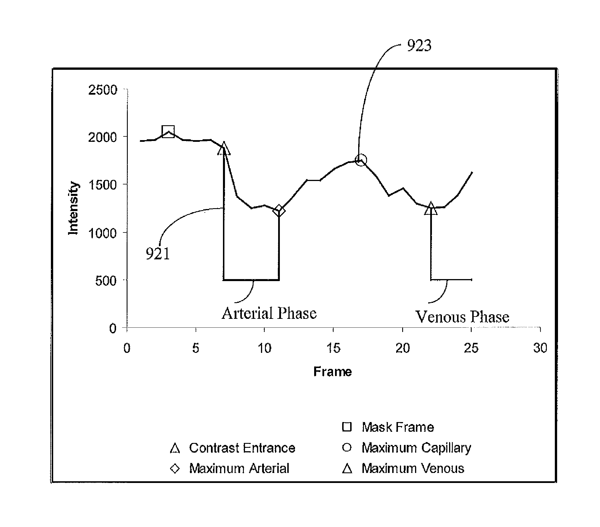
[0023]A system provides automatic detection and classification of the vessels as arteries, veins, and capillaries within a DSA image sequence and displays classified vessels, in response to a contrast agent bolus injection into a patient. The system identifies different phases of blood flow depicted by flow of contrast agent in patient anatomy X-ray images. Blood flow phases in the tissues of the body include arterial, capillary, and venous phases. In the capillary phase contrast agent is located almost entirely in the capillaries, within the tissue itself. In the arterial and venous phases, the contrast agent is located in the vessels supplying (arteries) blood to the tissue or draining (veins) blood from the tissue. The system automatically identifies, Arterial phase (start and end), Capillary phase (start, maximum blush, and end) and Venous phase (start and end). The system analyzes the change in image luminance intensity values over time to identify frames that specifically capt...


PUM


Abstract

An image data processing system automatically indicates an image of a digitally subtracted Angiography (DSA) image sequence is associated with at least one of, arterial, venous, or capillary phases of blood flow. The system includes an interface for acquiring data representing a DSA sequence of digitally subtracted images enhancing vessel structure. An image data processor automatically indicates an image of the DSA sequence is associated with at least one of, arterial, venous, or capillary phases of blood flow by determining individual minimum luminance intensity level values of individual images of the DSA sequence and using the determined individual minimum luminance intensity level values in identifying images of the DSA sequence are associated with at least one of, arterial, venous, or capillary phases of blood flow. An output processor automatically assigns an attribute to image data to identify vessel phase in response to the identifying images of the DSA sequence.

Description

[0001]This is a non-provisional application of provisional application Ser. No. 61 / 350,944 filed Jun. 3, 2010, by J. C. Rauch.FIELD OF THE INVENTION[0002]This invention concerns an image data processing system for automatically indicating an image of a Digitally Subtracted Angiography (DSA) image sequence is associated with at least one of arterial, venous, or capillary phases of blood flow.BACKGROUND OF THE INVENTION[0003]In diagnostic imaging it is desirable to be able to examine phases of blood flow to look for abnormalities. The ability to individually look at arterial, capillary, and venous phases simplifies a diagnosis and treatment workflow and produces improved consistency. There are some procedures in which it is advantageous to be able to visualize either the arteries or the veins independently of one another, as some ailments primarily affect either arteries or veins. It is also desirable to be able to visualize both the arteries and veins simultaneously, but with the art...

Claims


Application Information

Patent Timeline
Patent Type & Authority Patents(United States)
IPC IPC(8): G06K9/00
CPCG06T2207/20224G06T2207/10016G06T2207/30101G06T2207/10116A61B6/504G06T7/2053G06T7/254
Inventor RAUCH, JOHN, CHRISTOPHER
Owner SIEMENS HEALTHCARE GMBH
Why Eureka
Try Eureka