Method and apparatus for cone beam breast CT image-based computer-aided detection and diagnosis

a technology of computer-aided detection and diagnosis, applied in the field of breast imaging, can solve the problems of insufficient limited spatial resolution of bmri, and inability to achieve significant improvement of ct applied to mammograms, etc., and achieve no tissue overlap, superior soft tissue contrast, and high contrast resolution

Active Publication Date: 2016-07-19
UNIVERSITY OF ROCHESTER +1
View PDF8 Cites 15 Cited by
  • Summary
  • Abstract
  • Description
  • Claims
  • Application Information

AI Technical Summary

Benefits of technology

[0018]There is also a need in the art to improve accuracy and efficiency in radiologists' diagnostic performance.
[0021]It is another object of the invention to provide method to accurately evaluate breast density with CBBCT images.
[0028]To achieve the above and other objects, the present invention is directed to a CAD technique based on cone-beam breast computed tomography (CBBCT). Cone Beam Breast CT is a three-dimensional breast imaging modality with soft tissue contrast superior to that of mammography, high contrast resolution and no tissue overlap. With those advantages, it is possible to measure volumetric breast density accurately and quantitatively with CBBCT 3D images. Cone Beam Breast CT is a novel breast imaging technology, using cone beam CT technology and an ergonomically designed imaging system. CBBCT delivers true three-dimensional breast images without tissue overlap. The resolution of CBBCT images is 3D isotropic, with the voxel size as small as 155 μm. Even without intravenous contrast injection, the CBBCT soft tissue contrast-to-noise ratio is as high as 8. The breast coverage of CBBCT is comparable to and even larger than that of mammography. With those novel features, CBBCT images of the breast can be an ideal tool to quantitatively measure volumetric density of the breast.
[0029]CBBCT-based computer aided diagnosis (CBBCT-CAD) technology is a clinically useful tool for breast cancer detection and diagnosis that will help radiologists to make more efficient and accurate decisions. The CBBCT-CAD is able to: 1) use 3D algorithms for image artifact correction, mass and calcification detection and characterization, duct imaging and segmentation, vessel imaging and segmentation, and breast density measurement, 2) present composite information of the breast including mass and calcifications location and characteristics, duct structure, vascular structure and breast density to the radiologists to aid them in determining the probability of malignancy of a breast lesion.
[0030]The novelties of the CBBCT-CAD system include: 1) The first 3D CAD system based on CBBCT technology; 2) The first CAD system to provide composite breast information that is not available from other modalities-based CAD systems; 3) Prominent accuracy and efficiency improvements in radiologists' diagnostic performance.

Problems solved by technology

However, mammography and MRI have their inherent limitations.
Although it is reported that CAD (computer-aided detection and diagnosis) on mammography increases sensitivity, it is also reported that there is no significant improvement with the CAD applied to the mammograms.
Although it is a three-dimensional (3D) visualization of the breast, BMRI has limited spatial resolution (in-plane resolution of ˜1 mm and slice thickness of 2-3 mm).
Small lesions may be missed or incorrectly characterized due to the resolution limitation.
Although BMRI-based CAD has had some success in distinguishing benign from malignant masses, acknowledged limitations of the system include an inability to consider T2-weighted images.
Thus, the system potentially may miss useful additional morphology information available in those sequences.
In addition, current BMRI-based CAD cannot improve radiologist accuracy in distinguishing malignant from benign MRI screen-detected lesions because of its poor DCIS detection sensitivity.
However, DBT collects projections over limited angles, resulting in spatial resolution loss in the direction perpendicular to the detector.
Although CAD in DBT can help radiologists to handle a large amount of data, the resolution loss limits its ability to detect subtle breast lesions and calcification clusters, so that it is likely that DBT must be used with digital mammography for breast cancer screening and diagnosis.
Though ultrasound has considerable potential, it also has well known limitations.
The technique is difficult to teach and perform.
Also, differentiating between benign and malignant lesions can be challenging.
The addition of ultrasound could also lead to unnecessary biopsies of benign lesions.
Another major limitation of US-CAD is the fact that microcalcifications are difficult to image with ultrasound.
In addition, US-CAD is limited by the relatively poor spatial resolution of ultrasound.
However, breast tissue overlaps in two-dimensional mammography projection images.
The breast density measurement may be inaccurate using mammograms.

Method used


Image

  • Method and apparatus for cone beam breast CT image-based computer-aided detection and diagnosis
  • Method and apparatus for cone beam breast CT image-based computer-aided detection and diagnosis
  • Method and apparatus for cone beam breast CT image-based computer-aided detection and diagnosis

Examples


Embodiment Construction

[0060]A preferred embodiment will be set forth in detail with reference to the drawings, in which like reference numerals refer to like elements or steps throughout.

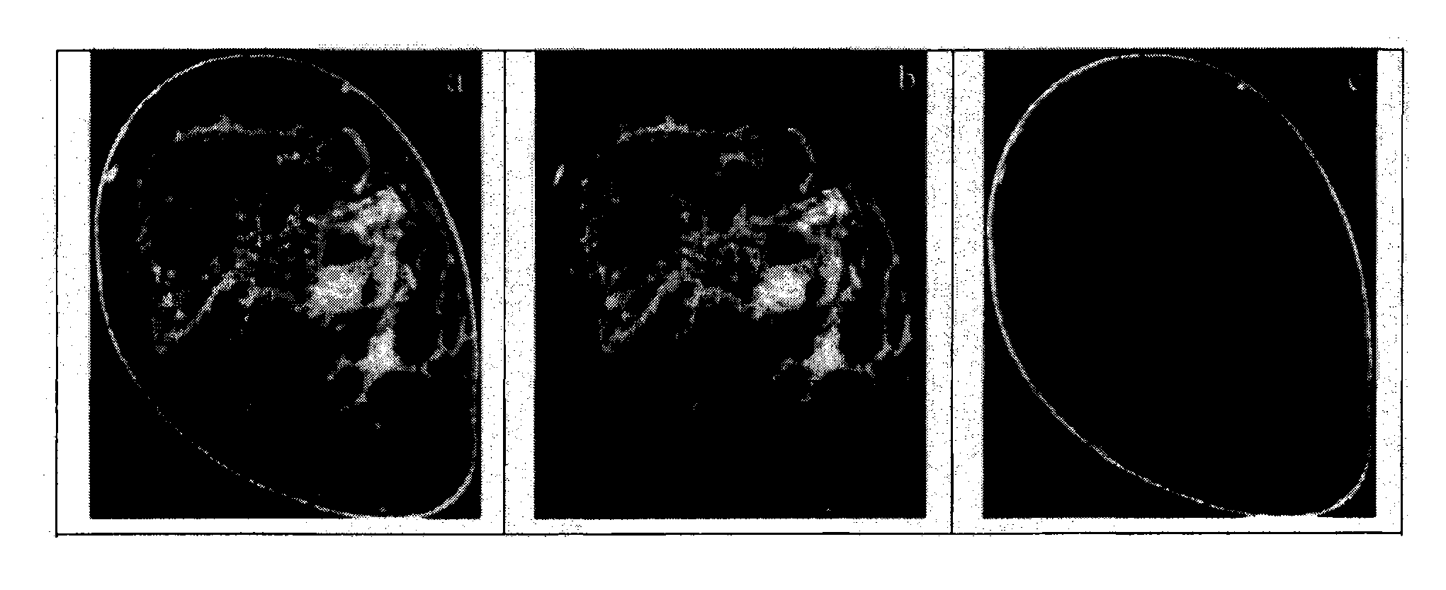
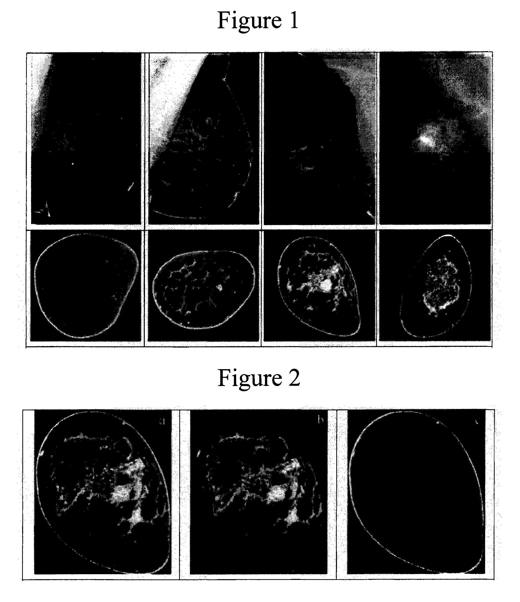
[0061]The first step is skin removal. Although the skin in CBBCT images has higher intensity than the fatty stroma, its intensity is only slightly higher than that of the glandular tissue. A simple thresholding cannot segment the skin from other tissue. Based on the fact that the skin is on the outermost layer of the whole breast, a morphological process is proposed to remove the skin from the breast in CBBCT images.

[0062]1. A histogram thresholding method is applied to the whole CBBCT image volume to separate the image into three distinct parts which have significant different intensities: air, fat and tissues (including skin and glands).

[0063]2. Based on the fact that the skin is between the inner breast and the air, a morphological 3D erosion operation is applied to erode the tissue between air and fat.

[0064]3. When t...


PUM


Abstract

Cone Beam Breast CT (CBBCT) is a three-dimensional breast imaging modality with high soft tissue contrast, high spatial resolution and no tissue overlap. CBBCT-based computer aided diagnosis (CBBCT-CAD) technology is a clinically useful tool for breast cancer detection and diagnosis that will help radiologists to make more efficient and accurate decisions. The CBBCT-CAD is able to: 1) use 3D algorithms for image artifact correction, mass and calcification detection and characterization, duct imaging and segmentation, vessel imaging and segmentation, and breast density measurement, 2) present composite information of the breast including mass and calcifications, duct structure, vascular structure and breast density to the radiologists to aid them in determining the probability of malignancy of a breast lesion.

Description

REFERENCE TO RELATED APPLICATION[0001]The present application is a national stage of PCT / US2012 / 025141, filed Feb. 14, 2012, which claims the benefit of U.S. Provisional Patent Application No. 61 / 442,559, filed Feb. 14, 2011, the entire disclosures of which are hereby incorporated by reference in their entirety into the present disclosure.STATEMENT OF GOVERNMENT INTEREST[0002]This invention was made with government support under Grant Nos. R01 9 HL078181 and 4 R33 CA94300 awarded by National Institutes of Health. The government has certain rights in the invention.FIELD OF THE INVENTION[0003]The present invention is directed to breast imaging and more particularly to the provision of three-dimensional (3D) breast information, including 3D location and measurement of mass and calcifications, 3D duct structure, 3D vascular structure, and volumetric breast density.DESCRIPTION OF RELATED ART[0004]Many researchers have made significant progress in breast lesion auto detection on mammogram...

Claims


Application Information

Patent Timeline
Patent Type & Authority Patents(United States)
IPC IPC(8): G06T7/00A61B6/00A61B6/03G06F19/00
CPCA61B6/502A61B6/032A61B6/5217G06T7/0012G06F19/321G06T2207/10081G06T2207/30068G16H50/30G16H30/20G16H30/40A61B6/0414A61B6/4085A61B6/4435A61B6/463A61B6/481
Inventor NING, RUOLAZHANG, XIAOHUACONOVER, DAVID L.LIU, SHAOHUA
Owner UNIVERSITY OF ROCHESTER
Why Eureka
Try Eureka