
Endogenous optical imaging method for automatically separating arteries and veins

A technology of automatic separation and optical imaging, applied in the field of biomedical imaging

Active Publication Date: 2011-04-06
NAT UNIV OF DEFENSE TECH
View PDF4 Cites 14 Cited by
  • Summary
  • Abstract
  • Description
  • Claims
  • Application Information

AI Technical Summary

Problems solved by technology

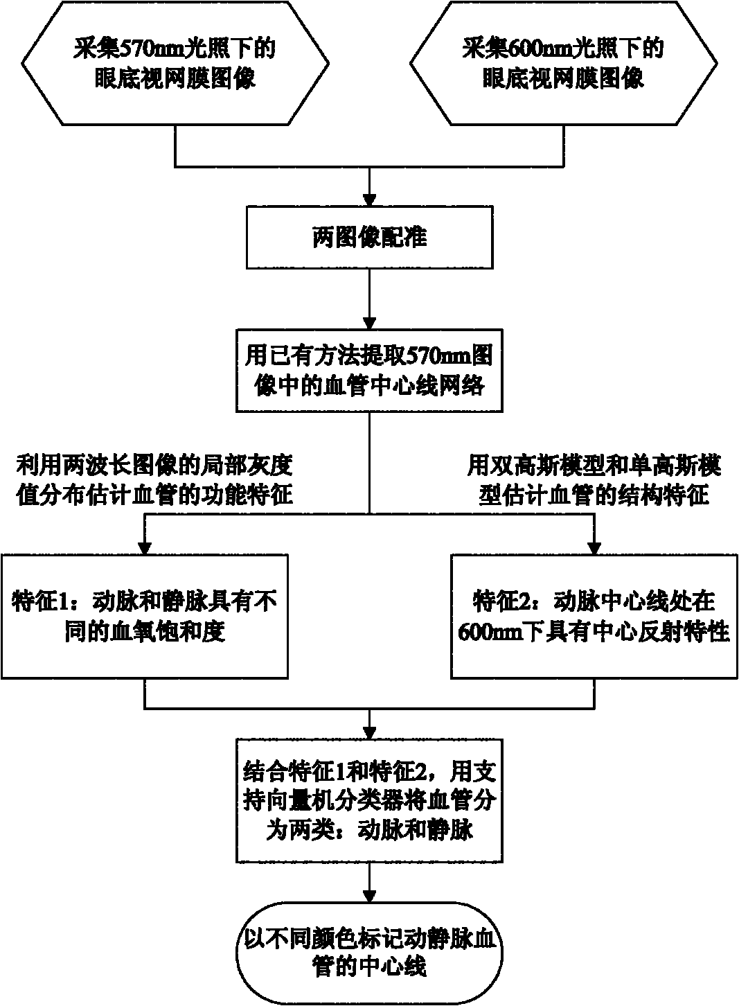
However, the above method has a poor separation effect on small blood vessels, and is limited by the pre-extraction of the vascular network structure. Under the existing vascular network extraction method, it is impossible to separate small arteries and small veins.
The method of arteriovenous separation using MRA imaging (magnetic resonance imaging) mainly relies on the opposite characteristics of blood flow in arteries and veins (Svensson, J et al., 2002. Separation of arteries and veins using flow-induced phase effects in contrast-enhanced MRA of the lower extremities (arteriovenous separation based on blood flow phase characteristics in lower extremity MRA imaging), Magnetic Resonance Imaging, 20, 49–57), but this feature only exists in the extremities of the human body and some cardiovascular In the system, it is absent in the complex cerebral vascular network, so it cannot be applied to the separation of arteries and veins in the cerebral cortex
However, there is no public report on the automatic separation of arteries and veins using the spectral value characteristics of low-frequency oscillation signals in biological tissues.

Method used


Image

  • Endogenous optical imaging method for automatically separating arteries and veins
  • Endogenous optical imaging method for automatically separating arteries and veins
  • Endogenous optical imaging method for automatically separating arteries and veins

Examples


Embodiment Construction

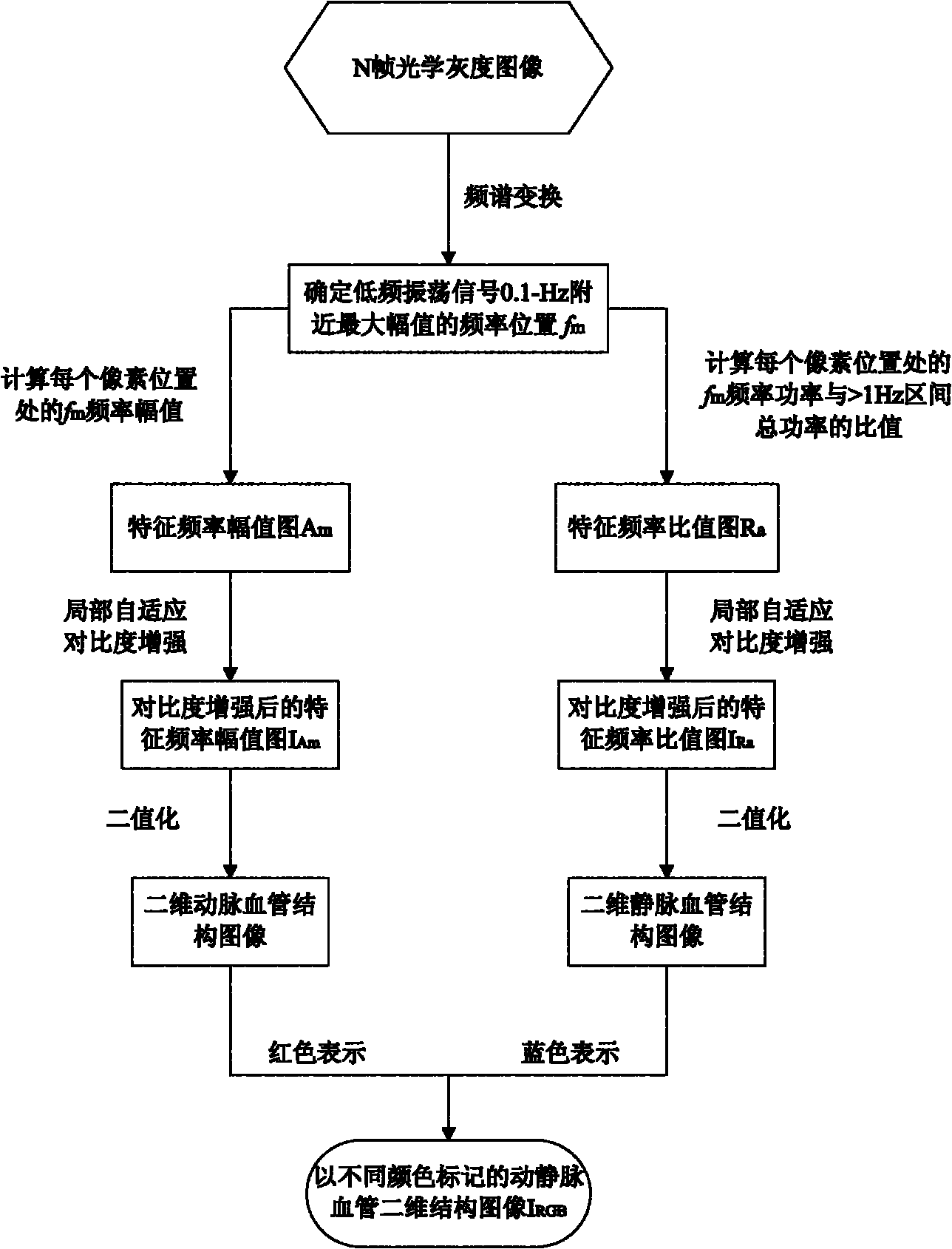
[0033] figure 2 For the general flow chart of the present invention, the present invention comprises the following steps:

[0034] 1. Irradiate narrow-band quasi-monochromatic light onto the measured object, and use a CCD or CMOS camera to continuously collect N frames of optical images reflected by the measured object with the same exposure time and frame interval through the optical imaging system;

[0035] 2. Calculate the average time series according to formula 1 The average gray value of the nth point in :

[0036] formula one

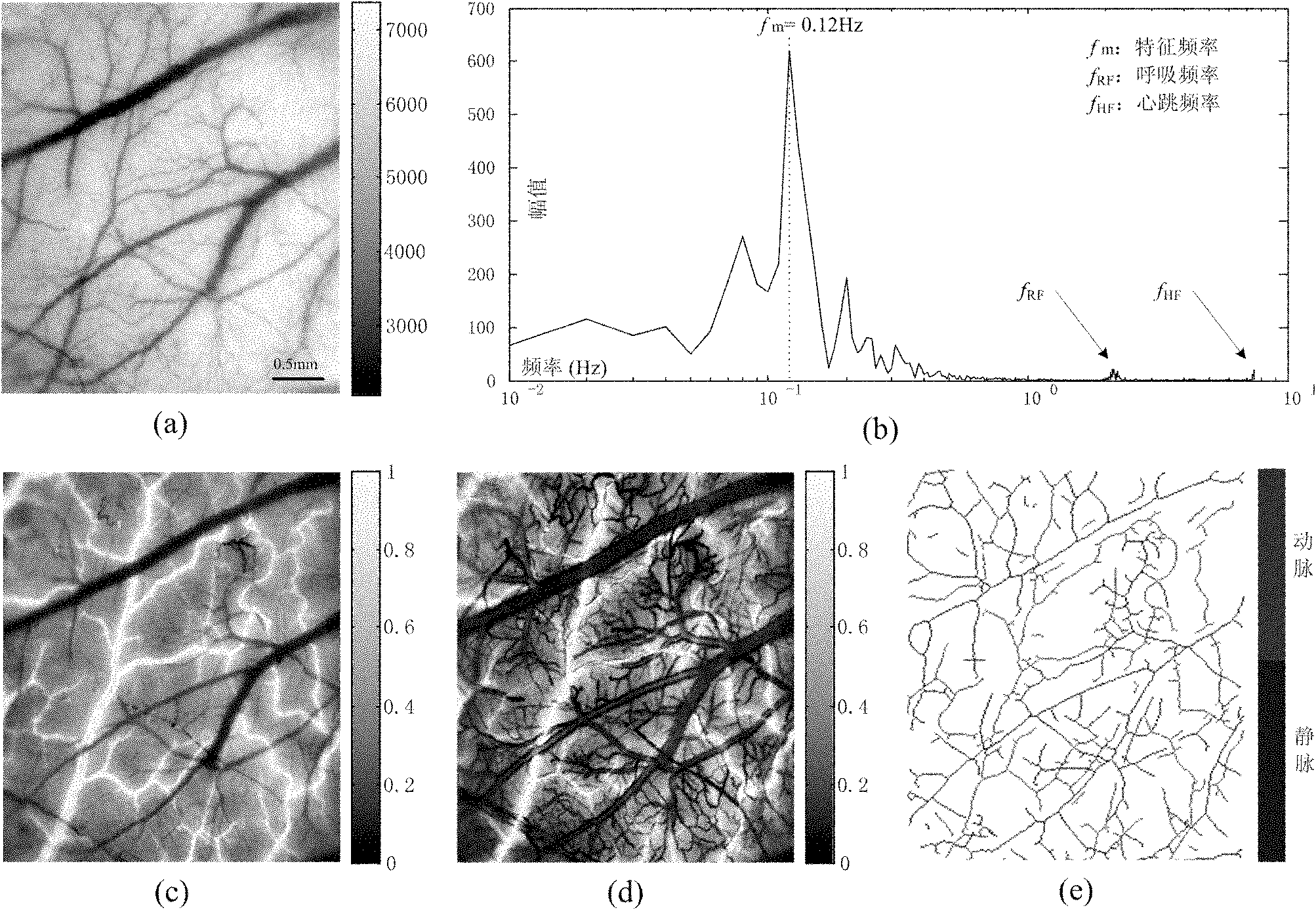
[0037] 3. Use the fast Fourier transform algorithm to convert the average pixel time series obtained in the second step Transform to the spectral domain and determine the frequency value with the largest magnitude in the frequency interval (0.05Hz, 1Hz) f m ;

[0038] 4. Use the fast Fourier transform algorithm to transform each pixel time series into the spectral domain, and calculate the characteristic frequency of each pixel tim...


PUM


Abstract

The invention discloses an endogenous optical imaging method for automatically separating arteries and veins and aims at providing an effective endogenous optical imaging method for effectively and automatically separating arteries and veins of organisms. In the technical scheme, the endogenous optical imaging method comprises the following steps of: carrying out spectrum analysis to a collected image sequence by adopting narrow-band quasi monochromatic light for illumination, and then realizing the identification and automatic separation of the arteries and veins by utilizing the amplitude distribution features of low-frequency oscillating signals of the image sequence through the operations of frequency spectrum transformation, enhancement of a local self-adaption contrast, automatic threshold segmentation and the like. The endogenous optical imaging method can be used for automatically separating the arteries and the veins and highlighting and separating the vessel structures of many arterioles and veinlets, thereby avoiding the design complexity of the imaging device using light with various central wavelengths for illumination.

Description

Technical field: [0001] The invention belongs to a biomedical imaging method, in particular to a method for automatically separating arteriovenous blood vessels by using narrow-band quasi-monochromatic light illumination and utilizing endogenous physiological signal spectral value characteristics. Background technique: [0002] The automatic separation of arteriovenous vessels in the cerebral cortex has important practical significance for basic biomedical research and clinical diagnosis and treatment of vascular-related diseases. Vascular diseases such as arterial tumors and arteriosclerosis are increasingly threatening people's lives. Vascular analysis methods can be used to diagnose and prevent such diseases in a timely and reliable manner. Currently, there are limited reports on methods for automatic separation of arteriovenous vessels. According to the difference in spectral absorption characteristics of arterial blood and venous blood oxygen saturation, the blood ox...

Claims


Application Information

Patent Timeline
Patent Type & Authority Applications(China)
IPC IPC(8): A61B5/00
Inventor 胡德文王玉成刘亚东李明
Owner NAT UNIV OF DEFENSE TECH
Learn More