Method of imaging of in vivo retina haemodynamics and measuring of absolute flow velocity

A hemodynamic and flow velocity measurement technology, applied in the field of ophthalmic imaging, which can solve the problems of complex hardware modification and difficult transplantation.

Active Publication Date: 2016-02-03
WENZHOU MEDICAL UNIV
View PDF7 Cites 14 Cited by
  • Summary
  • Abstract
  • Description
  • Claims
  • Application Information

AI Technical Summary

Problems solved by technology

This method uses two beams with different incident light directions to measure their respective Doppler flow velocity components, and then calculates the absolute flow velocity through the triangular relationshi

Method used


Image

  • Method of imaging of in vivo retina haemodynamics and measuring of absolute flow velocity
  • Method of imaging of in vivo retina haemodynamics and measuring of absolute flow velocity
  • Method of imaging of in vivo retina haemodynamics and measuring of absolute flow velocity

Examples


Embodiment Construction

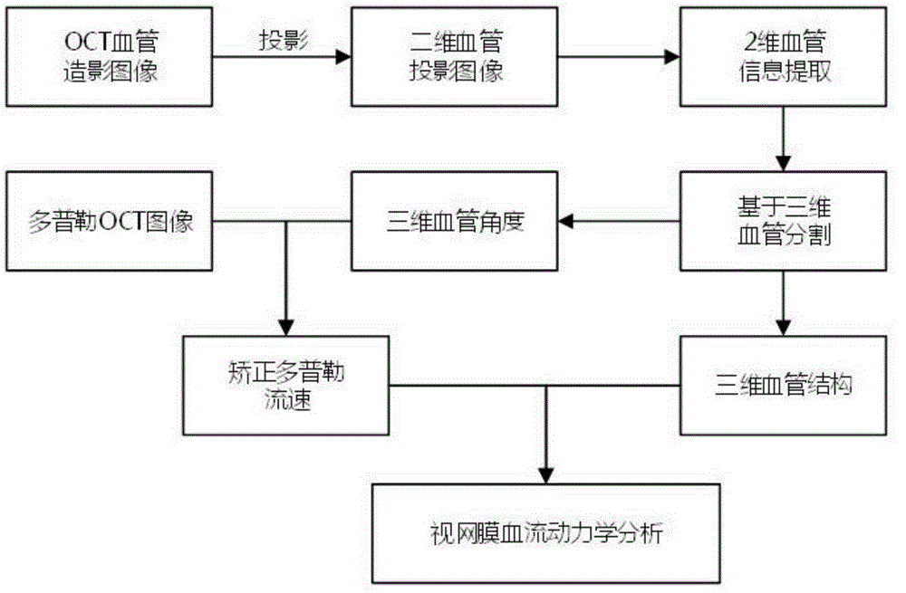
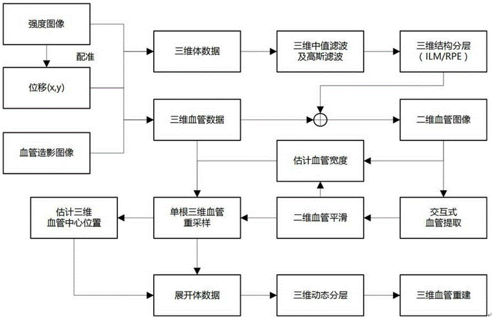
[0046] The invention integrates OCT angiography and Doppler OCT flow velocity detection technology, and proposes an imaging and measurement method of retinal hemodynamics in vivo. It includes the following steps:

[0047] OCT angiography;

[0048] 1. Use FD-OCT to obtain the three-dimensional structural data of the retinal optic disc (ONH). The FD-OCT involved has an image longitudinal resolution of 3 μm and an acquisition rate of not less than 50 kHz. The three-dimensional angiographic data consisted of 512×128×8 A-scans, which were obtained by block scanning. Each 3D data consists of 128 tomograms, and each tomogram consists of 8 repeated B-scans at the same position. The Doppler variance method based on intensity information realizes the imaging of microvessels by performing repeated scans at the same position and using the intensity change between two adjacent B-scans. The algorithm of the Doppler variance method based on intensity information is shown in the following...


PUM


Abstract

Provided is a method of imaging of in vivo retina haemodynamics and measuring of the absolute flow velocity. On the basis of conventional FD.OCT, an OCT angiography technology and a Doppler OCT imaging technology are integrated, and measurement of the retinal vessel absolute flow velocity and the haemodynamics is achieved. According to the method of imaging of the in vivo retina haemodynamics and measuring of the absolute flow velocity, distribution of a three-dimensional blood vessel geometric structure is rebuilt through the OCT angiography technology, an included angle of a blood vessel and a light beam is calculated and serves as a Doppler angle according to the geometric structure of a three-dimensional blood vessel, a Doppler frequency shift of a blood flow is obtained through a Doppler OCT technology, correction is conducted according to a calculated Doppler angle, and the absolute flow velocity is obtained, and the problem of the retinal vessel Doppler angle is solved. According to the method of imaging of the in vivo retina haemodynamics and measuring of the absolute flow velocity, the three-dimensional blood vessel structure rebuilt through the OCT angiography technology is combined with the absolute flow velocity value of a particular location, and measurement of the in vivo retina haemodynamics can be achieved by simulating the flow of the blood in the blood vessel.

Description

technical field [0001] The invention relates to the field of ophthalmic imaging, in particular to an imaging and absolute flow velocity measurement method of retinal hemodynamics in vivo. Background technique [0002] Changes in retinal perfusion or flow velocity play an important role in the pathogenesis of ophthalmic diseases such as glaucoma and diabetic retinopathy. Therefore, the measurement of retinal hemodynamics in vivo is a very important research content in the field of ophthalmic imaging. Quantitative assessment of retinal hemodynamics can provide very useful information for pathological changes and early diagnosis and treatment of eye diseases. [0003] Currently, a variety of imaging techniques are available to assess retinal vascular velocity, such as video fluorescein angiography, laser speckle velocity imaging, laser Doppler velocimetry, laser scanning Doppler velocity, ultrasound color Doppler imaging technology, etc. However, each of these existing clinic...

Claims


Application Information

Patent Timeline
IPC IPC(8): A61B3/12A61B3/14A61B5/0265A61B5/0285
Inventor 黄胜海吕帆沈梅晓朱德喜
Owner WENZHOU MEDICAL UNIV
Why Eureka
Try Eureka