
Diffusion-weighted magnetic resonance imaging using 3D mosaic segmentation and 3D navigator phase correction

a magnetic resonance imaging and mosaic segmentation technology, applied in the field of diffusion-weighted magnetic resonance imaging, can solve the problems of inability to achieve, sensitive to phase variations, severe image artefacts in the final image, etc., and achieve the effect of reducing image artefacts and speeding up scan times

Active Publication Date: 2014-06-24
SIEMENS HEALTHCARE GMBH +1
View PDF5 Cites 2 Cited by
  • Summary
  • Abstract
  • Description
  • Claims
  • Application Information

AI Technical Summary

Benefits of technology

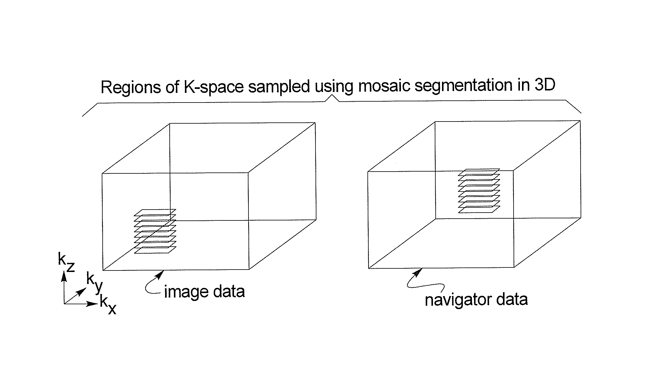
[0016]The method in accordance with the invention provides a way to acquire true 3D navigator data, which can be used to correct the spatially varying phase errors in the corresponding imaging data at each shot. To make this possible, a ‘mosaic’ sampling scheme is used that samples (at each shot) a contiguous set of k-space points in 3D k-space, which form a subset of the full range of k-space required. The navigator data are acquired from a fixed region at the center of k-space and the imaging data are acquired from a region of k-space that may be offset from the k-space centre along all three axes. At each shot the imaging data offsets are varied, so that the whole of k-space is sampled by the multi-shot acquisition. Because the data points are contiguous, the Nyquist sampling condition is now fulfilled in all three dimensions and 3D phase correction can be applied without problems from aliased signals.
[0017]The extent of the sampled region in each dimension would typically be less than that of the full range of k-space points required for the chosen image resolution. However, with some scanning protocols it might be possible to sample the complete set of k-space points along one or two of the k-space axes at each shot. In some cases, this approach might reduce image artefacts or allow faster scan times.

Problems solved by technology

This leads to severe image artefacts in the final image if no correction is applied.
This means that it is now necessary to apply a correction for the spatial phase variation in all three dimensions, which is not possible using the existing acquisition and navigator correction schemes used in 2D multi-shot imaging.
As there is no navigator phase correction applied in this sequence, it is susceptible to phase variations from one echo-train to the next.
Also the combination of the stimulated echo acquisition and the low flip-angle detection pulses is likely to result in a relatively low signal-to-noise ratio (SNR).
In addition, data acquired during early systole, when pulsatile effects are most significant, were rejected and not used during image reconstruction.

Method used


Image

  • Diffusion-weighted magnetic resonance imaging using 3D mosaic segmentation and 3D navigator phase correction
  • Diffusion-weighted magnetic resonance imaging using 3D mosaic segmentation and 3D navigator phase correction
  • Diffusion-weighted magnetic resonance imaging using 3D mosaic segmentation and 3D navigator phase correction

Examples


Embodiment Construction

[0023]FIG. 5 schematically illustrates a magnetic resonance tomography apparatus in which gradient pulses according to the present invention are generated. The design of the magnetic resonance tomography apparatus corresponds a conventional tomography apparatus, with the exceptions discussed below. A basic field magnet 1 generates a temporally constant strong magnetic field for polarization or alignment of the nuclear spins in the examination region of the subject such as, for example, a part of a human body to be examined. The high homogeneity of the basic magnetic field necessary for the magnetic resonance data acquisition is defined in a spherical measurement volume M in which the parts of the human body to be examined are introduced. For support of the homogeneity requirements, and in particular for elimination of temporally invariable influences, shim plates made from ferromagnetic material are mounted at a suitable location. Temporally variable influences are eliminated by shi...


PUM


Abstract

In a magnetic resonance apparatus and operating method therefore, 3D navigator data are acquired and are used to correct spatially varying phase errors in contemporaneously acquired imaging data in each shot of a multi-shot data acquisition sequence. A mosaic sampling scheme is used to enter the diffusion-weighted magnetic resonance data and the navigator data into k-space respectively in blocks that each form a subset of the entirety of k-space. The navigator data in each shot are entered into a block that is located at the center of k-space, and, in each shot, the corresponding image data are entered into an offset block in k-space, that is offset in at least one spatial direction from the navigator data block. The offset is varied from shot-to-shot.

Description

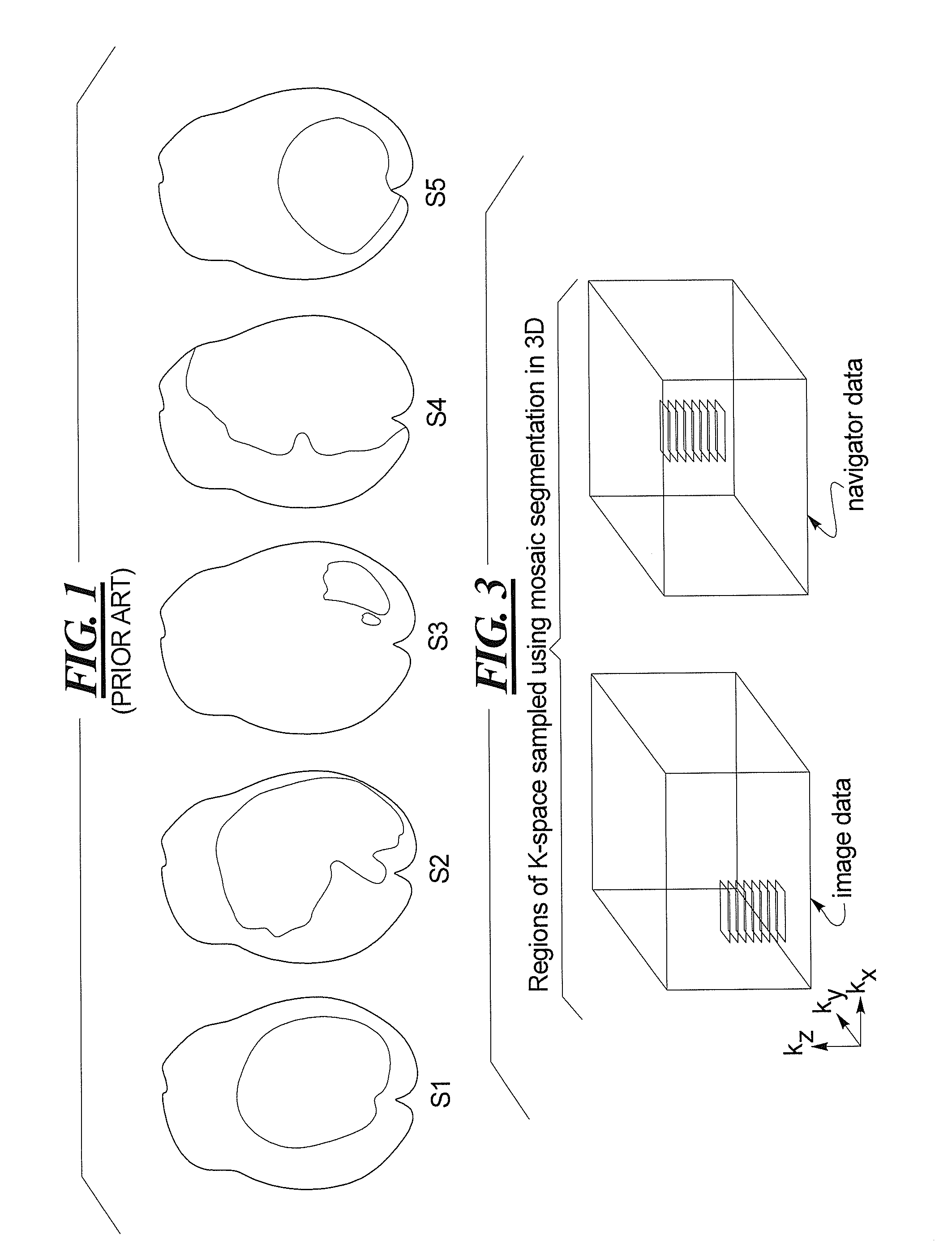
BACKGROUND OF THE INVENTION[0001]1. Field of the Invention[0002]The present invention relates to diffusion-weighted magnetic resonance imaging, and in particular to a method and magnetic resonance apparatus wherein phase errors in the diffusion-weighted magnetic resonance data are corrected.[0003]2. Description of the Prior Art[0004]Multi-shot, diffusion-weighted imaging (DWI) of the brain is sensitive to spatially dependent, non-linear phase errors that are caused by the effect of CSF pulsation and cardiac-related pulsatile motion on the brain during the diffusion-encoding gradients. The phase images in FIG. 1 shows how these phase errors vary substantially from one shot to the next, as illustrated for exemplary shots S1, S2, S3, S4 and S5. This leads to severe image artefacts in the final image if no correction is applied.[0005]For 2D acquisitions, in which each image plane is excited by a separate slice-selective radiofrequency (RF) pulse, a number of techniques have been demonst...

Claims


Application Information

Patent Timeline
Patent Type & Authority Patents(United States)
IPC IPC(8): G01V3/00
CPCG01R33/56341G01R33/56536G01R33/5676A61B5/055G01R33/5608G01R33/5611G01R33/5619G01R33/56333
Inventor FROST, ROBERTJEZZARD, PETERMILLER, KARLAPORTER, DAVID ANDREW
Owner SIEMENS HEALTHCARE GMBH
Learn More