System and method for detecting and tracking change in dental X-rays and dental images
a technology of applied in the field of system and method for detecting and tracking changes in dental x-rays, can solve problems such as poor or misaligned bridge work, tooth loss to infection, and the onset of bone loss can be quite sudden
- Summary
- Abstract
- Description
- Claims
- Application Information
AI Technical Summary
Benefits of technology
Problems solved by technology
Method used
Image
Examples
first embodiment
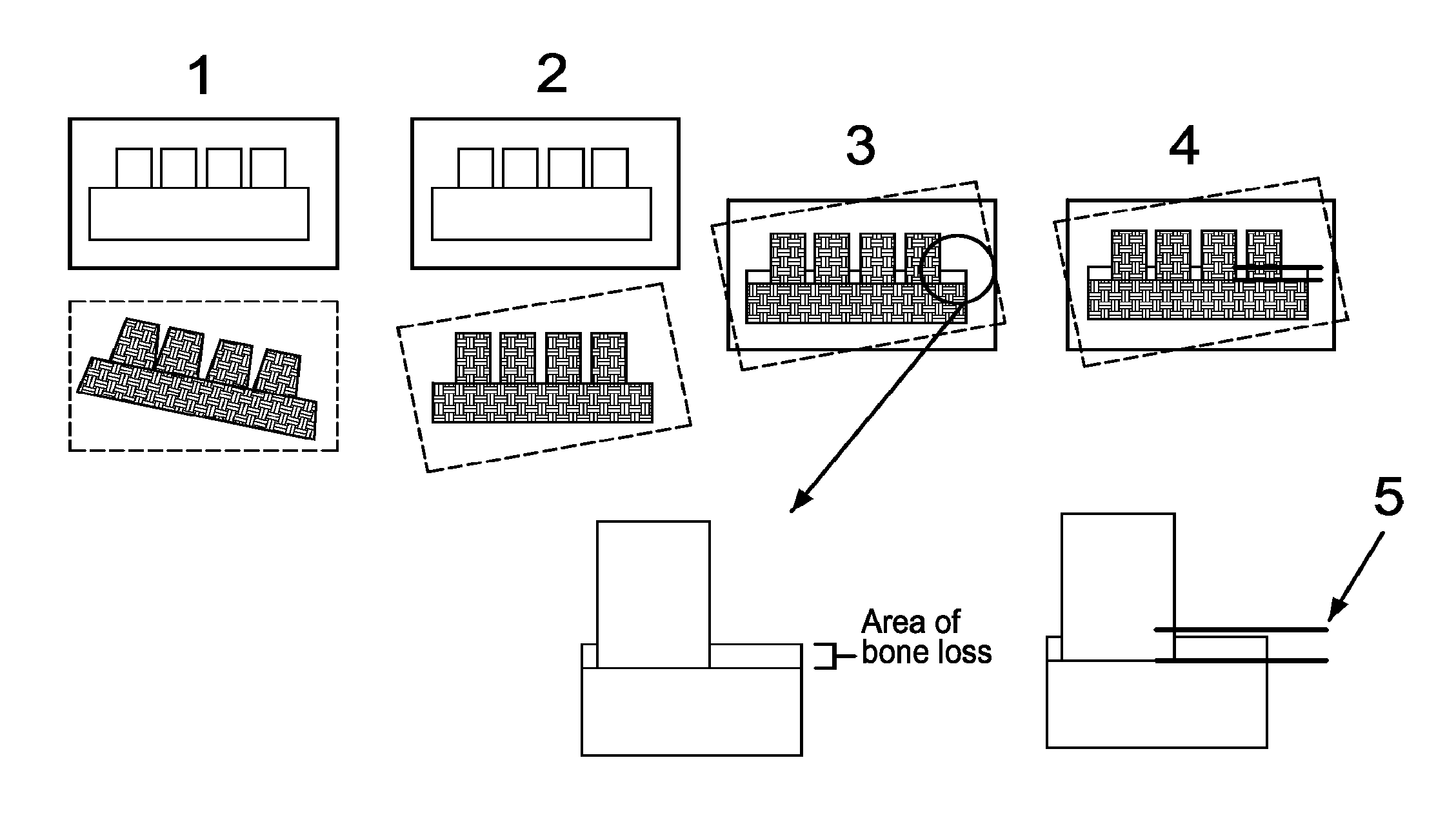
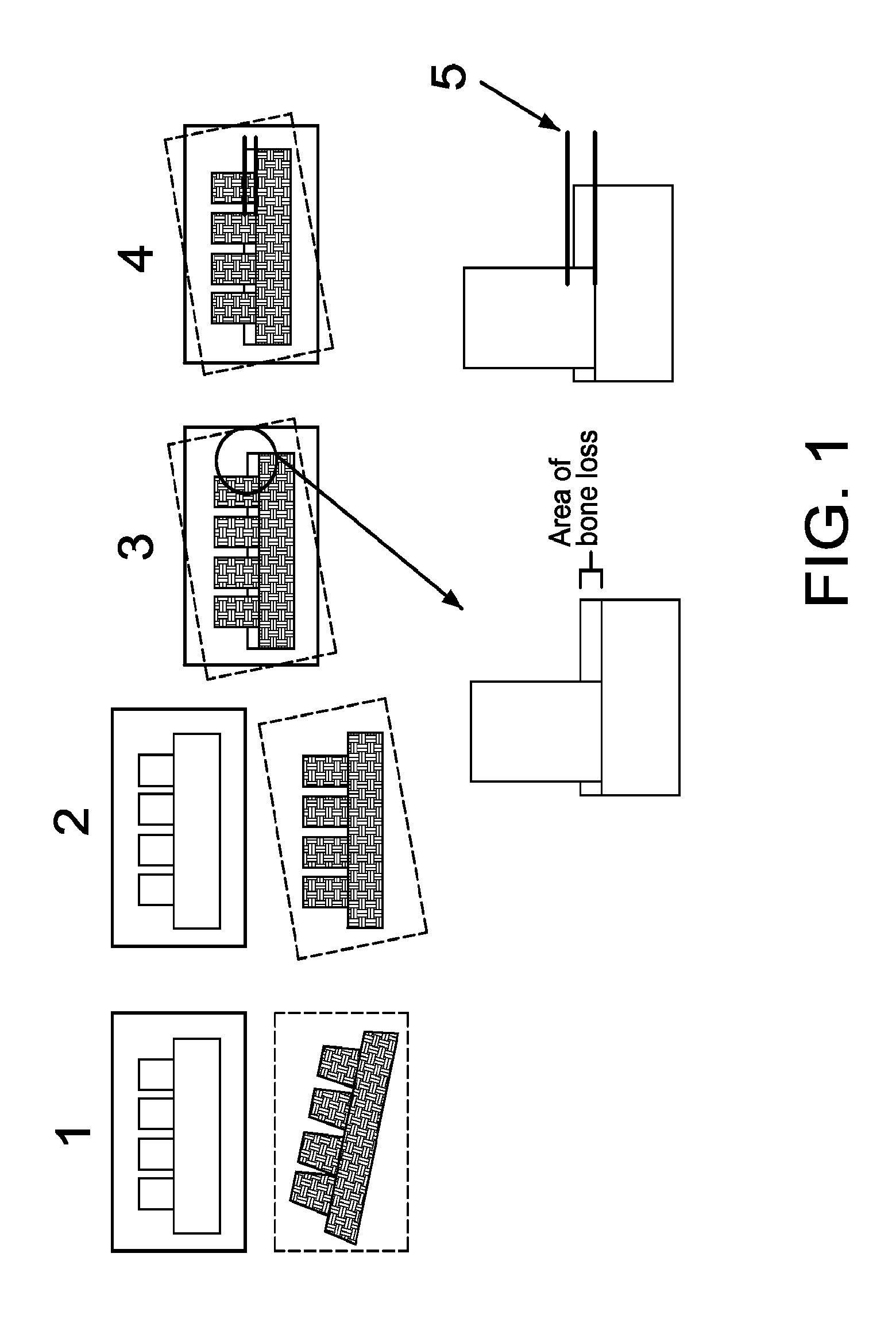
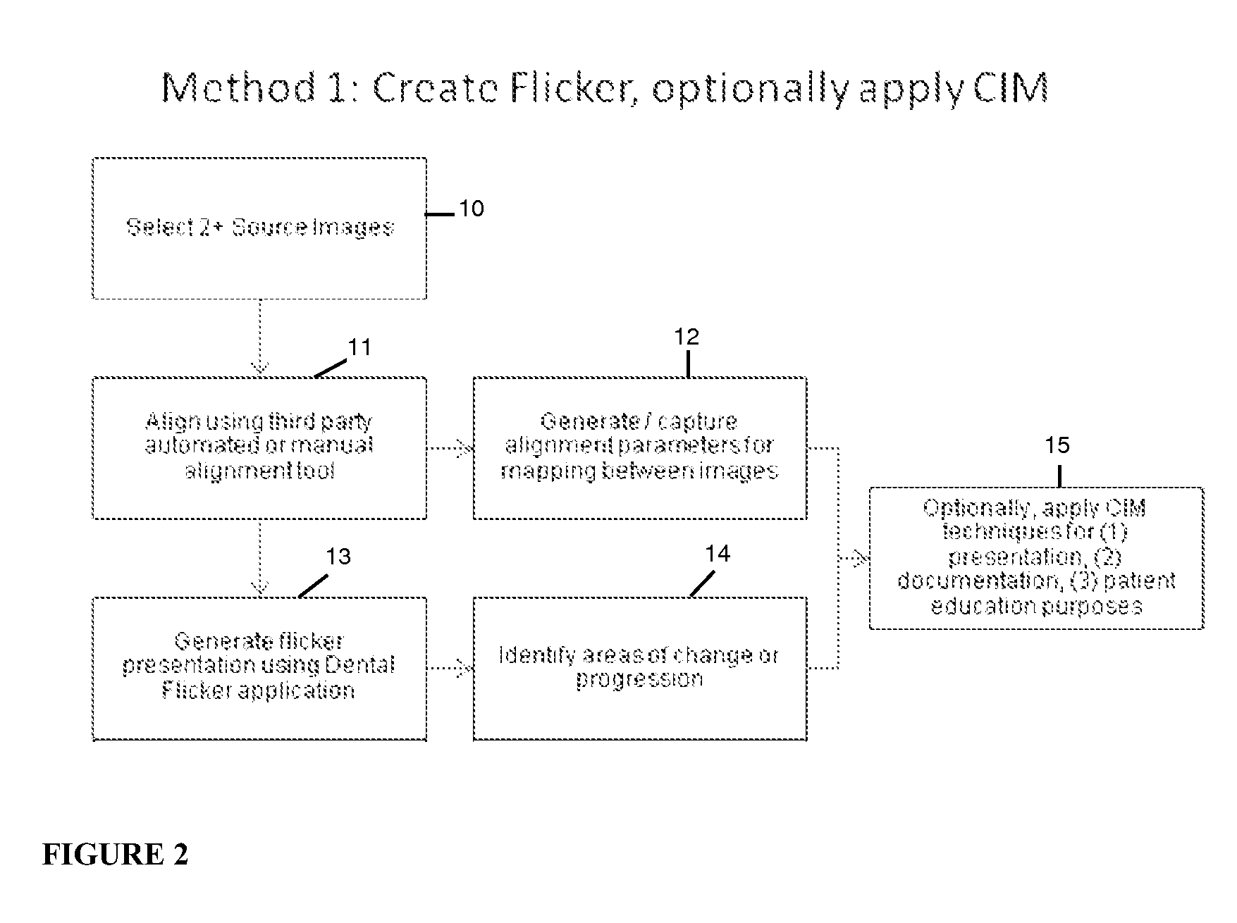
[0035]In accordance with a first embodiment of the invention, computer-aided registration and alignment is used to create a “flicker.” Such a method in accordance with the first embodiment includes establishing a baseline using a historical (existing) x-ray or by creating a new one. Virtually any x-ray (including those from digital sources as well as analog sources which have been scanned) can serve as a baseline, as long as it shows the features or landmarks to be examined, and the perspective, image type and rough image attributes can be approximated in subsequently prepared images. FIG. 1 shows two x-rays of the same area captured from a similar perspective. As illustrated at (1), two source images of the patient from a similar perspective are taken at different points in time, where, in this case, the first image A is taken straight on the tooth and the second image B is taken from a slightly lower perspective of the jaw, with a slight rotation. The technique is most effective w...
second embodiment
[0041]In accordance with a second embodiment of the invention, Cross-Image Mapping (CIM) or comparable techniques are used to apply “before and after” indicators to a single image. Such a method in accordance with the second embodiment allows a DF software user (e.g., a doctor, nurse or technician) to map two or more images using manual or automated registration. These aligned images do not need to be displayed for this technique to be effective, so actual modifications to the image are necessary only if called for by the mapping approach employed. As in the first embodiment, the first step includes acquiring two images of the patient from similar perspectives taken at two different points in time. As illustrated at (1) in FIG. 3, image A is taken straight on and image B is taken from a slightly lower perspective of the jaw with a slight rotation. The images are manually registered by the user as illustrated at (2) in FIG. 3. The CIM module of the DF application then creates formula...
third embodiment
[0044]In accordance with a third embodiment of Dental Flicker techniques, images of different types (for example photos and x-rays) but sharing certain landmarks (for example, teeth) in common, are aligned and / or mapped to allow location of a specific spot in one image on another. For example, this embodiment might be used to allow dentists and patients to relate a specific spot on the gums to the bones and / or teeth below that spot on the gums.
[0045]In such cases it may be necessary to align the images using only selected portions of one or both unaligned (source) images. FIG. 5 illustrates the flickering of two image types, in this case an x-ray and a photograph of the same areas. As illustrated in FIG. 5, the user circles or otherwise identifies the common landmarks (e.g., visible portion of teeth). The system would then align the entire area of the images using only the identified portions as the basis of this alignment. It is noted that the area can be designated in one image (a...
PUM
 Abstract
Description
Claims
Application Information
 - R&D
- Intellectual Property
- Life Sciences
- Materials
- Tech Scout
- Unparalleled Data Quality
- Higher Quality Content
- 60% Fewer Hallucinations
Browse by: Latest US Patents, China's latest patents, Technical Efficacy Thesaurus, Application Domain, Technology Topic, Popular Technical Reports.
© 2025 PatSnap. All rights reserved.Legal|Privacy policy|Modern Slavery Act Transparency Statement|Sitemap|About US| Contact US: help@patsnap.com


