
Noninvasive method and system for measuring pulmonary ventilation

a pulmonary ventilation and non-invasive technology, applied in the field of measuring respiratory air volume in humans, can solve the problems of restricting breathing, difficult to use the mouthpiece for long-term subject monitoring, and employing a mouthpiece, and achieve the effect of expanding the rib cage and accurately detecting respiratory events

Active Publication Date: 2014-07-29
ADIDAS
View PDF10 Cites 6 Cited by
  • Summary
  • Abstract
  • Description
  • Claims
  • Application Information

AI Technical Summary

Benefits of technology

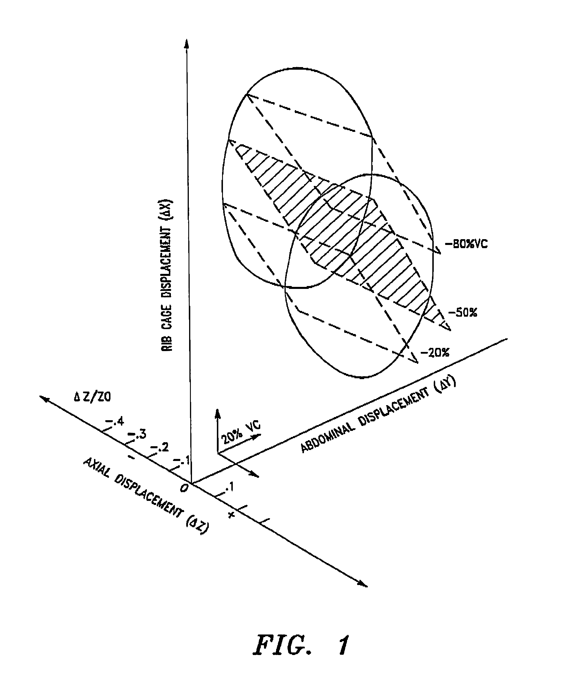
The present invention offers noninvasive methods and systems for determining pulmonary ventilation which offer significant improvements over conventional methods. These noninvasive methods can accurately measure breathing while awake and during sleep, and can detect sleep apnea and hypopnea. The invention can help in the diagnosis and management of sleep disorders and offers a reliable tool for monitoring breathing during sleep. These methods are calibrated using a two-degrees-of-freedom model which is derived from changes in the circumference of the rib cage and abdomen. The invention can help improve the accuracy of respiratory inductive plythysmograph (RIP) derived tidal volume during sleep and minimize the impact of slippage of the rip belts and changes in posture.

Problems solved by technology

There are, however, several drawbacks and disadvantages associated with employing a mouthpiece.
A mouthpiece is difficult to use for long term subject monitoring, especially for ill, sleeping or anesthetized subjects.
Further, it is uncomfortable for the subject, tends to restrict breathing, and is generally inconvenient for the physician or technician to use.
Although the noted systems eliminate most of the disadvantages associated with a mouthpiece, the systems do not, in general, provide an accurate measurement of air volume.
Further, the systems are typically only used to signal an attendant when a subject's breathing activity changes sharply or stops.
A potential limitation of the use of such coils or magnetometers is presented in environments that contain large metal structures or electric motors.
Such devices produce extraneous electromagnetic fields and consequently affect the magnetometer voltage output.
A limitation of any approach that uses chest wall motion to assess ventilation is, however, that the overall volume change of the chest wall being measured includes not only changes in lung volume, but also blood volume shifts into and out of the thoracoabdominal cavity.
Another limitation is related to distortion that can occur within the rib cage or abdomen (e.g. between the upper and lower rib cage or between the lower transverse and AP rib cage).
As is well known in the art, the accuracy of “two-degrees-of-freedom” model and, hence, methods employing same to determine volume-motion coefficients of the rib cage and abdomen, is further limited by virtue of changes in spinal flexion that can accompany changes in posture.
The second contributing cause of the error is due to posturally-induced changes in volume-motion coefficients.
There are, however, similarly several drawbacks and disadvantages associated with the “three-degrees-of-freedom” model.
A major drawback is that the “three-degrees-of-freedom” model reflected in Eq.
3 above is still limited in accuracy to about 15% of actual ventilation in individuals who are doing freely moving postural tasks, such as bending, sitting or standing, due to spinal flexion.

Method used


Image

  • Noninvasive method and system for measuring pulmonary ventilation
  • Noninvasive method and system for measuring pulmonary ventilation
  • Noninvasive method and system for measuring pulmonary ventilation

Examples


example 1

Subjects

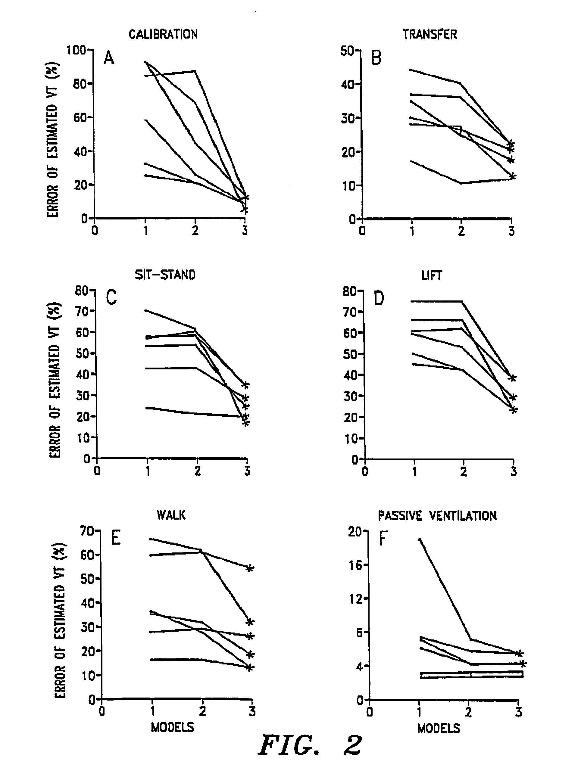
[0218]Thirty (30) subjects were selected to assess the accuracy of the “three-degrees-of-freedom” model of the invention. As reflected in Table 1, ten (10) healthy subjects with no known history of sleep-disordered breathing and ten (10) obese subjects were studied while awake in the supine, right and left lateral decubitus positions. Ten (10) obese subjects with obstructive sleep apnea (obese sleeping subjects) were studied while unrestrained during a daytime nap.

[0219]

TABLE 1Non-Obese AwakeObese AwakeObese SubjectsSubjectsSubjectsDaytime Nap+Subject(BMI)(BMI)(BMI)12052512304561327614942849405275934629403872334518273839921505610 253938Mean ± SD25.7 ± 3.446.7 ± 9.145.7 ± 1.0BMI: Body Mass Index

[0220]The non-obese, awake subjects comprised seven (7) males and three (3) females, age 28-49 yr. (mean age 30.2 yr). The obese, awake subjects comprised six (6) males and four (4) females, age 24-64 yr. (mean age 44.4 yr). The obese, napping subjects comprised five (5) males and five...

example 2

Subjects

[0252]The following study was conducted to assess the accuracy of the “three-degrees-of-freedom” model of the invention and a pulmonary ventilation or magnetometer system employing the subject model to detect apneas and hypopneas during sleep.

[0253]As reflected in Table 6, fifteen (15) subjects (10 males and 5 females) with variable degrees of clinical suspicion for obstructive sleep apnea (OSA) were selected for the study. The mean age for the group was 45.47±12.5 years (mean±SD). The mean body mass index for the group was 35.00±5.7 kg / m2.

[0254]

TABLE 6Body Mass IndexSubjectAge(BMI)15032271283343343931530366484174029849289744310 343911 443412 383313 394614 464115 4631Mean ± SD35.00 ± 5.745.47 ± 12.5

Device and Measurements

[0255]The anteroposterior displacements of the rib cage and abdomen, as well as the axial displacements of the chest wall (i.e. Xi) were similarly measured using a light-weight, portable pulmonary ventilation system of the invention (also referred to herein ...


PUM


Abstract

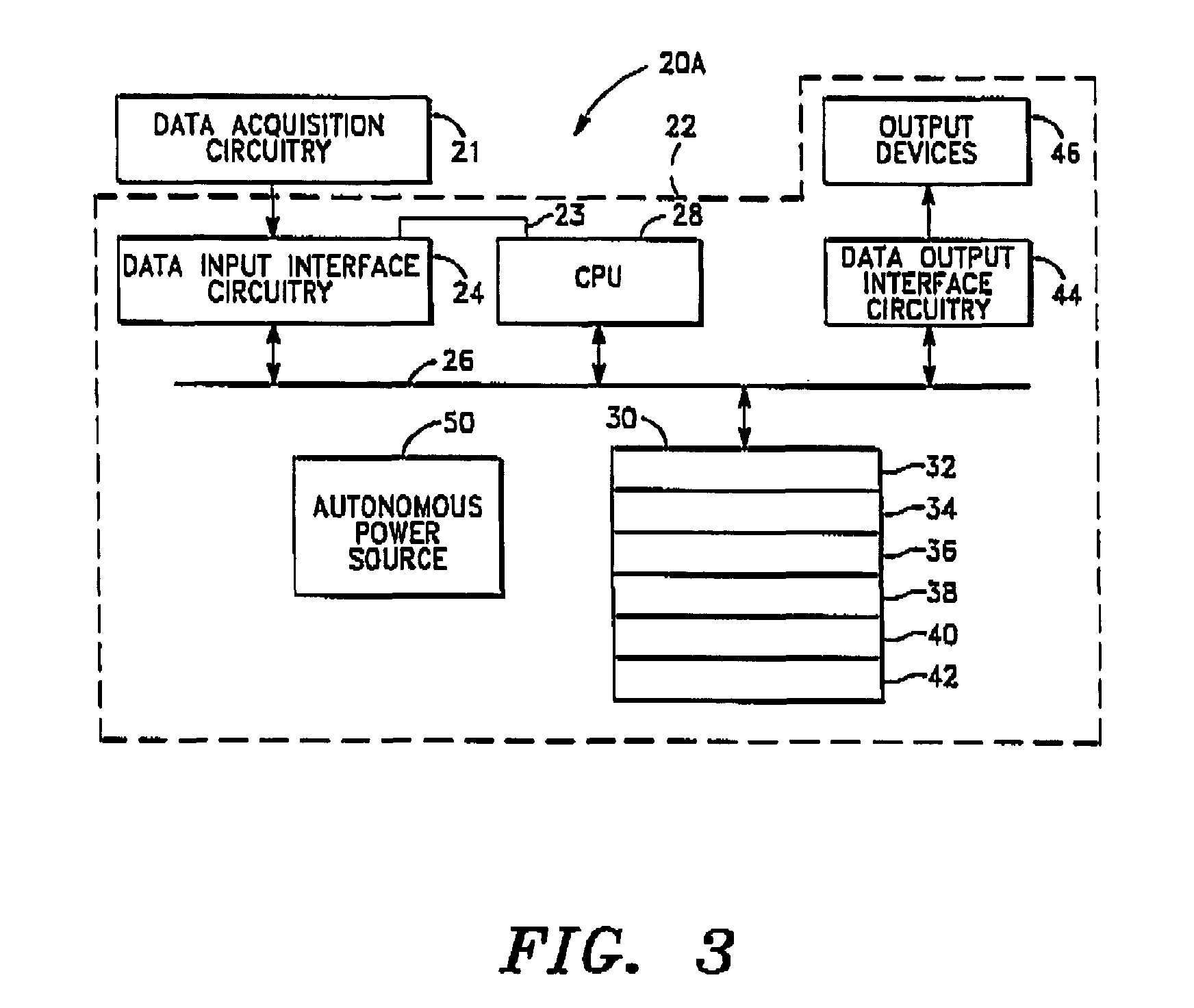
A pulmonary ventilation system comprising a method to determine at least one pulmonary ventilation parameter as a function of a plurality of measured anatomical distances and volume-motion coefficients, sensors for acquiring the anatomical distances, a device for determining the plurality of motion coefficients, and a device for determining the ventilation parameter based on the acquired anatomical distances and determined plurality of volume-motion coefficients. In one embodiment, the system further includes a method for acquiring base-line ventilation characteristics and a method for correlating the base-line ventilation characteristics to the ventilation parameter determined with the empirical relationship.

Description

FIELD OF THE PRESENT INVENTION[0001]The present invention relates generally to measuring respiratory air volume in humans. More particularly, the invention relates to a noninvasive method for quantitatively measuring pulmonary ventilation and a ventilation system employing same.BACKGROUND OF THE INVENTION[0002]As is well known in the art, in medical diagnosis and treatment, it is often desirable to quantitatively measure over time the respiratory air volume or pulmonary ventilation. This has conventionally been done by having the patient or subject breathe into a mouthpiece connected to a flow rate measuring device. Flow rate is then integrated to provide air volume change.[0003]There are, however, several drawbacks and disadvantages associated with employing a mouthpiece. A mouthpiece is difficult to use for long term subject monitoring, especially for ill, sleeping or anesthetized subjects. Further, it is uncomfortable for the subject, tends to restrict breathing, and is generally...

Claims


Application Information

Patent Timeline
Patent Type & Authority Patents(United States)
IPC IPC(8): G01R33/02A61B5/02A61B5/091A61B5/08A61B5/04A61B5/103G01B7/14
CPCA61B5/091A61B5/1135
Inventor MCCOOL, FRANKLIN, DENNIS
Owner ADIDAS
Learn More