Pneumovirus compositions and methods for using the same
a technology of pneumovirus and composition, applied in the field of newly discovered viruses, can solve the problems of difficult diagnosis and treatment, rapid transmission of diseases, and difficult elimination
- Summary
- Abstract
- Description
- Claims
- Application Information
AI Technical Summary
Benefits of technology
Problems solved by technology
Method used
Image
Examples
example 1
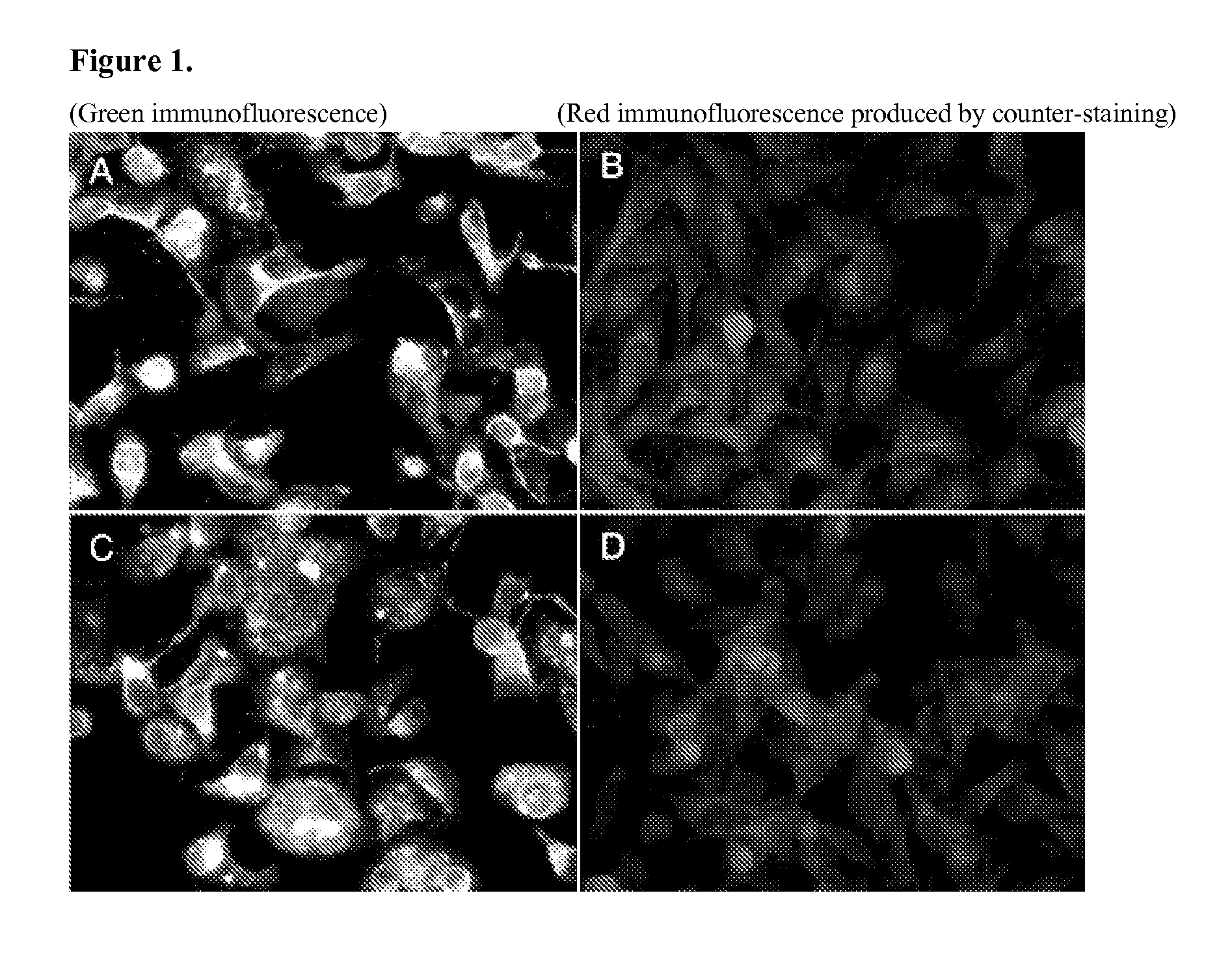
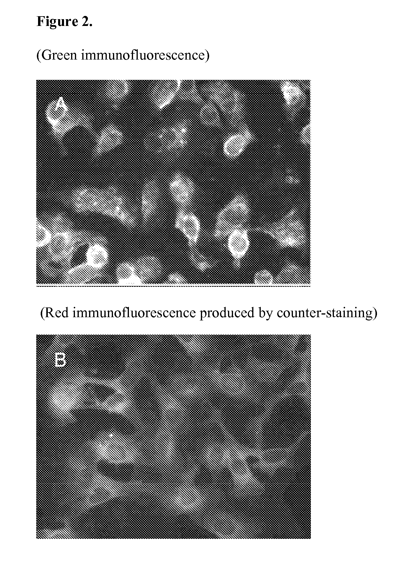
[0044]We obtained samples from nasal and pharyngeal swabs from mixed-breed dogs and used to them to inoculate cultures of canine A72 cells (American Type Culture Collection, CRL-1542) to isolate respiratory viruses. At a late stage of passage in culture corresponding to approximately 21 days post-inoculation, some cultures showed subtle cytopathic changes. After continued passage the cultures exhibited small foci of rounded cells followed by rapid cell death throughout the culture. This pattern was subjectively judged to be uncharacteristic of the viruses commonly associated with ARDC. Thirteen individual viral isolates were obtained and are described in this Example. Testing with a panel of diagnostic reagents specific to common canine respiratory agents failed to identify a known virus. Further testing with additional reagents for other viruses ultimately revealed positive recognition by immunofluorescence assay (IFA) using a monoclonal antibody (Mab) pool against human respirator...
example 2
[0051]The materials and methods presented in this Example were used to obtain the results described in the remaining Examples.
Virus Isolation
[0052]Nasal and pharyngeal specimens were collected from mixed breed dogs with respiratory disease signs using moist swabs and were processed within 24 h of receipt at the Animal Heath Diagnostic Center at Cornell University. Prior to inoculation, the swabs were immersed in 3 ml minimum essential medium (MEM) with Earle's salts (Gibco 10370, Invitrogen, Carlsbad, Calif., USA), 0.5% bovine serum albumin, penicillin (200 U / ml), streptomycin (200 μg / ml), and fungizone (2.5 μg / ml) for 30 min and then mechanically agitated for 10 s. Aliquots of the nasal and pharyngeal swab extracts, 0.5 ml each, were combined and used to inoculate a single T25 flask of semi-confluent A72 cells (Binn et al., 1980) (CRL-1542, American Type Culture Collection, Manassas, Va., USA) without additional medium. The extract was allowed to remain on the monolayer for 1-3 h a...
example 3
Isolation and Identification
[0057]Nasal and pharyngeal swab eluates were used to inoculate A72 cells. After a relatively long period in culture, usually appearing after the third or fourth passage, a limited CPE was observed. Initial CPE typically included scattered small foci of rounded cells, sometimes with small syncytia and vacuolization. This CPE progressed slowly over several days in stationary cultures. Subculturing often led to monolayers that maintained this low-grade CPE but in some cultures there was rapid progression and disintegration of the monolayer within 24-48 hours. This pattern, although similar to some herpesvirus infections, did not test positive for canine herpesvirus and did not appear to be typical of any of the canine respiratory viruses that are commonly isolated using similar procedures. Several of the early passage isolates replicated poorly in culture, showing little or no CPE following the transfer of supernatant to fresh cells. However, with continued ...
PUM
| Property | Measurement | Unit |
|---|---|---|
| volume | aaaaa | aaaaa |
| length | aaaaa | aaaaa |
| length | aaaaa | aaaaa |
Abstract
Description
Claims
Application Information
 - R&D
- Intellectual Property
- Life Sciences
- Materials
- Tech Scout
- Unparalleled Data Quality
- Higher Quality Content
- 60% Fewer Hallucinations
Browse by: Latest US Patents, China's latest patents, Technical Efficacy Thesaurus, Application Domain, Technology Topic, Popular Technical Reports.
© 2025 PatSnap. All rights reserved.Legal|Privacy policy|Modern Slavery Act Transparency Statement|Sitemap|About US| Contact US: help@patsnap.com


