Image quality for diagnostic resolution digital slide images

a digital slide and diagnostic resolution technology, applied in the field of digital pathology, can solve the problems of exacerbated challenges associated with assessing image quality and/or interpretability, difficult to examine every area of a digital slide at full resolution to determine its quality and/or interpretability, and relative difficulty in objectively assessing the quality or interpretability of imagery data generated by such devices.

Active Publication Date: 2015-08-04
LEICA BIOSYST IMAGING
View PDF26 Cites 1 Cited by
  • Summary
  • Abstract
  • Description
  • Claims
  • Application Information

AI Technical Summary

Benefits of technology

[0010]In some embodiments the image adjustments include digital recolorization of a stain on a single stain image. This can result in higher quality digital images for more objective diagnosis and improve quality of image analysis algorithm results. In other embodiments, the one or more of the plurality of single stain images can be processed with one or more image analysis algorithms or image quality algorithm. The image quality algorithm can successfully separate the individual stains, digitally adjust each stain independently and recombine them in pairs to present the digital slides. The use of the IQ algorithm can produce digitally enhanced stains, a reduction in samples used, automatic co-location of multiple stains, for example hematoxylin and eosin (“H&E”) with H&DAB, thereby accelerating review, saving preparation time and time spent requeuing the number of glass slides and storage. The image quality algorithm can also configure for any two or three stained sample, for example H&E with H&DAB or H&E&DAB. The image quality algorithm also performs digital stain adjustments for stain normalization including dilution / concentration, removal of background staining, cellular detail emphasis, chromogen tuning and remapping.
[0012]A significant advantage of the described systems and methods is that they provide a pathologist or clinician with improved image quality digital slides that facilitate certain types of diagnostic or interpretative tasks. For example, the system and method provides for single stain digital adjustments such as boosting weak stains, diluting strong stains, removing background staining, standardization of digital image characteristics of stains, recolorization of stains (e.g., to primary colors having higher contrast than actual stains used), personalized staining (e.g., based on preference of an individual pathologist), spatial filtering on single stain images, segmentation and quantification on single stain images (e.g., counting nuclei in a Progesterone_receptor (“PR”), support new multiplex reagent with multichannel detection or spectral imaging detector (e.g., ER / PR on same slide and other dual or triple stains—reduces the need for two or more physical slides to be stained, provides advantage over separate slides with inexact physical samples) and novel methods of visualization.
[0014]Development of New Reagents (Stains): In one embodiment, the reagents can be optimized for digital scanning at a diagnostic resolution. An example of an optimized reagent can be achieved by developing new protocols for use of conventional and new reagents to optimize the sample for being digitally scanned at a diagnostic resolution (e.g., multiplex reagents or single reagent). In an embodiment new reagent systems (for example, stains) can be optimized for digital pathology, e.g., produce better quality digital images when scanned at diagnostic resolution. Among other IQ elements, the process can include deconvolving the new reagent and also New / optimized reagent protocols in combination with deconvolution. In one embodiment the process can include linking the properties of the reagent and the light passing through the stained tissue into the pixel sensors on the camera (e.g., optimizing the reagent(s) into the RGB channels.

Problems solved by technology

While scientific measures such as modulation transfer function (“MTF”), resolution and the signal to noise ratio (“SNR”) can be used to objectively quantify image capture devices, it is relatively difficult to objectively assess the quality or interpretability of the imagery data that is generated by such devices, particularly when the imagery data is destined to be subjectively examined by a human, for example in the evaluation of digital slide images in pathology.
Furthermore, the challenges associated with assessing image quality and / or interpretability are exacerbated for digital slide images that are read by pathologists because digital slides are extraordinarily large, typically multiple gigabytes in size.
This significant amount of imagery data makes it very difficult to examine every area of a digital slide at full resolution to determine its quality and / or interpretability.
Additionally, the patterns (i.e., the features and clues) that pathologists look for when making a diagnosis vary between tissue types and this is further confounded because there is no formal agreement among pathologists about the criteria and tasks used to make diagnoses of specific tissue types.
Further, analysis of digital slide images and diagnosis based on digital slide images is made difficult by weak stains that result in faint coloring, strong stains that result in heavy coloring, non-standardized digital image characteristics of stains, similarly colored stains in a single slide, stains with low contrast to each other on a single slide, and poorly physical slide preparation.

Method used


Image

  • Image quality for diagnostic resolution digital slide images
  • Image quality for diagnostic resolution digital slide images
  • Image quality for diagnostic resolution digital slide images

Examples


Embodiment Construction

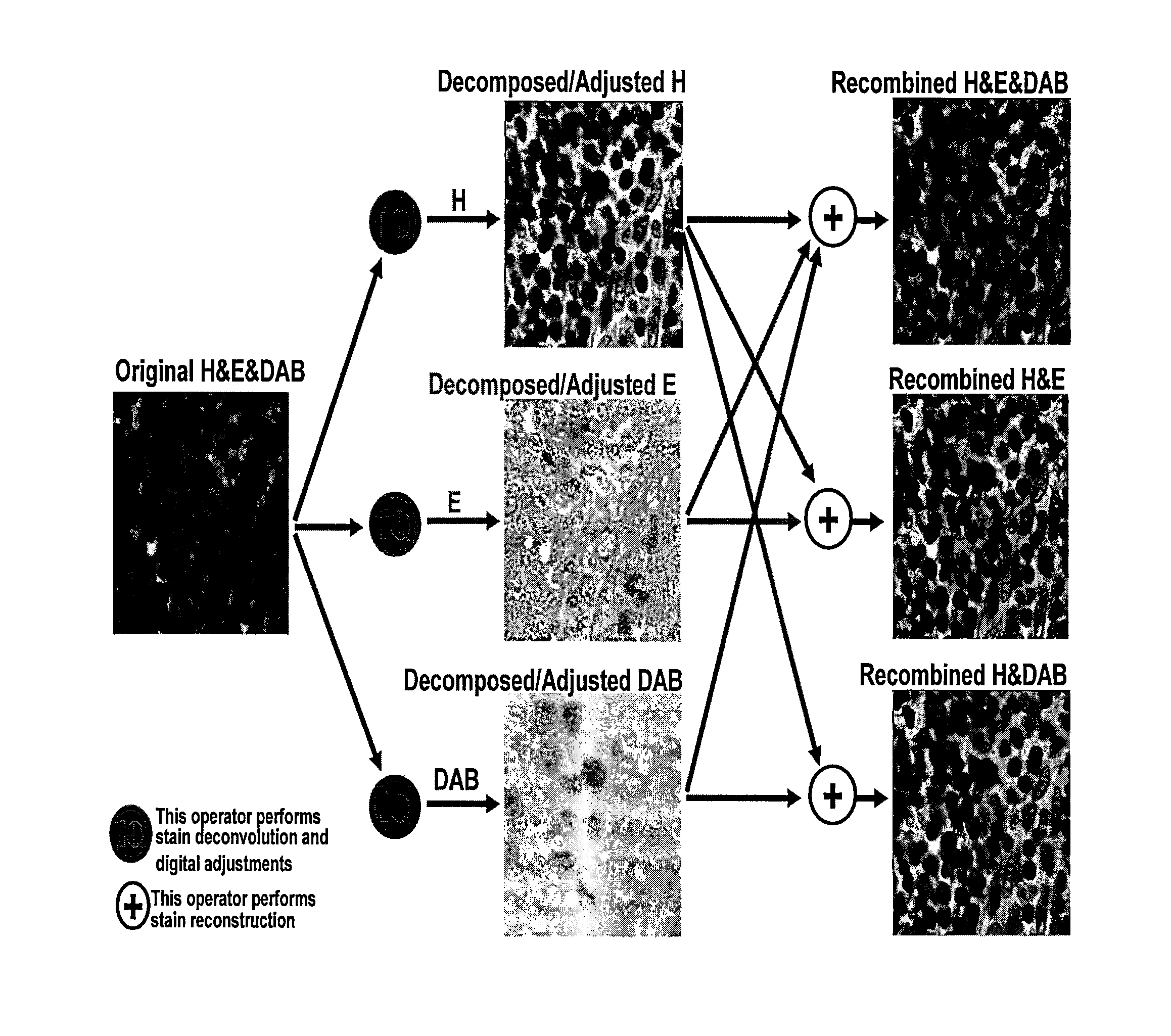
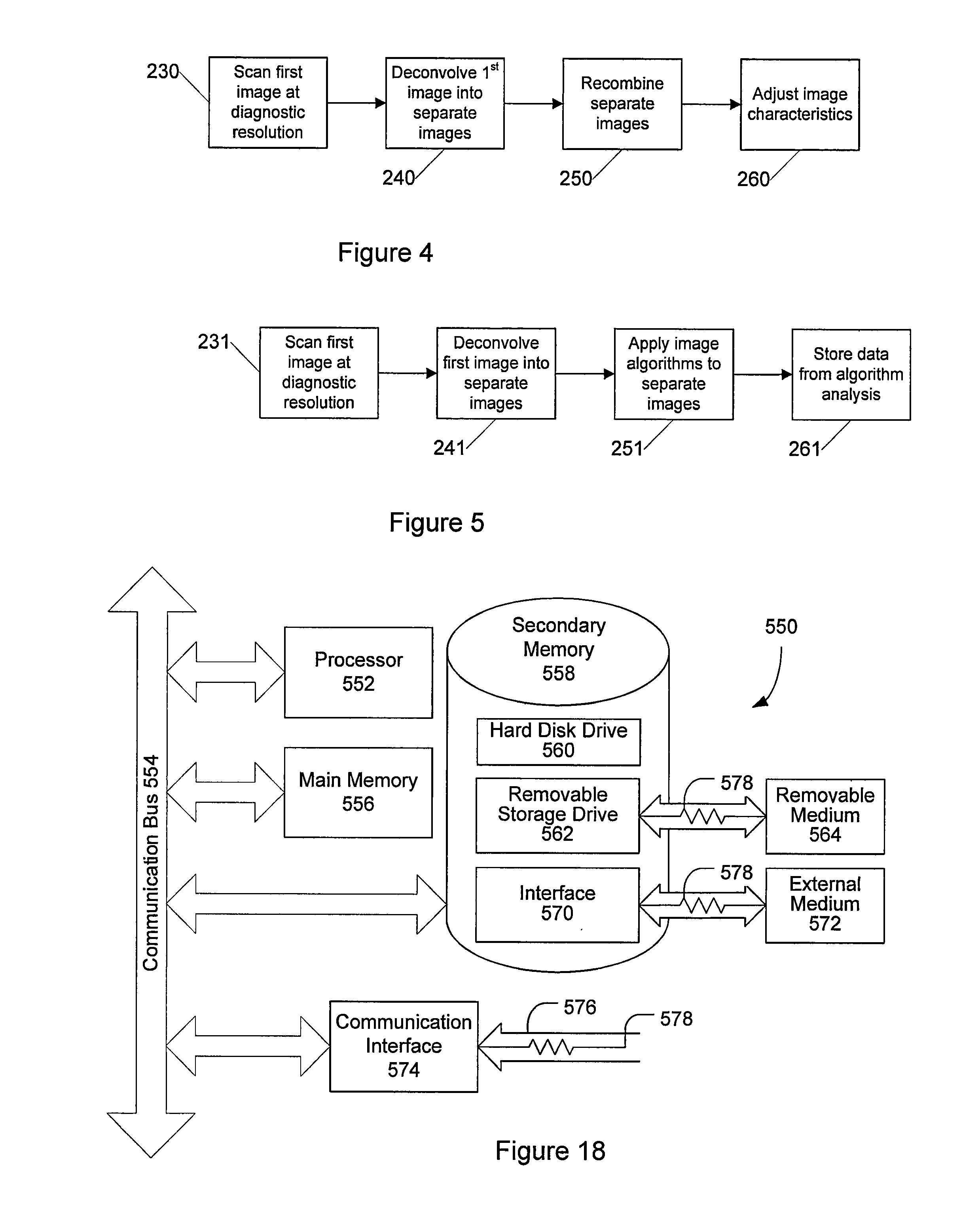
[0028]Certain embodiments as disclosed herein provide for systems and methods for improving diagnostic resolution of digital slide images. After reading this description it will become apparent to one skilled in the art how to implement the invention in various alternative embodiments and alternative applications. However, although various embodiments of the present invention will be described herein, it is understood that these embodiments are presented by way of example only, and not limitation. As such, this detailed description of various alternative embodiments should not be construed to limit the scope or breadth of the present invention as set forth in the appended claims.

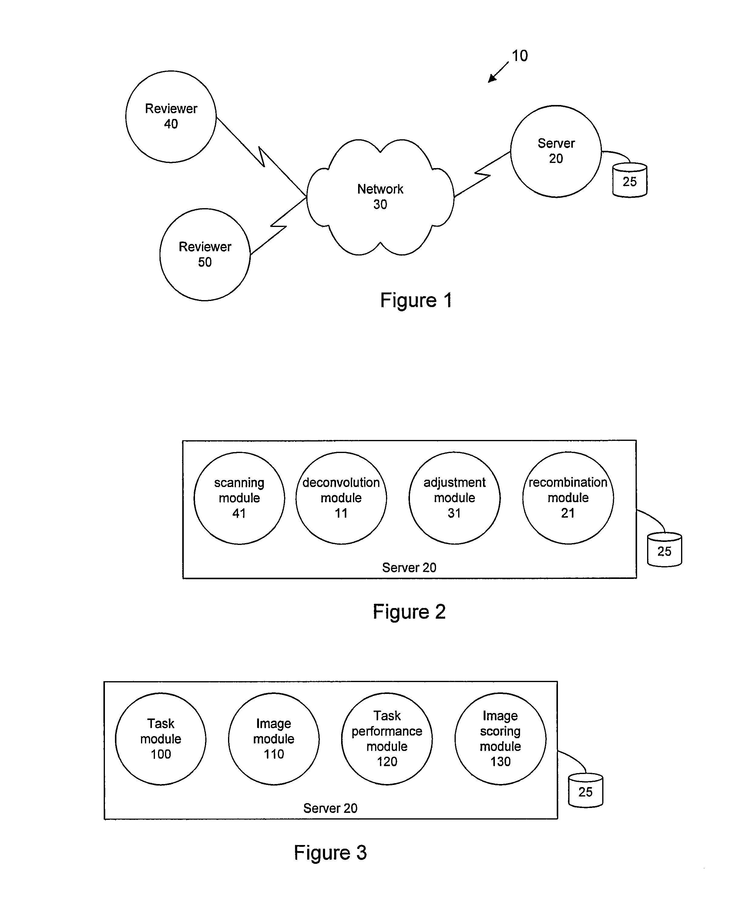
[0029]FIG. 1 is a network diagram illustrating an example system 10 for image interpretability assessment according to an embodiment of the present invention. In the illustrated embodiment, the system 10 comprises an image server 20 that is communicatively coupled with reviewer device 40 and reviewer device ...


PUM


Abstract

An improved diagnostic resolution of digital slide images is obtained by scanning a first digital slide image at diagnostic resolution that is then deconvolved into separate images with one stain per image. The single stain images are then enhanced with image adjustments and / or processed with image analysis algorithms. The resulting single image data sets from the image analysis algorithms can then be stored. Additionally, the resulting enhanced single images can be recombined into a second digital slide image at diagnostic resolution that is also enhanced.

Description

RELATED APPLICATION[0001]The present application claims priority to U.S. provisional patent application Ser. No. 60 / 974,337 filed Sep. 21, 2007, which is incorporated herein by reference in its entirety.BACKGROUND[0002]1. Field of the Invention[0003]The present invention generally relates to the field of digital pathology and more specifically relates to systems and methods for improving the image quality of a digital slide image for diagnostic resolution.[0004]2. Related Art[0005]While scientific measures such as modulation transfer function (“MTF”), resolution and the signal to noise ratio (“SNR”) can be used to objectively quantify image capture devices, it is relatively difficult to objectively assess the quality or interpretability of the imagery data that is generated by such devices, particularly when the imagery data is destined to be subjectively examined by a human, for example in the evaluation of digital slide images in pathology.[0006]Furthermore, the challenges associa...

Claims


Application Information

Patent Timeline
Patent Type & Authority Patents(United States)
IPC IPC(8): G06K9/00G06T5/00G06T5/50
CPCG06K9/0014G06T5/003G06T5/50G06T2207/10024G06T2207/30024G02B21/241G06V20/695G06V20/69G06T7/0012G06T2207/10056
Inventor SOENKSEN, DIRK G.PERZ, CINDY
Owner LEICA BIOSYST IMAGING
Why Eureka
Try Eureka