Treatment for dupuytren's disease

a dupuytren's disease and treatment technology, applied in the field of dupuytren's disease, can solve the problems of unknowable dupuytren's disease, uncurable underlying disease, and inability to cure the disease, so as to avoid surgical intervention and the associated recovery time

Active Publication Date: 2015-09-22
OXFORD UNIV INNOVATION LTD
View PDF21 Cites 5 Cited by
  • Summary
  • Abstract
  • Description
  • Claims
  • Application Information

AI Technical Summary

Problems solved by technology

The commonest manifestation is progressive flexion contracture of the digits of the hand, resulting in significantly compromised function.
The causes of Dupuytren's disease are not well understood and underlying disease is not currently curable.
Bacterial collagenases suffer from certain disadvantages: for example lack non-selective cleaving of various collagen materials including collagen type IV associated with blood vessels; and, in the case of Xiaflex™, possible allergic reactions and potential immunogenicity; and administration may cause haemorrhage whilst the prolonged activity of collagenase limits the dose that can be administered locally due to risk of side effects as the drug disperses.
To date collagenase therapies have appeared relatively effective in treatment of contracture of the metacarpophalangel joint, whilst the correction of proximal interphalangeal joints has been much less satisfactory.
There has been very little conclusive insight into potential treatments gained from studies into the biochemical pathway of Dupuytren's disease.

Method used


Image

  • Treatment for dupuytren's disease
  • Treatment for dupuytren's disease
  • Treatment for dupuytren's disease

Examples


example 1

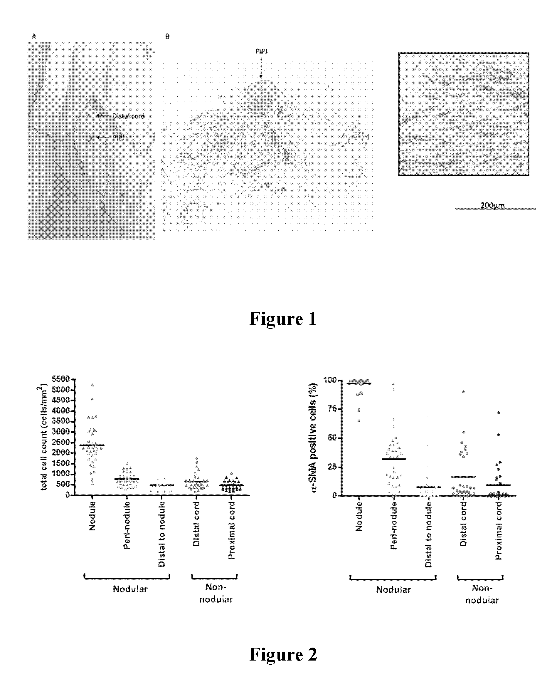
[0131]Over 100 Dupuytren's patient samples were collected to examine myofibroblast distribution. Our data on >100 Dupuytren's cords show that in the majority of patients, myofibroblasts are concentrated in nodules, located in the palm and at the level of the affected joints (see FIG. 1). According to FIG. 1, nodules rich in myofibroblasts are located in the vicinity of the finger joints. FIG. 1 shows: A: intraoperative view of Dupuytren's cord, with location of proximal interphalangeal joint (PIPJ; 1) marked; B: Low magnification photomicrograph of histological section stained for α-smooth muscle actin. A collection of α-SMA rich cells in a nodule is located in the vicinity of the PIPJ; C: High magnification view of nodular area, showing α-SMA positive cells (myofibroblasts).

[0132]Of over 100 cords analysed, more than 60% contained nodules. Although there was marked heterogeneity, nodules were very cellular, with approximately 2.5 thousand cells per mm2 arranged in whorls. On averag...

example 2

Role of Inflammation in Dupuytren's Disease

[0142]The nodules were then examined for the presence of other cell types, specifically inflammatory cells. We found that large numbers of both macrophages and mast cells were present in nodules but not in non-nodular regions of the cords.

[0143]FIG. 7 shows inflammatory cells in Dupuytren's nodule and cord. Digital cord sections were serially stained for α-SMA, CD68 positive macrophages and mast cell tryptase. The images are representative of 15 patient samples.

[0144]We systematically quantified the number of inflammatory cells observed throughout excised Dupuytren's cord tissue in 10 patient samples. For each region (nodule, cord distal to nodule and non-nodular cord), the total number of cells, the number of α-SMA positive cells and cells stained for neutrophil elastase, mast cell tryptase, CD3positive T cells, CD 4 positive T cells, CD68 positive macrophages were counted (×20 magnification) (Table 1).

[0145]

TABLE 1Quantification of total ...

example 3

Advanced Glycation End Products and their Receptor

[0147]We examined the distribution of RAGE in Dupuytren's tissue and both palmar and non-palmar skin. We found abundant staining for RAGE in Dupuytren's nodules, where it co-localised with the myofibroblasts (seeFIG. 8). Digital cord samples were longitudinally bisected and fixed in formalin. Histological sections were taken from the cut surface of cord and serial sections were stained for (A, C) α-SMA and (B, D) RAGE antibodies. Scale bars as shown. Images are representative from 15 patient samples. RAGE co-localises with α-SMA distribution in Dupuytren's nodules.

[0148]We also found increased staining for RAGE in the superficial layers of the epidermis in palmar skin compared to non-palmar skin and FACS staining showed significantly higher RAGE expression by dermal fibroblasts from palmar skin compared to non-palmar skin. See FIG. 9.

[0149]Non-palmar and palmar skin samples were fixed in formalin. Histological sections were stained f...


PUM

PropertyMeasurementUnit
timeaaaaaaaaaa
timeaaaaaaaaaa
timeaaaaaaaaaa

Abstract

Musculoskeletal fibroproliferative disorders, such as Dupuytren's disease may be treated by administering locally a TNF-α antagonist. TNF-α antagonists find particular utility in inhibiting the progression of early disease state Dupuytren's disease and other musculoskeletal fibroproliferative disorders and, in combination with extracellular matrix degradation agents (such ascollagenase or matrix metalloproteinase I), treating advanced disease state Dupuytren's disease and, in particular inhibiting recurrence.

Description

[0001]This is a national stage application of PCT / EP2011 / 069147, filed internationally on Oct. 31, 2011, which claims priority to GB 1018325.9, filed Oct. 30, 2010; GB 1018362.2, filed Nov. 1, 2010 and GB 1113718.9, filed Aug. 10, 2011.FIELD OF THE INVENTION[0002]This invention relates to the treatment of musculoskeletal fibroproliferative disorders such as fibromatosis and, in particular, Dupuytren's disease. In particular it relates to a composition or therapeutic agent or to a combination of such compositions or therapeutic agents for the treatment, prophylaxis or prevention of progression of musculoskeletal fibroproliferative disorders, especially Dupuytren's disease, to the use of such composition / therapeutic agent or combination of compositions / therapeutic agents for the treatment, prophylaxis or prevention of progression of musculoskeletal fibroproliferative disorders, especially Dupuytren's disease and to a method of treating musculoskeletal fibroproliferative disorders, esp...

Claims


Application Information

Patent Timeline
Patent Type & Authority Patents(United States)
IPC IPC(8): A61K38/17A61K9/00A61K9/06C07K16/24A61K45/06A61K31/00A61K9/08A61K38/00
CPCA61K38/1709A61K9/0014A61K9/0019A61K9/06A61K9/08A61K31/00A61K45/06C07K16/241A61K38/00A61P19/02A61P19/04A61P21/00A61P35/00A61P43/00C07K2317/21C07K2317/24C07K2317/55C07K2317/76C07K2319/30
Inventor NANCHAHAL, JAGDEEPMIDWOOD, KIM SUZANNE
Owner OXFORD UNIV INNOVATION LTD
Why Eureka
Try Eureka