
Flexible passive acoustic driver for magnetic resonance elastography

a passive acoustic driver and magnetic resonance technology, applied in the field of devices for implementing magnetic resonance elastography, can solve the problems of many diseases that go undetected, valuable diagnostic tools are limited to those tissues and organs which the physician can prescribe, and many diseased internal organs go undiagnosed

Active Publication Date: 2015-10-06
MAYO FOUND FOR MEDICAL EDUCATION & RES
View PDF28 Cites 2 Cited by
  • Summary
  • Abstract
  • Description
  • Claims
  • Application Information

AI Technical Summary

Benefits of technology

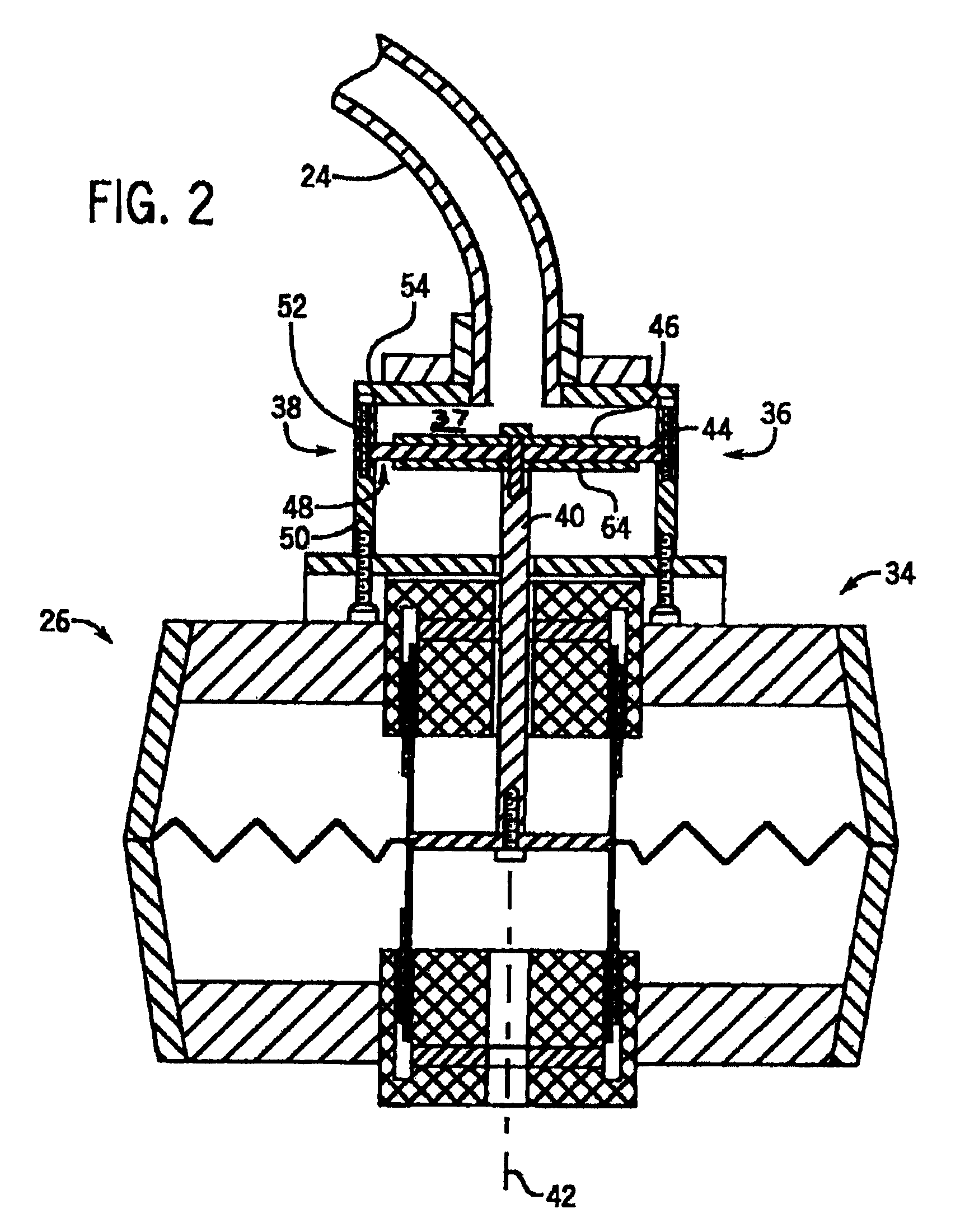
[0012]The present invention provides a passive acoustic driver that receives acoustic pressure waves through a tube and imparts pressure waves to a subject of an imaging procedure. In one implementation, the passive driver includes a flexible bag forming walls of an acoustic cavity, and engages the subject of the examination. Also, the driver includes an intake pipe which is connectable to the tube and conveys acoustic pressure waves into the acoustic cavity, and a structure filling material located inside the acoustic cavity provides support for the flexible bag. The flexible bag is designed to conform to the shape of the subject and may be coupled with an elastic band to be affixed to the subject This flexible passive acoustic driver is patient-friendly, comfortable and safe.
[0014]One aspect of the invention is to improve the patient comfort when using a passive acoustic driver. The driver may flex to follow the general contour of the subject being imaged. The driver compresses as needed to uniformly distribute the force holding the passive driver in place. Pressure points are thus avoided.

Problems solved by technology

It is fortuitous that many diseased tissues are detected by the physical properties measured by these imaging modalities, but it should not be surprising that many diseases go undetected.
Unfortunately, this valuable diagnostic tool is limited to those tissues and organs which the physician can feel, and many diseased internal organs go undiagnosed unless the disease happens to be detectable by one of the above imaging modalities.
Palpation is the most common means of detecting tumors of the prostate gland and the breast, but unfortunately, deeper portions of these structures are not accessible for such evaluation.
In addition, the current flowing in the driver coil produces a magnetic field which can alter the magnetic fields during the magnetic resonance pulse sequence resulting in undesirable image artifacts.
Such drivers do not produce troublesome disturbances in the scanner magnetic fields when operated, but they are limited in the forces they can produce, particularly at larger displacements.
There are clinical situations where existing passive drivers cannot reliably or comfortably be positioned to adequately vibrate, or illuminate, tissues in the region of interest.
Existing MRE passive drivers, such as the prior art driver 100 shown in FIG. 3, are rigid and cylindrical and consequently do not always conform well to the anatomical shape of a subject.
As a result, incomplete surface area contact between the subject and the passive driver causes reduced MRE driver efficiency and a reduced MRE signal.
These problems are most problematic in, but not limited to, situations where the mass loading of the region of interest is high or where there is a need to vibrate tissue deeper within the body.
While this rigid back plate may impart motion to the subject with high efficiency, there are many situations where the rigid back plate can interfere with subject placement and comfort, such as when a subject is lying upon the driver.

Method used


Image

  • Flexible passive acoustic driver for magnetic resonance elastography
  • Flexible passive acoustic driver for magnetic resonance elastography
  • Flexible passive acoustic driver for magnetic resonance elastography

Examples


Embodiment Construction

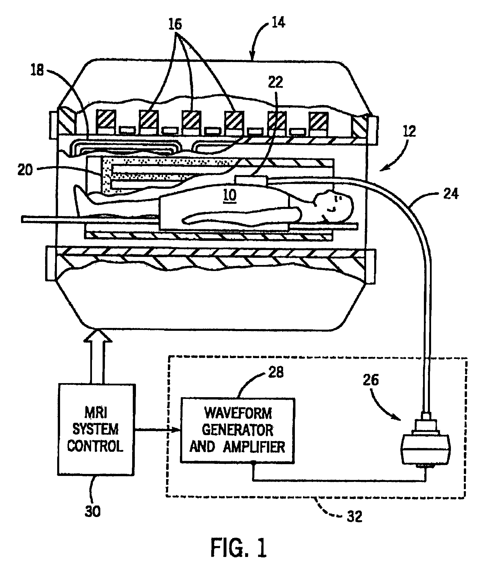
[0022]The present invention is employed in a system such as that described in the previously-cited U.S. Pat. No. 5,592,085, which provides a means for measuring the strain in gyromagnetic materials, such as tissues, using MR methods and apparatus. The present invention may also be employed with other medical imaging modalities including, but not limited to, ultrasound. Referring to FIG. 1, a subject to be examined 10 is placed in the bore 12 of an MRI system magnet 14 and is subjected to magnetic fields produced by a polarizing coil 16, gradient coils 18 and an RF coil 20 during the acquisition of MR data from a region of interest in the subject 10. The homogeneity of these magnetic fields is important and any objects placed in the bore 12 must be carefully constructed of materials that will not perturb them.

[0023]The present invention is a passive driver system that may be placed on the subject 10 and energized to produce an oscillating, or vibratory, stress. It includes a passive ...


PUM


Abstract

A flexible passive acoustic driver for use in an acoustic driver system which applies an oscillating stress to a subject undergoing a magnetic resonance elastography (MRE) examination which includes receiving acoustic pressure waves from an active driver through a tube and imparts pressure waves to a subject of an imaging procedure. In one configuration, the passive driver includes a flexible bag that forms the walls of an acoustic cavity, and a structure filling material located inside the acoustic cavity provides support for the flexible bag. The flexible bag conforms to the shape of the subject and may be held in place by an elastic band. The passive driver can have an integrated or detachable non-active push-on compartment which is rigid or semi-flexible to improve the human-driver mechanical coupling and the driver energy efficiency of converting acoustic pressure to mechanical vibration applied to a subject.

Description

CROSS-REFERENCE TO RELATED APPLICATIONS[0001]This application claims the benefit of U.S. Provisional Patent Application No. 61 / 478,295 filed Apr. 22, 2011, the disclosure of which is hereby incorporated by reference in its entirety.STATEMENT REGARDING FEDERALLY SPONSORED RESEARCH[0002]This invention was made with government support under EB001981 awarded by the National Institutes of Health. The government has certain rights in this invention.FIELD OF THE INVENTION[0003]The field of the invention is nuclear magnetic resonance imaging methods and systems. More particularly, the invention relates to devices for implementing magnetic resonance elastography (MRE).BACKGROUND OF THE INVENTION[0004]The physician has many diagnostic tools at his or her disposal which enable detection and localization of diseased tissues. These include x-ray systems that measure and produce images indicative of the x-ray attenuation of the tissues and ultrasound systems that detect and produce images indicat...

Claims


Application Information

Patent Timeline
Patent Type & Authority Patents(United States)
IPC IPC(8): A61B5/055A61B5/00A61B8/08
CPCA61B5/055A61B5/0051A61B8/485
Inventor EHMAN, RICHARD L.CHEN, JUNROSSMAN, PHILLIP J.
Owner MAYO FOUND FOR MEDICAL EDUCATION & RES
Learn More