
Method and system for filtering image data and use thereof in virtual endoscopy

a technology of image data and filtering, applied in the field of image processing, can solve the problems of generating blur, invasive, painful, and inconvenient colonoscopy procedures, and achieve the effects of enhanced filtering, accurate colorectal cancer screening, and enhanced 3d representation of any potential lesion

Active Publication Date: 2015-10-20
CADENS MEDICAL IMAGING
View PDF22 Cites 3 Cited by
  • Summary
  • Abstract
  • Description
  • Claims
  • Application Information

AI Technical Summary

Benefits of technology

The patent describes a method for removing unnecessary parts from image data that is used for electronic colon cleansing. This helps to improve the accuracy and efficiency of the process. Additionally, the method may provide faster results than other segmenting methods, which is another advantage.

Problems solved by technology

Conventional colonoscopies using this procedure, although reliable, are both costly in money and time.
Moreover, it is an invasive, uncomfortable and sometimes painful procedure for the patient.
However, the imaging techniques used to obtain the image data typically present a spatial resolution which generates a blur at each anatomical interface.
Consequently, when removing the tagging material, these algorithms may artificially smooth the surface between the removed material and the colon's inner wall and polyps or other abnormalities present in the colon may be inadvertently removed or wrongly reduced to an acceptable level, which is a major concern.
However, since the different anatomical structures may be very different from each other, a simple threshold may not allow to conveniently segment a structure in a single pass.
This may lead an operator to a wrong diagnosis, which is a major issue.

Method used


Image

  • Method and system for filtering image data and use thereof in virtual endoscopy
  • Method and system for filtering image data and use thereof in virtual endoscopy
  • Method and system for filtering image data and use thereof in virtual endoscopy

Examples


Embodiment Construction

[0098]In the following description of the embodiments, references to the accompanying drawings are by way of illustration of examples by which the invention may be practiced. It will be understood that various other embodiments may be made and used without departing from the scope of the invention disclosed.

[0099]The invention concerns a method and a system for filtering image data that are particularly useful for filtering medical images. Throughout the present description, the method will be described for the particular application of electronic colon cleansing in virtual colonoscopy but the skilled addressee will appreciate that the method is not limited to this specific application and that many other applications may be considered, as it will become apparent upon reading of the present description.

[0100]The method for filtering image data of the invention may be generally useful for facilitating the subsequent segmentation of an anatomical structure but is also well adapted for...


PUM


Abstract

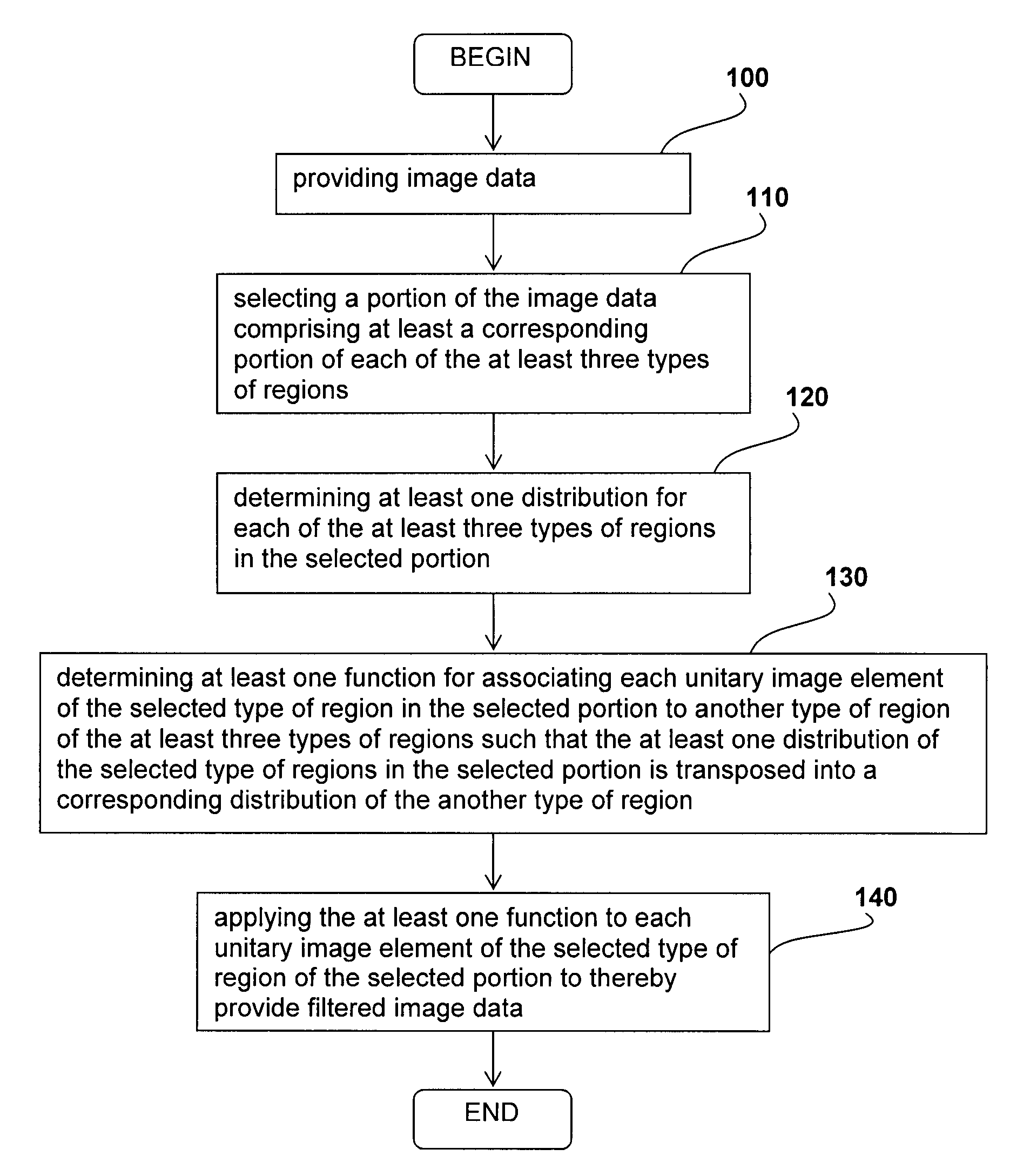
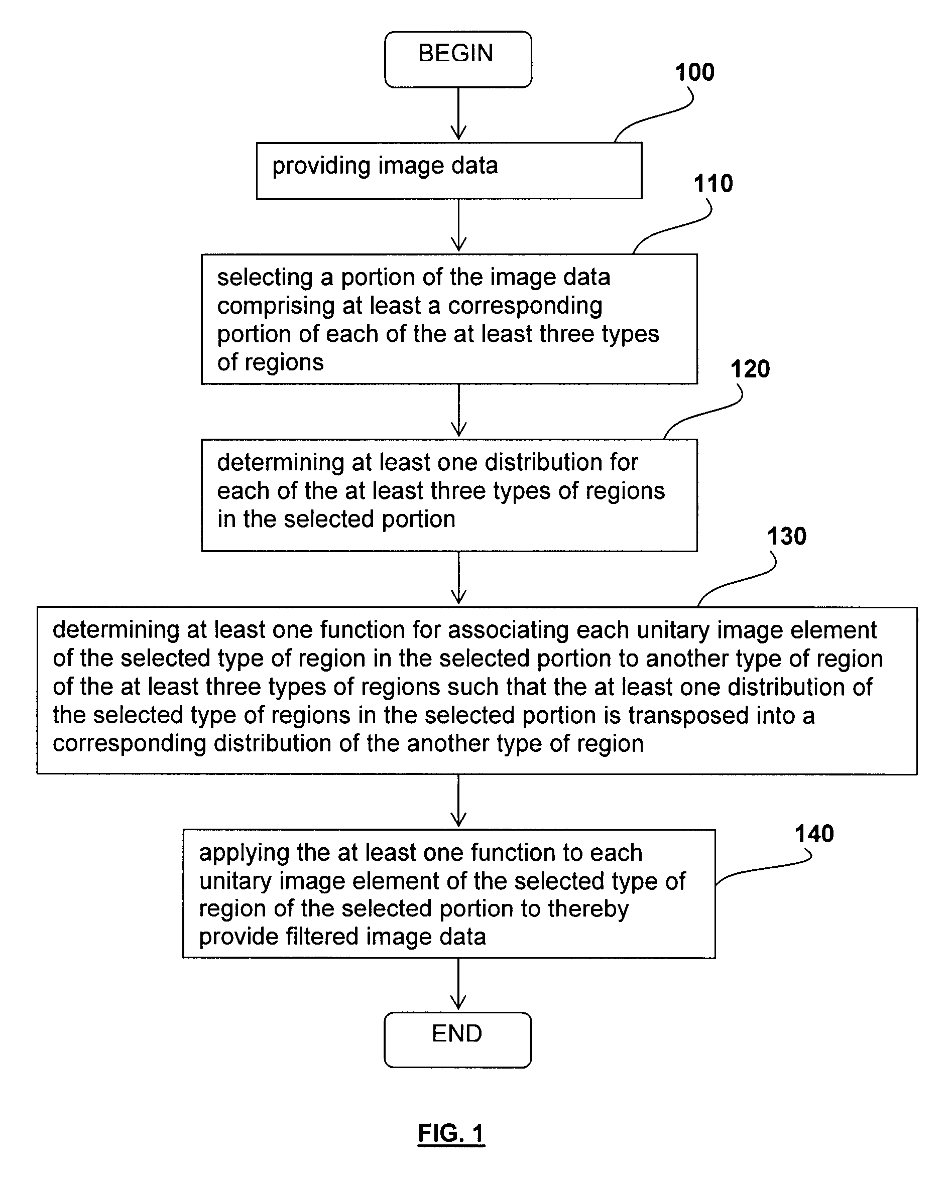
A method for filtering image data having at least three types of distinct regions from a selected type of region, each of the at least three types of distinct regions being characterized by a plurality of unitary image elements, the method comprising providing the image data; selecting a portion of the image data comprising at least a corresponding portion of each of the at least three types of regions; determining at least one distribution for each of the at least three types of regions in the selected portion; determining at least one function for associating each unitary image element of the selected type of region in the selected portion to another type of region of the at least three types of regions such that the at least one distribution of the selected type of regions in the selected portion is transposed into a corresponding distribution of the another type of region; and applying the at least one function to each unitary image element of the selected type of region of the selected portion to thereby provide filtered image data. Applications of the method for suppressing tagged material in virtual colonoscopy are also disclosed.

Description

CROSS-REFERENCE TO RELATED APPLICATIONS[0001]This application is a National Phase Application of PCT International Patent Application No. PCT / CA2009 / 001743, International Filing Date Nov. 27, 2009, which is incorporated herein by reference.CROSS REFERENCE TO RELATED APPLICATION[0002]The present application relates to PCT application entitled “Method and system for determining an estimation of a topological support of a tubular structure and use thereof in virtual endoscopy”, the specification of which is incorporated herein by reference.FIELD OF THE INVENTION[0003]The invention generally relates to image processing and more particularly relates to a method and a system for filtering image data. It also relates to applications of the method for suppressing tagged material in virtual colonoscopy.BACKGROUND OF THE INVENTION[0004]Conventional endoscopic procedures typically rely on the use of a flexible fiber optic tube which is inserted in the patient's body to visually examine an inne...

Claims


Application Information

Patent Timeline
Patent Type & Authority Patents(United States)
IPC IPC(8): G06K9/00A61B5/055G06T7/00A61B5/00A61B6/00A61B8/13
CPCA61B6/50A61B5/055A61B5/4255A61B6/506G06T7/0081G06T7/0087A61B6/505A61B8/13G06T2207/10081G06T2207/30028G06T7/11G06T7/143G06T5/90G06T5/10A61B6/03
Inventor VINCENT, THOMAS BERNARD PASCALCHANDELIER, FLORENT ANDRE ROBERT
Owner CADENS MEDICAL IMAGING
Learn More