Method for intraoperative generation of an updated volume data set

a technology for volume data and intraoperative generation, applied in the field of intraoperative generation of updated volume data set of patients, can solve problems such as inability to be used in the current situation

Active Publication Date: 2005-06-28
SIEMENS HEATHCARE GMBH
View PDF5 Cites 24 Cited by
  • Summary
  • Abstract
  • Description
  • Claims
  • Application Information

AI Technical Summary

Benefits of technology

[0006]An object of the present invention is to provide a method to intraoperatively gener

Problems solved by technology

This is not currently possible because the 3D image data do not represent an image that is sufficiently current, since a frequent or even continuous complete intraoperative upd

Method used


Image

  • Method for intraoperative generation of an updated volume data set
  • Method for intraoperative generation of an updated volume data set
  • Method for intraoperative generation of an updated volume data set

Examples


Embodiment Construction

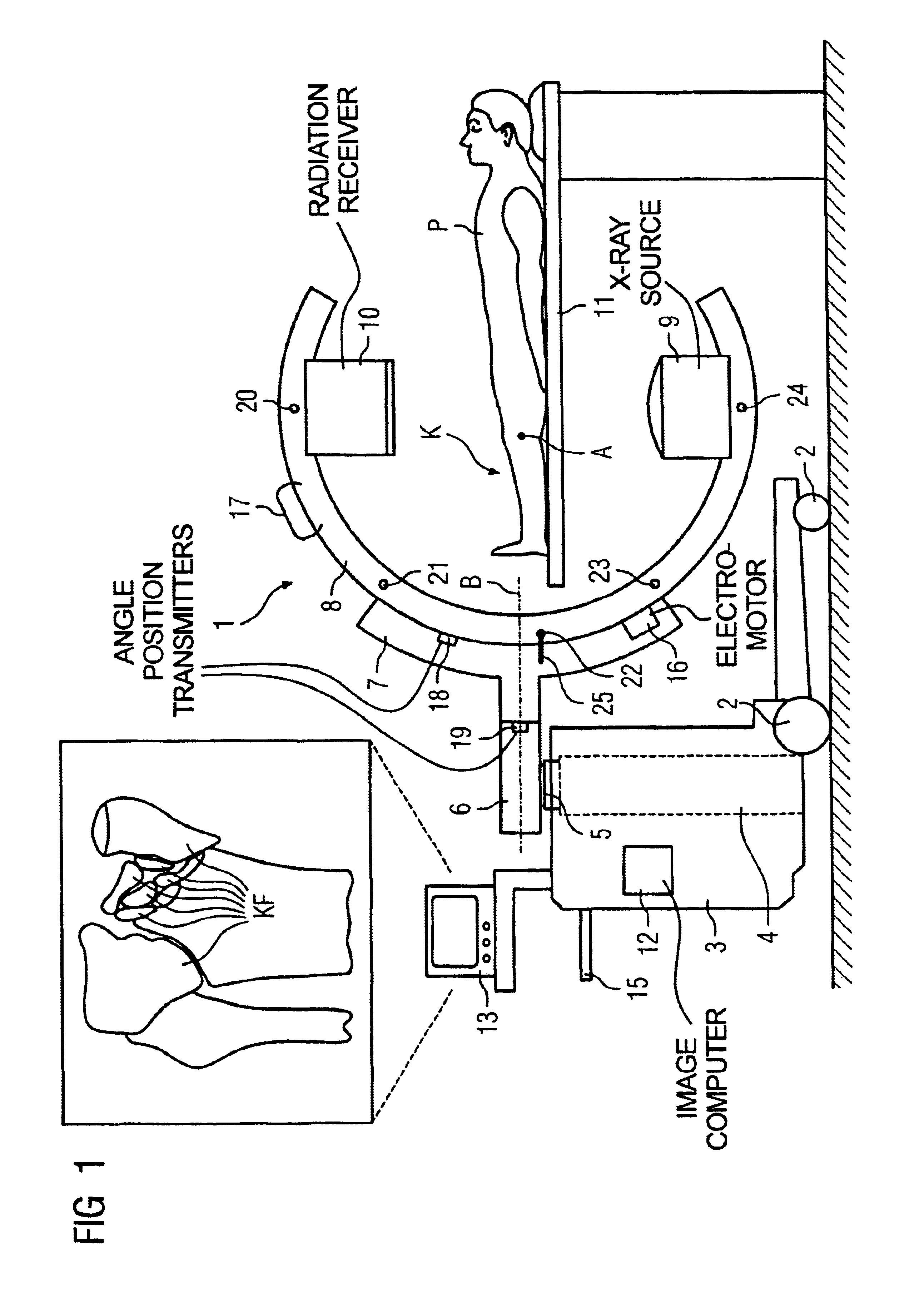
[0021]The C-arm x-ray device 1 shown in FIG. 1 is suitable for implementing the inventive method and has a device cart 3, provided with wheels, in which is arranged a lifting device 4 (shown only schematically in FIG. 1) having a column 5. A holder 6 is arranged at the column 5 at which a positioner 7 is present to position a C-arm 8. An x-ray source 9 and an x-ray radiation receiver 10 are arranged opposite one another on the C-arm 8. The x-ray source 9 preferably emits a conical x-ray beam in the direction of the x-ray radiation receiver 10 (having a planar receiver surface), which can, for example, be an x-ray intensifier or a flat image detector. In the exemplary embodiment, the C-arm 8 can be isocentrically adjusted both around its orbital axis A (schematically indicated in the FIG. 1) and around its angulation axis B (schematically indicated in the FIG. 1).

[0022]Volume data sets, for example voxel volumes, can be generated with the C-arm x-ray device 1 representing body parts ...


PUM


Abstract

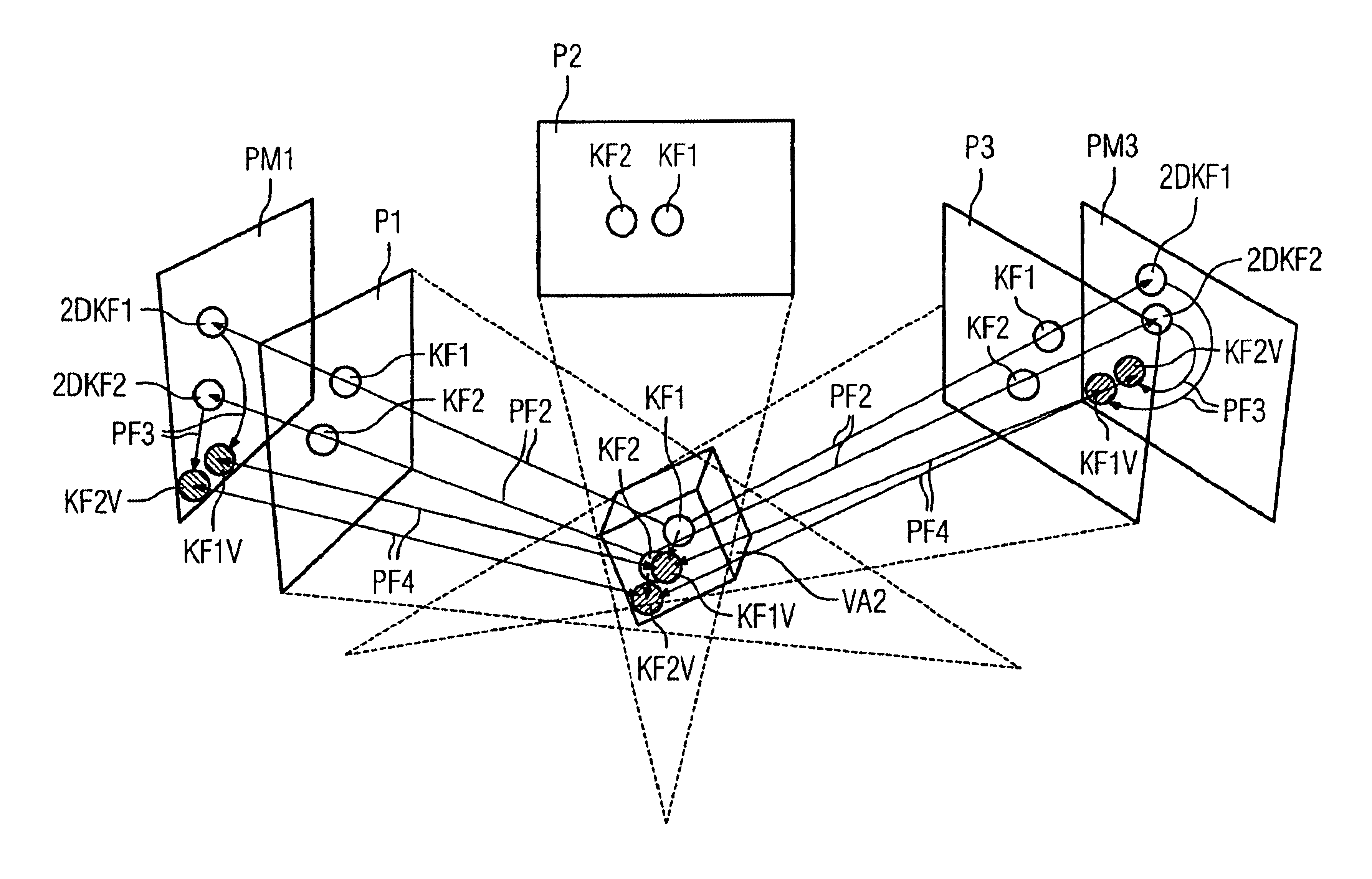
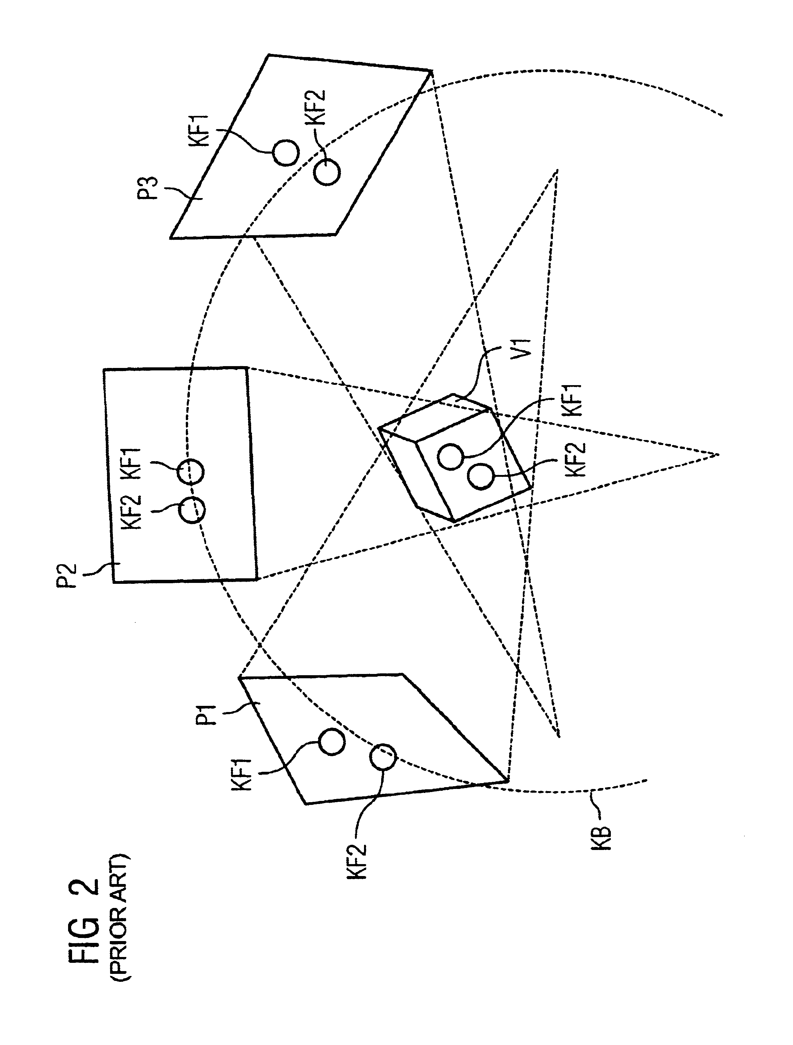
To intraoperatively generate an updated volume data set in which a image information of biological tissue is volume data set representing reconstructed from a series of n 2D biological tissue x-ray projections, one version, in intraoperatively acquired m 2D biological tissue x-ray projections the biological tissue 2D contour is segmented and this is back-projected in the reconstructed volume data set. In another version, in the volume data set the biological tissue 3D contour is segmented and this is projected in intraoperatively acquired m biological tissue 2D x-ray projections. The 3D contour is visually repositioned by projection in the m 2D x-ray projections in the volume data set, until its projection is substantially congruent with the biological tissue image information in the respective 2D x-ray projections. In both versions, a volume data set updated around the intraoperatively determined biological tissue 3D contour is generated.

Description

BACKGROUND OF THE INVENTION[0001]1. Field of the Invention[0002]The invention concerns methods for intraoperative generation of an updated volume data set of a patient.[0003]2. Description of the Prior Art[0004]In the course of a medical procedure on a patient, in particular a minimally invasive procedure, it is frequently necessary to monitor position changes of biological tissue in the patient by means of diagnostic x-ray imaging. Such a requirement exists, for example, in the case of complicated fractures, in particular multiple fractures with a number of small bone fragments with complex morphology, which in the osteosynthesis must be aligned and set relative to one another. Conventionally, the repositioning of the bone fragments and their subsequent setting ensue using individual 2D x-ray projections, and the surgeon during the minimally invasive procedure must mentally reconstruct the three-dimensional anatomical situation in reality from the 2D x-ray projections acquired at d...

Claims


Application Information

Patent Timeline
IPC IPC(8): A61B6/03A61B6/00G06T1/00G06F17/00G06T15/00H05G1/00A61B6/02A61B19/00G06T11/00G06T15/08
CPCA61B19/52A61B6/584G06T11/003A61B2019/5238Y10S378/901A61B2019/5289A61B90/36A61B2090/364A61B2090/376
Inventor HEBECKER, AXELMITSCHKE, MATTHIASRAHN, NORBERTRITTER, DIETER
Owner SIEMENS HEATHCARE GMBH
Why Eureka
Try Eureka