Systems and methods for simulation-based radiation estimation and protection for medical procedures

a radiation estimation and simulation technology, applied in the field of simulation, can solve the problems of microscopic damage to living tissue, serious side effects, and tissue damage type that is a risk to both the patient and the medical team

Active Publication Date: 2016-04-26
MENTICE
View PDF7 Cites 10 Cited by
  • Summary
  • Abstract
  • Description
  • Claims
  • Application Information

AI Technical Summary

Benefits of technology

[0015]Methods and systems for simulating an x-ray guided medical procedure on a human and calculating the radiation exposure to one or more humans during the x-ray guided medical procedure are disclosed. The methods and systems disclosed enable a user to determine a change in radiation metrics based on a corresponding change to an input parameter to an x-ray equipment model or human exposure model. By determining the change in radiation metrics, the methods and systems may output current and cumulative radiation metrics live, during the x-ray guided procedure. The updated metrics further enable the methods and systems to provide feedback and performance evaluations to the user. By providing information about the amount of radiation being exposed to the patient or medical team, the user may adjust the input parameters to achieve an optimized balance between radiation exposure, x-ray image quality, and risks associated with the exposure during the procedure. For example, during an endovascular procedure, the user may adjust the input parameters in response to radiation metrics indicating a dangerous level of exposure.
[0017]The radiation metric processor calculates radiation exposure metrics to one or more humans located at the simulated x-ray guided medical procedure. The calculation of the radiation exposure metrics occurs during the x-ray guided medical procedure, and is based on the input parameters to the x-ray equipment and human exposure models. The radiation metrics are calculated with a model that associates a change in the input parameters with a change in the radiation exposure metric. In this way, when a user changes an input parameter, the methods and systems may output the change in radiation metrics during the procedure, without interruption.
[0018]The feedback system includes an x-ray imaging simulator and radiation metric display. The radiation metric display shows the first and second radiation exposure metrics to a user during the x-ray guided procedure. The x-ray imaging simulator generates an x-ray image of the human based on the x-ray equipment model and human exposure model at each point in time. The x-ray imaging simulator applies the noise pattern to the x-ray image to create a simulated noisy x-ray image. The x-ray imaging simulator then displays the noisy x-ray image to the user. In this way, the x-ray imagery shown to the user may be generated without using ionizing radiation; thus a user can train on a specific medical procedure in a realistic operating room environment, without exposing a patient, or him- or herself, or his or her team, to harmful radiation.

Problems solved by technology

However, exposure to radiation typically results in serious side effects, including microscopic damage to living tissue.
This type of tissue damage is a risk to both the patient, as well as the medical teams that work in these environments, because of secondary “scatter” radiation.
Secondary scatter radiation is harmful radiation that the medical team is exposed to as a result of scattering off of a patient or other objects in the environment.
Fluoroscopic procedures are expected to pose a higher risk of radiation exposure in comparison to CT-based procedures, in part due to recent advancements that have lowered the exposure in CT-based procedures.
Further, legislation and guidelines have recently been passed that limit the utilization of CT scans.
Since the team is performing an operation on the patient during interventional fluoroscopy, it is not possible for them to reduce their own exposure by maintaining a large distance to the x-ray source while imaging, such as is the practice for diagnostic CT scans.
However, it is very difficult for a medical professional to develop a good understanding of the harmful effects of such changes, since radiation is neither visible nor otherwise noticeable to humans.
Further, changes to a single parameter or combination of parameters may change the quality of the x-ray image produced by the x-ray machine, something which may influence correct decision making in the delivery of treatment.
Because these parameters may be interrelated or functionally dependent, accurately determining how a change to a parameter affects radiation dose rate or image quality is computationally complex.
Currently, medical teams do not receive training that shows how a change to an operation setting during a fluoroscopic procedure causes a change to the radiation exposed to the patient or medical team.
Training modules typically do not include any hands-on components, because there is currently no effective way of providing realistic hands-on training without using real radiation.
Although some training programs use empty operating rooms and “phantoms” as substitutes for patients, several drawbacks exist.
Specifically, these training programs still expose medical teams to secondary radiation and they block the operating room from being used for real procedures.
Furthermore, they do not show how changes to operating settings during an operation cause changes in radiation exposure and image quality, or how operating settings may need to be changed at different points of an x-ray guided medical procedure to balance the trade-off between radiation exposure and image quality.
Although techniques and systems for measuring, estimating, and visualizing radiation exposure have been developed, none of the presently known art describes a comprehensive solution for training medical professionals or teams on the effects of operating setting adjustments and their impact on radiation exposure and image quality in a highly realistic and completely radiation-free simulated environment.
Further, systems for measuring, estimating, and visualizing radiation exposure do not show how changes in a simulation parameter affect radiation dynamically during the course of a procedure.
Simulations that take into account multiple different simulation parameters are often computationally complex, and generally executed in a time- and resource-intensive Monte Carlo-style fashion.
Further, to change a set of parameters, the simulation is generally re-executed, and thus, unable to effectively show how changes to a parameter affect radiation during a live procedure.
Moreover, systems for measuring, estimating, and visualizing radiation exposure do not provide any meaningful information about the risk levels associated with different levels of radiation exposure.

Method used


Image

  • Systems and methods for simulation-based radiation estimation and protection for medical procedures
  • Systems and methods for simulation-based radiation estimation and protection for medical procedures
  • Systems and methods for simulation-based radiation estimation and protection for medical procedures

Examples


Embodiment Construction

[0036]The invention described herein provides systems and methods for the realistic simulation of x-ray guided medical procedures in a radiation-free environment. The system and methods may be used to train and certify professionals to reduce medical and occupational radiation exposure, and educate operators of x-ray systems how to use x-ray equipment in the least harmful way. Embodiments of the invention may also be used for therapeutic device design, development and testing.

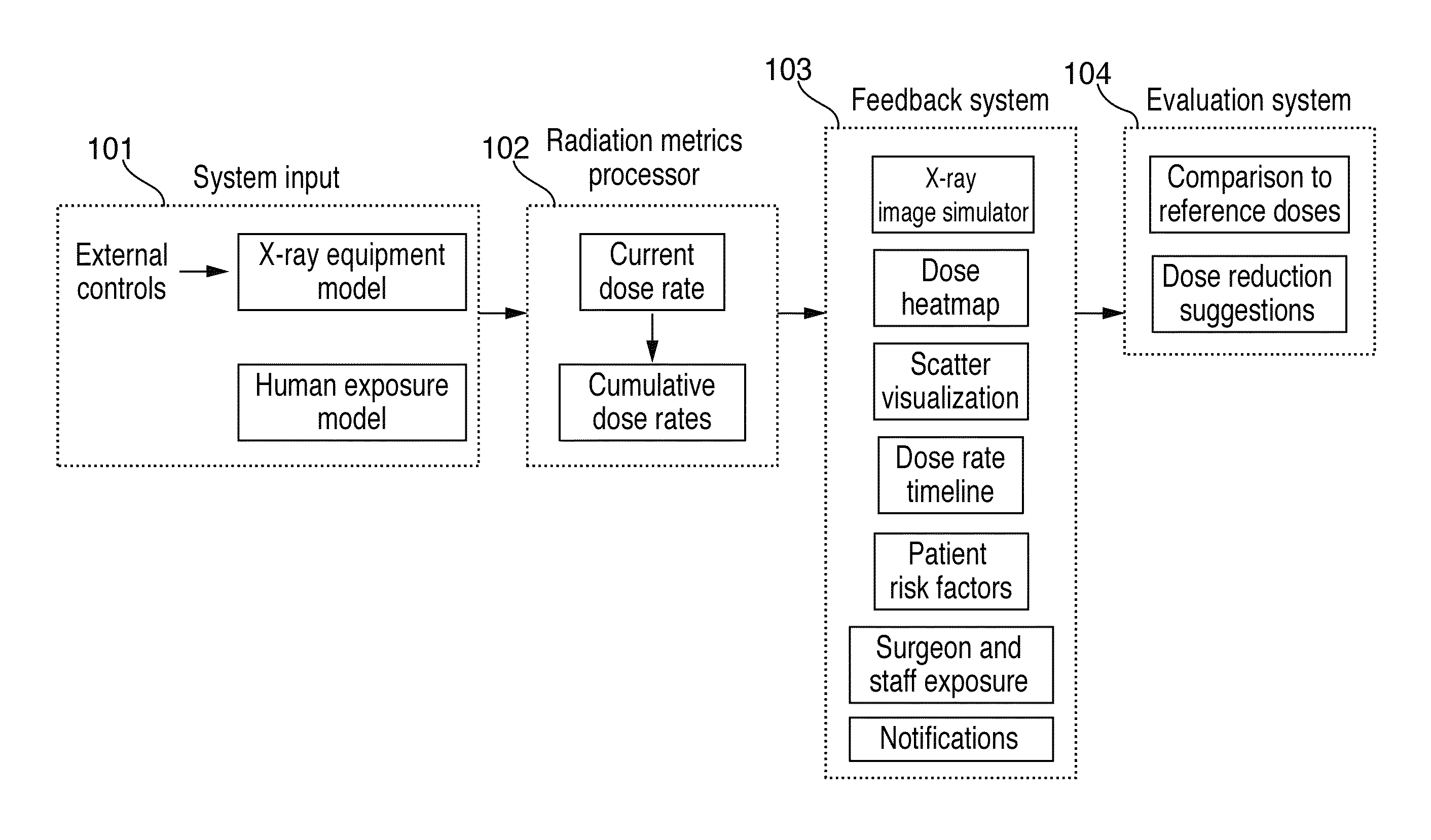
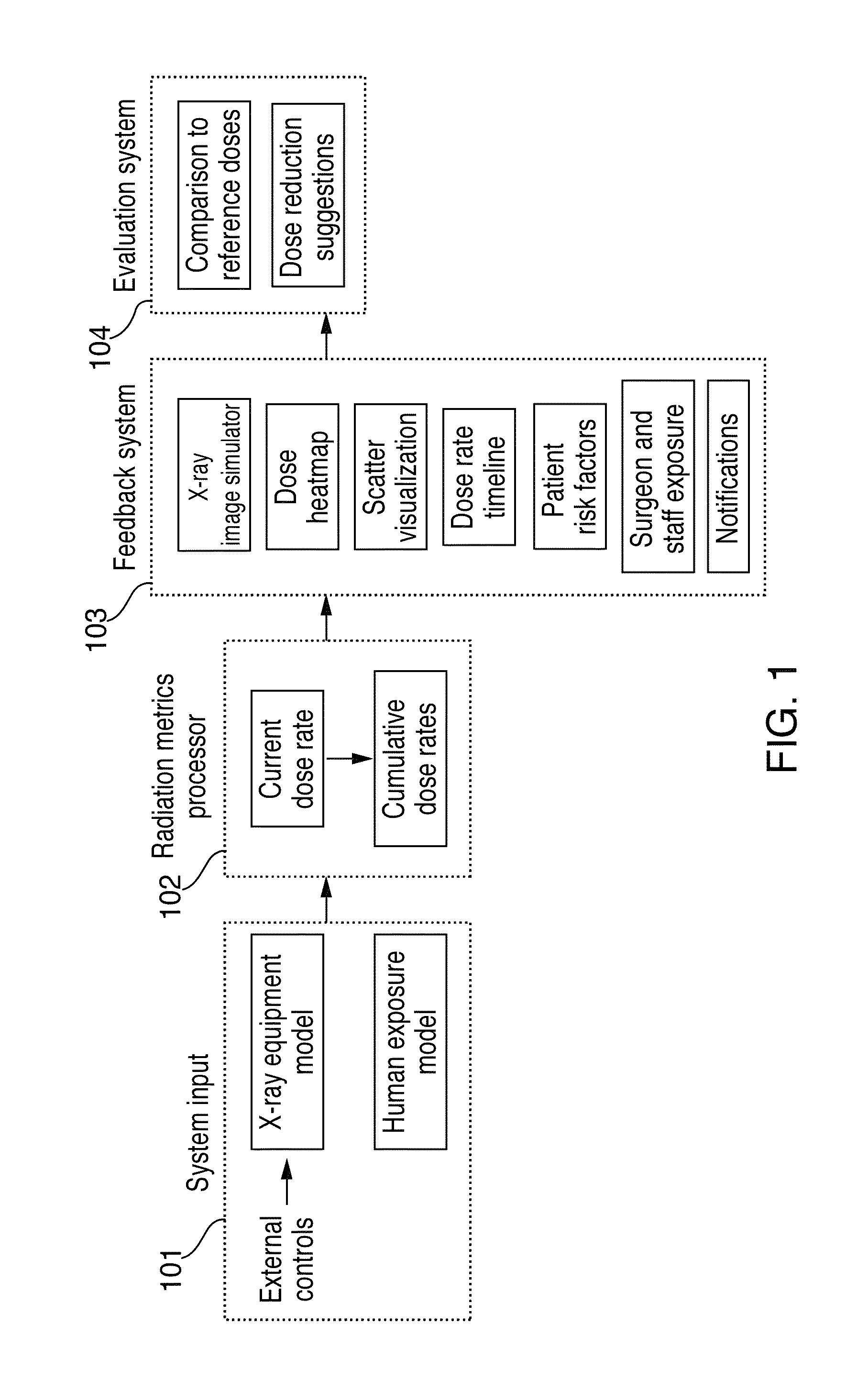
[0037]FIG. 1 shows a diagram of several components of the radiation protection simulation system according to one embodiment of the invention. These components enable a user to simulate an x-ray guided procedure, and visualize the changes to radiation exposure and image quality as the user adjusts operating settings during the procedure. The components of the radiation protection simulation system may include a system input 101, radiation metrics processor 102, feedback system 103, and evaluation system 104. Us...


PUM

PropertyMeasurementUnit
timeaaaaaaaaaa
thicknessaaaaaaaaaa
thicknessaaaaaaaaaa

Abstract

Systems and methods for determining radiation exposure during an x-ray guided medical procedure are disclosed. In some embodiments, the system includes an x-ray equipment model that simulates the emission of radiation from x-ray equipment during the x-ray guided medical procedure, a human exposure model that simulates one or more human anatomies during the x-ray guided medical procedure, a radiation metric processor that calculates at least one radiation exposure metric, and a feedback system for outputting information based on the at least one radiation exposure metric. The radiation metric processor calculates radiation exposure metrics based on input parameters that correspond to operating settings as well as the location and structure of one or more human anatomies.

Description

CROSS-REFERENCE TO RELATED APPLICATIONS[0001]The present application claims priority to U.S. Provisional Patent Application No. 61 / 887,835 filed on Oct. 7, 2013, which is incorporated herein by reference in its entirety.FIELD OF THE INVENTION[0002]The invention is related to systems and methods for simulating radiation exposure to patients or medical teams during an image-guided medical procedure, and in particular to simulating the radiation exposure of an x-ray based image-guided procedure, such as fluoroscopy.BACKGROUND OF THE INVENTION[0003]Medical equipment that uses ionizing radiation has found widespread application in the healthcare industry today. Allowing medical teams to diagnose and treat patients effectively, ionizing radiation has been used in different branches of medicine including, radiology, cardiology, neurology, oncology, trauma care, orthopedic surgery, endovascular intervention. The benefits of using x-ray imaging as a diagnostic tool and as a treatment option ...

Claims


Application Information

Patent Timeline
Patent Type & Authority Patents(United States)
IPC IPC(8): G06G7/48A61B6/10A61N5/10G01N33/48G01N33/50G06F19/00G16B5/00G16B45/00
CPCA61B6/10A61N2005/1094A61B6/107G09B23/286G16H40/63G16H50/50A61N5/1075G16B50/00G16B5/00G16B45/00
Inventor FALT, EDVARD PER GOSTALONN, LARS BIRGER
Owner MENTICE
Why Eureka
Try Eureka