Color x-ray histology for multi-stained biologic sample

a multi-stained, biologic technology, applied in the field of x-ray imaging, can solve the problems of difficult to achieve true 3d information and high throughput, laborious imaging physical slices of specimens, and inability to achieve satisfactory 3d representations

Active Publication Date: 2016-12-06
UNIVERSITY OF CHICAGO +2
View PDF12 Cites 1 Cited by
  • Summary
  • Abstract
  • Description
  • Claims
  • Application Information

AI Technical Summary

Problems solved by technology

But, with such techniques it is difficult to achieve true 3D information and high throughput.
However, the mismatch between histological slice thickness (typically 5 microns) and in-plane resolution (typically 200-500 nm) makes the generation of satisfactory 3D representations impractical.
Moreover, the entire process of sectioning, staining, and imaging physical slices of the specimen is labor intensive.
There are 3D imaging modalities, such as confocal microscopy, optical projection tomography, or magnetic resonance imaging, but they either lack the required 1-2 micron spatial resolution (MRI) or lack the ability to image in optically opaque whole specimens.
The natural X-ray contrast of most model organisms is not sufficiently high to allow subtle phenotyping.
Thus far, however, no one has developed a multiple stain process available for effective 3D phenotype imaging.

Method used


Image

  • Color x-ray histology for multi-stained biologic sample
  • Color x-ray histology for multi-stained biologic sample
  • Color x-ray histology for multi-stained biologic sample

Examples


Embodiment Construction

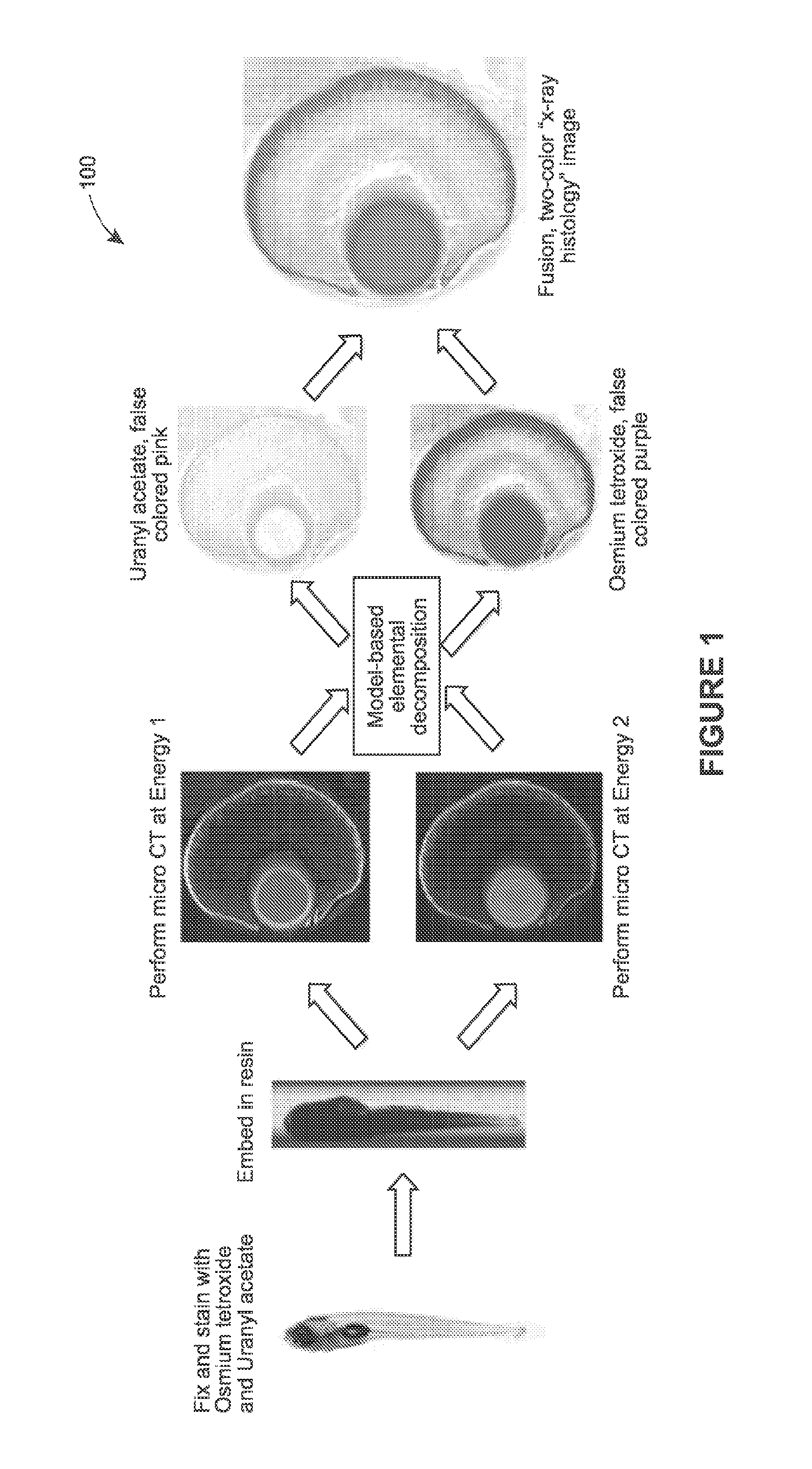
[0030]Various examples are provided of 3D X-ray imaging techniques that use multiple heavy-metal stains of model organisms or other biologic targets and multiple captured datasets to produce stain-based images, where each dataset contains different X-ray energy information.

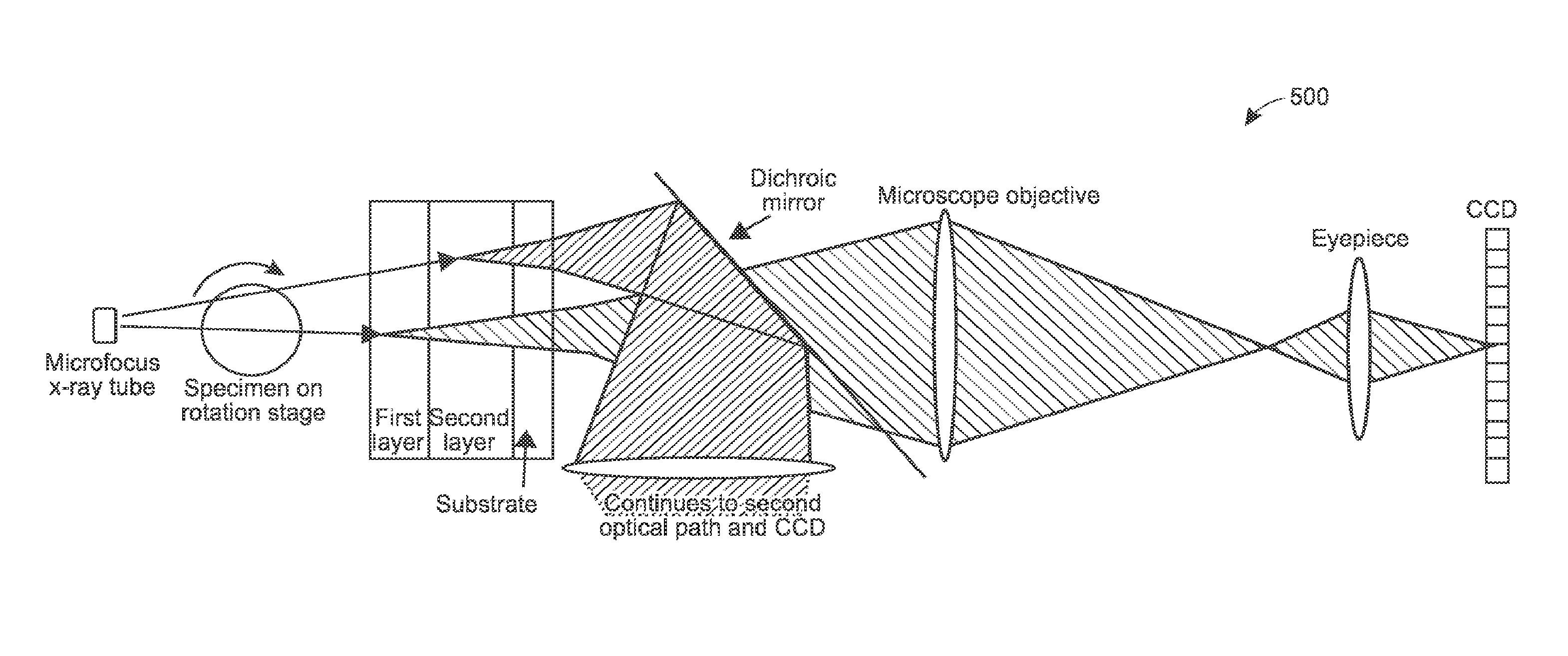
[0031]Generally, these techniques may be implemented using different types of X-ray illumination sources, including monochromatic X-ray energies, such as those generated by synchrotrons, and polychromatic (wide spectrum) X-ray energies, such as generated by X-ray tubes. Samples may be illuminated multiple times or a single time, depending on the type of X-ray energy and the type of X-ray detector. Numerous different examples are provided, and persons of ordinary skill will appreciate yet others in light of the following.

[0032]FIG. 1 provides process 100, in accordance with an example of the present techniques. In this example, the sample is a model organism, although the process (as with any process herein) may be...


PUM

PropertyMeasurementUnit
thicknessaaaaaaaaaa
thicknessaaaaaaaaaa
thicknessaaaaaaaaaa

Abstract

Systems and methods are provided for staining tissue with multiple biologically specific heavy metal stains and then performing X-ray imaging, either in projection or tomography modes, using either a plurality of illumination energies or an energy sensitive detection scheme. The resulting energy-weighted measurements can then be used to decompose the resulting images into quantitative images of the distribution of stains. The decomposed images may be false-colored and recombined to make virtual X-ray histology images. The techniques thereby allow for effective differentiation between two or more X-ray dyes, which had previously been unattainable in 3D imaging, particularly 3D imaging of features at the micron resolution scale. While techniques are described in certain example implementations, such as with microtomography, the techniques are scalable to larger fields of view, allowing for use in 3D color, X-ray virtual histology of pathology specimens.

Description

CROSS REFERENCE TO RELATED APPLICATION[0001]The present application claims the benefit of U.S. Provisional Application No. 61 / 553,066, entitled “Color X-Ray Histology for Multi-Stained Biologic Sample”, filed on Oct. 28, 2011, which is hereby incorporated by reference herein in its entirety.FIELD OF THE INVENTION[0002]The invention generally relates to X-ray imaging and, more particularly, to a system for X-ray imaging using multiple illumination energies.BACKGROUND[0003]The background description provided herein and throughout the application is for the purpose of generally presenting the context of the disclosure. Work of the presently named inventors, to the extent it is described in this background section, as well as aspects of the description that may not otherwise qualify as prior art at the time of filing, are neither expressly nor impliedly admitted as prior art against the present disclosure.[0004]With the completion of genome projects for numerous model organisms, includi...

Claims


Application Information

Patent Timeline
Patent Type & Authority Patents(United States)
IPC IPC(8): A61B6/00G01N23/223G01N23/04G06T19/00B82Y10/00G01N23/225A61B6/03
CPCG01N23/04B82Y10/00G01N23/046G01N23/223G01N23/2251G06T19/00A61B6/032A61B6/482A61B6/508G01N2223/0766G01N2223/6126G06T2210/41G21K2201/061G21K2201/064
Inventor LA RIVIERE, PATRICKWANG, YUXIN STEVECLARK, DARINCHENG, KEITH
Owner UNIVERSITY OF CHICAGO
Why Eureka
Try Eureka