

HSF1 as a marker in tumor prognosis and treatment
a tumor prognosis and tumor technology, applied in the field of hsf1 as a tumor prognosis and treatment, can solve the problems of complex development of individual patient treatment plans, many of the most common types of cancer remain difficult to manage, and are often incurable, and achieve the effect of increasing likelihood and increasing likelihood
- Summary
- Abstract
- Description
- Claims
- Application Information
AI Technical Summary
Benefits of technology
Problems solved by technology
Method used
Image

Examples
example 1
Characterization of HSF1 Antibody and HSF1 Expression in Breast Cancer and Various Other Cancer Types
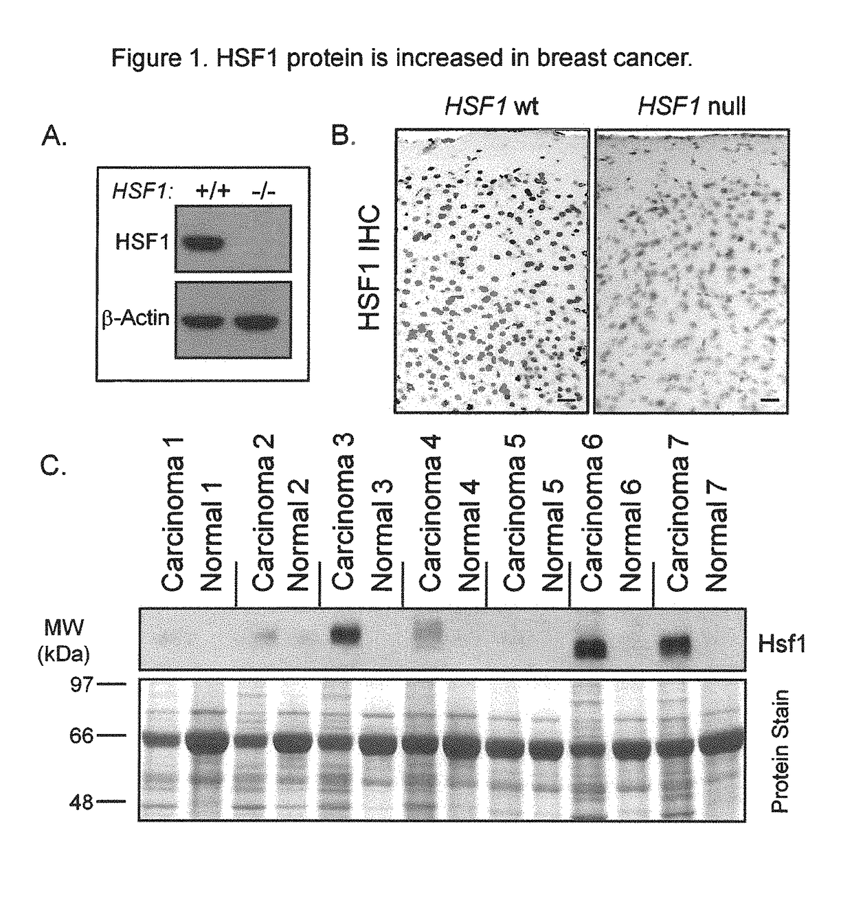
[0171]To facilitate our studies of HSF1, we verified the specificity of a commercially-available HSF1 antibody cocktail on samples from HSF1 wild-type and null mice. A strong immunoreactive band of the expected size for HSF1 was present in wild-type lysates but was absent in lysates null for HSF1 (FIG. 1A). Strong nuclear staining was observed by immunohistochemistry (IHC) in wild-type mouse tissues but not in corresponding tissues from HSF1 null mice (FIG. 1B) validating this antibody cocktail for IHC applications.
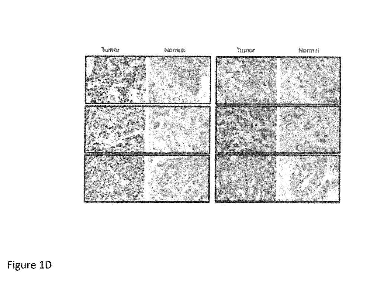
[0172]We examined the expression of HSF1 in invasive carcinoma and matched normal adjacent breast tissue from seven patients by immunoblot (FIG. 1C). More HSF1 was present in the tumors than the matched controls in all cases. Interestingly, there was a strong HSF1 band in three of seven samples obtained from the tumors and moderate to weak bands in the remaining tumors. Th...
example 2
Nuclear HSF1 is Highest in High-grade Breast Cancer and is Associated with Advanced Clinical Stage at Diagnosis
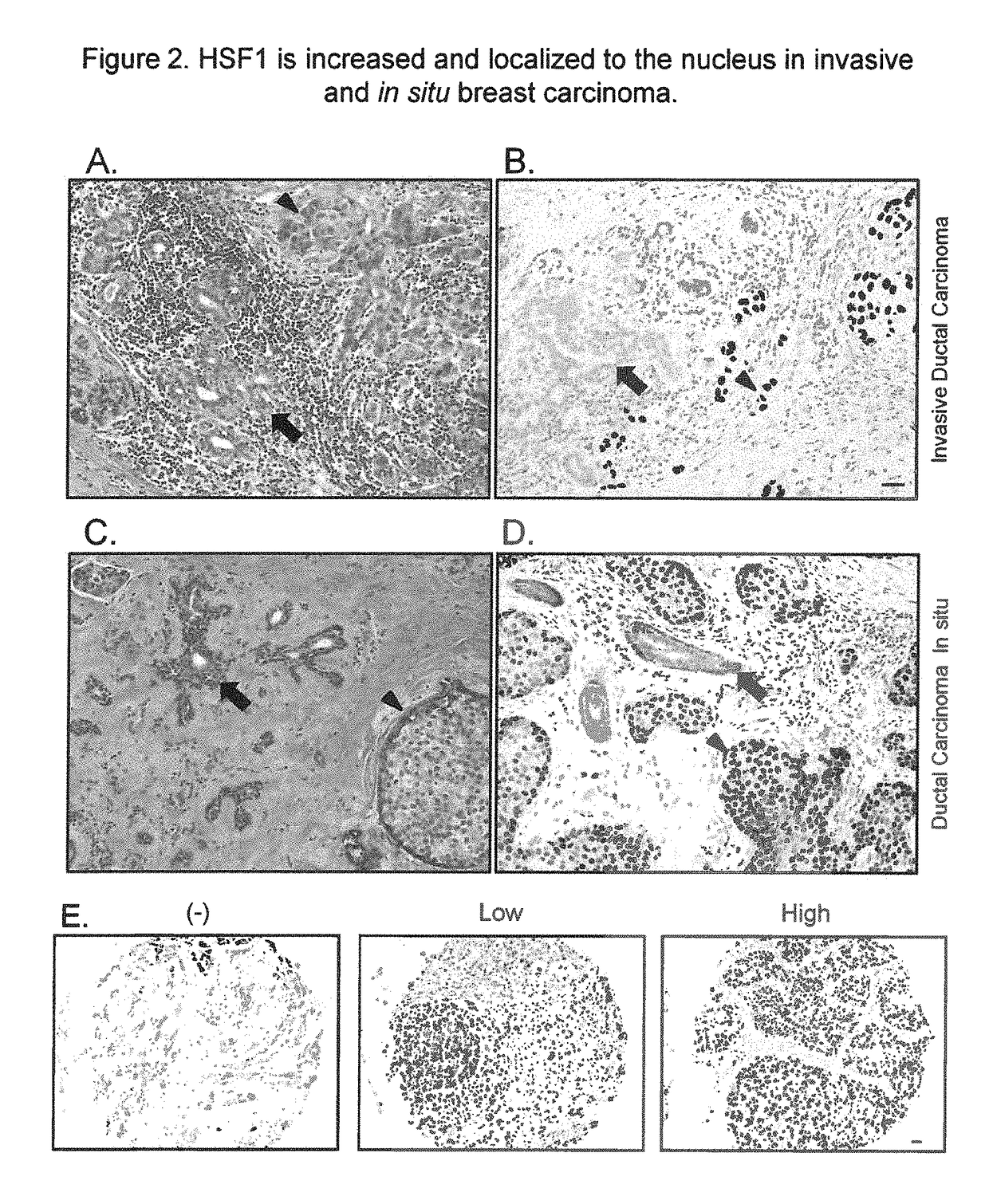
[0179]We next performed an in-depth analysis of HSF1 protein expression in a large breast cancer cohort. 1,841 invasive breast cancer cases from the Nurses' Health Study (NHS) were evaluated for HSF1 localization and expression (FIG. 2E). 404 (21.9%) were negative for nuclear HSF1 and 1437 had detectable nuclear HSF1 (78.1%) with 882 (47.9%) demonstrating low and 555 (30.2%) high HSF1. Levels of HSF1 expression differed by histological-grade (P<0.0001). 40.5% of well-differentiated low-grade carcinomas were HSF1-negative and only 14.4% showed high nuclear HSF1 (Table 1). Conversely, in poorly-differentiated high-grade cancers, only 13.0% were HSF1-negative and 48.1% showed high HSF1 expression. Levels of HSF1 also differed by clinical parameters. Compared with HSF1-negative tumors, those with nuclear HSF1 expression were more likely to be diagnosed at a more advanced clinic...
example 3
HSF1 Accumulates in the Nuclei of In Situ Carcinomas
[0181]Nuclear HSF1 was detected in 84.5% of the DCIS cases. The frequency and levels of HSF1 expression were similar between DCIS and invasive cancer, confirming our earlier observations on a smaller number of tumor sections. No statistically significant association was found between HSF1 expression and DCIS nuclear grade, however (Table 6). Our limited sample size of DCIS cases (n=200) may have limited the power to detect such an association. Nonetheless, these observations highlight that HSF1 is activated before malignant cells gain the ability to invade across the basement membrane.
[0182]
TABLE 6Frequency of HSF1 expression in DCIS according to tumor grade, Nurses' Health Study (1976 to 1996). Number of cases and (%). Chi-square analysis.HSF1 ExpressionNoneLowHighP-valueDCIS0.4907DCIS, low nuclear grade 4 (22.2)11 (61.1) 3 (16.7)DCIS, intermediate grade16 (16.8)54 (56.8)25 (26.3)DCIS, high nuclear grade11 (12.6) 46 (52.9)30 (34.5...
PUM
| Property | Measurement | Unit |
|---|---|---|
| mass | aaaaa | aaaaa |
| mass | aaaaa | aaaaa |
| mass | aaaaa | aaaaa |
Abstract
Description
Claims
Application Information