Protection of cells from degeneration and treatment of geographic atrophy
Active Publication Date: 2017-07-18
UNIV OF KENTUCKY RES FOUND
5 Cites 10 Cited by
AI-Extracted Technical Summary
Problems solved by technology
Although there are multiple approved therapies for the neovascular form of AMD (Ferrara, 2010), there is no effective treatment for the far more common atrophic form of the disease.
GA, the advanced stage of atrophic AMD, is characterized by degeneration and death of the RPE, and is the leading cause of untreatable visio...
View moreBenefits of technology
[0012]In some embodiments of the method, the inhibiting of P2X7 activation includes contacting the cell with a P2X7 activation inhibitor, e.g., P2X7 receptor antagonist A438079.
[0013]In some embodiments of the method, inhibiting IRAK1 and/or IRAK 4 includes contacting the cell with an IRAK1 and/or IRAK4 inhibitor (e.g., N-(2-Morpholinylethyl)-2-(3-nitrobenzoylamido)-benzimidazole).
[0014]In some embodiments the method can also include inhibiting MyD88 associated with or in the cell; inhibiting IL-18 associated with or in the cell; inhibiting an inflammasome associated with or in the cell; increasing levels of a DICER polypeptide associated with or in the cell; and/or inhibiting AluRNA associated with or in the cell.
[0015]The presently-disclose d subject matter also includes a method of treating macular degeneration, which involves identifying a subject in need of treatment for macular degeneration; and inhibiting P2X7 activation associated with or in a cell of the subject; inhibiting IRAK1 associated with or in a cell of the subject; inhibiting IRAK4 associated with or in a cell of the subject; inhibiting MyD88 associated with or in the cell...
View moreAbstract
Therapeutic uses P2X7 inhibition and inhibition of IRAK1 and/or IRAK4, methods protecting a cell, and screening methods for identifying inhibitors are described herein.
Application Domain
Technology Topic
Image
Examples
- Experimental program(2)
Example
Example 1
[0062]Subretinal Injection and Imaging.
[0063]Subretinal injections (1 μL) in mice were performed using a Pico-Injector (PLI-100, Harvard Apparatus). In vivo transfection of plasmids was achieved using 10% Neuroporter (Genlantis). Fundus imaging was performed on a TRC-50 IX camera (Topcon) linked to a digital imaging system (Sony). Immunolabeling of RPE flat mounts was performed using antibodies against human zonula occludens-1 (Invitrogen).
[0064]Cell Viability.
[0065]Cell viability measurements were performed using the CellTiter 96 AQueous One Solution Cell Proliferation Assay (Promega) in according to the manufacturer's instructions.
[0066]mRNA Abundance.
[0067]Transcript abundance was quantified by real-time RT-PCR using an Applied Biosystems 7900 HT Fast Real-Time PCR system using the 2−ΔΔct method.
[0068]Protein Abundance and Activity.
[0069]Protein abundance was assessed by Western blot analysis using primary antibodies recognizing Caspase-1 (1:500; Invitrogen), pIRAK1 (1:500; Thermo Scientific), pIRAK4 (1:500, Abbomax, Inc), PYCARD (1:200, Santa Cruz Biotechnology), NLRP3 (1:500, Enzo Life Sciences) and Vinculin (1:1,000; Sigma-Aldrich). Caspase-1 activity was visualized using Caspalux1 E1D2 (OncoImmunin) reagent according to the manufacturer's instructions.
[0070]Mice.
[0071]All animal experiments were approved by institutional review committees and in accordance with the Association for Research in Vision and Ophthalmology Statement for the Use of Animals in Ophthalmic and Visual Research. Wild-type C57BL/6J, Tlr3−/−, Tlr4−/− (C57BL/10ScNJ), Trif−/− (Ticam1Lps2), Ifng−/−, Ifngr1−/−, Il1r1−/−, Il18r1−/−, P2rx7−/−, Myd88f/f, and Dicer1f/f mice were purchased from The Jackson Laboratory. Casp1−/−, Nlrp3−/−, and Pycard−/− mice have been previously described (Kanneganti et al., 2006). Unc93b1 mutant mice were generously provided by B. A. Beutler via K. Fitzgerald. Myd88−/− and Tlr7−/− mice were generously provided by S. Akira via T. Hawn and D. T. Golenbock. Mda5−/− mice were generously provided by M. Colonna. Prkr−/− mice were generously provided by B. R. Williams and R. L. Silverman. Mavs−/− mice were generously provided by Z. Chen via K. Fitzgerald. For all procedures, anesthesia was achieved by intraperitoneal injection of 100 mg/kg ketamine hydrochloride (Ft. Dodge Animal Health) and 10 mg/kg xylazine (Phoenix Scientific), and pupils were dilated with topical 1% tropicamide (Alcon Laboratories).
[0072]Fundus Photography.
[0073]Retinal photographs of dilated mouse eyes were taken with a TRC-50 IX camera (Topcon) linked to a digital imaging system (Sony).
[0074]Human Tissue.
[0075]Donor eyes or ocular tissues from patients with geographic atrophy due to AMD or age-matched patients without AMD were obtained from various eye banks. These diagnoses were confirmed by dilated ophthalmic examination prior to acquisition of the tissues or eyes or upon examination of the eye globes post mortem. The study followed the guidelines of the Declaration of Helsinki. Institutional review boards granted approval for allocation and histological analysis of specimens.
[0076]Immunolabeling.
[0077]Human eyes fixed in 2-4% paraformaldehyde were prepared as eyecups, cryoprotected in 30% sucrose, embedded in optimal cutting temperature compound (Tissue-Tek OCT; Sakura Finetek), and cryosectioned into 10 μm sections. Depigmentation was achieved using 0.25% potassium permanganate and 0.1% oxalic acid. Immunohistochemical staining was performed with the rabbit antibody against NLRP3 (1:100, Sigma Aldrich) or rabbit antibody against Caspase-1 (prediluted, AbCam). Isotype IgG was substituted for the primary antibody to assess the specificity of the staining. Bound antibody was detected with biotin-conjugated secondary antibodies, followed by incubation with ABC reagent and visualized by Vector Blue (Vector Laboratories). Levamisole (Vector Laboratories) was used to block endogenous alkaline phosphatase activity. Slides were washed in PBS, counterstained with neutral red (Fisher Scientific), rinsed with deionized water, air dried, and then mounted in Vectamount (Vector Laboratories). Fluorescent labeling of human tissue was performed with the rabbit antibody against PYCARD (1:50, Clone N-15, Santa Cruz Biotechnology). Immunolabeling was visualized by fluorescently conjugated anti-rabbit secondary antibody (Invitrogen). Tissue autofluorescence was quenched by incubating the sections in 0.3% Sudan black (Fisher Scientific). Sections were mounted in Vectashield with DAPI (Vector Laboratories). Mouse RPE/choroid flat mounts were fixed with 4% paraformaldehyde or 100% methanol, stained with rabbit antibodies against human zonula occludens-1 (1:100, Invitrogen) and visualized with Alexa594 (Invitrogen). All images were obtained using the Leica SP-5 or Zeiss Axio Observer Z1 microscopes.
[0078]Subretinal Injection.
[0079]Subretinal injections (1 μL) in mice were performed using a Pico-Injector (PLI-100, Harvard Apparatus). In vivo transfection of plasmids coding for two different Alu sequences (pAlu) or empty control vector (pNull) (Bennett et al., 2008; Kaneko et al., 2011; Shaikh et al., 1997) was achieved using 10% Neuroporter (Genlantis). AAV1-BEST1-Cre (Alexander and Hauswirth, 2008) or AAV1-BEST1-GFP were injected at 1.0×10¹¹ pfu/mL and in vitro transcribed Alu RNA was injected at 0.3 mg/mL.
[0080]Drug Treatments.
[0081]siRNAs formulated in siRNA buffer (20 mM KCL, 0.2 mM MgCl2 in HEPES buffer at pH 7.5; Dharmacon) or phosphate buffered saline (PBS; Sigma-Aldrich); the TLR4 antagonist Ultra Pure Rhodobacter sphaeroides LPS (LPS-RS, InvivoGen), a peptide inhibitor of MyD88 homodimerization IMG-2005 (IMGENEX), control inhibitor (IMGENEX), recombinant IL-18 (Medical & Biological Laboratories), neutralizing rat antibodies against mouse IL-1β (IMGENEX), neutralizing rat antibodies against mouse IL-18 (Medical & Biological Laboratories), isotype control IgGs (R&D Systems or eBioscience as appropriate), Caspase-1 inhibitor Z-WEHD-FMK (R&D Systems), Caspase-3 inhibitor Z-DEVD-FMK (R&D Systems), Caspase control inhibitor Z-FA-FMK (R&D Systems), (2R,4R)-APDC (Enzo Life Sciences), Glipizide (Sigma-Aldrich), Glyburide (Sigma-Aldrich), and P2X7 receptor antagonist A438079 (Tocris Bioscience) were dissolved in phosphate buffered saline (PBS; Sigma-Aldrich) or dimethyl sulfoxide (DMSO; Sigma-Aldrich), and injected into the vitreous humor in a total volume of 1 μL with a 33-gauge Exmire microsyringe (Ito Corporation). To assess the effect of MyD88 blockade on pAlu-induced RPE degeneration, 1 μL of cholesterol (chol) conjugated MyD88 siRNA (17+2 nt; 2 μg/μL) was intravitreously injected 1 day after pAlu injection. As a control, Luc siRNA-chol (17+2 nt) was used with identical dosages.
[0082]Bone Marrow Chimeras.
[0083]Bone marrow transplantation was used to create Myd88−/− chimera mice wherein the genetic deficiency of Myd88 was confined to either circulating cells (Myd88−/−→WT) or nonhematopoietic tissue (WT→Myd88−/−). Briefly, bone marrows were collected from femur and tibia of congenic WT or Myd88−/− donor mice by flushing with RPMI1640. After two washing steps, cells were resuspended in RPMI1640. 1×10⁷ cells in 150 μL of RPMI1640 were injected into the tail vein of irradiated donor mice. Two chimera groups were generated: WT→Myd88−/− (WT cells into Myd88−/− mice) and Myd88−/− →WT (Myd88−/− cells into WT mice). 2 months after bone marrow transfer, mice were injected subretinally with Alu RNA, vehicle, pAlu, or pNull, and monitored for RPE degeneration 7 days later.
[0084]Real-Time PCR.
[0085]Total RNA was extracted from tissues or cells using Trizol reagent (Invitrogen) according to manufacturer's recommendations, DNase treated and reverse transcribed (QuantiTect, Qiagen). The RT products (cDNA) were amplified by real-time quantitative PCR (Applied Biosystems 7900 HT Fast Real-Time PCR system) with Power SYBR green Master Mix. Oligonucleotide primers specific for human IL1B, human NLRP3, human 18S rRNA, mouse Myd88, mouse Nlrp3, mouse 1118, and mouse 18S rRNA were used. The QPCR cycling conditions were 50° C. for 2 min, 95° C. for 10 min followed by 40 cycles of a two-step amplification program (95° C. for 15 s and 58° C. for 1 min). At the end of the amplification, melting curve analysis was applied using the dissociation protocol from the Sequence Detection system to exclude contamination with unspecific PCR products. The PCR products were also confirmed by agarose gel and showed only one specific band of the predicted size. For negative controls, no RT products were used as templates in the QPCR and verified by the absence of gel-detected bands. Relative expressions of target genes were determined by the 2−ΔΔct method.
[0086]miRNA Quantification.
[0087]Total RNA containing miRNAs was polyadenylated and reverse transcribed using universal primer using the All-In-One miRNA q-RT-PCR Detection Kit (GeneCopoeia) according to the manufacturer's specifications using a universal reverse primer in combination with forward primers for: mouse miR-184; mouse miR-221/222; mouse miR-320a; and mouse miR-484. miRNA levels were normalized to levels of U6 snRNA using the 2−ΔΔct method. Detection was achieved by SYBR green qPCR with the following conditions: 95° C. for 10 min followed by 40 cycles of 95° C. for 10 s, 60° C. for 20 s and 72° C. for 20 s. Amplicon specificity was assessed by melt curve analysis and unique bands by agarose gel electrophoresis.
[0088]Western Blotting.
[0089]Tissues or cells were homogenized in lysis buffer (10 mM Tris base, pH 7.4, 150 mM NaCl, 1 mM EDTA, 1 mM EGTA, 1% Triton X-100, 0.5% NP-40, protease and phosphatase inhibitor cocktail (Roche)). Protein concentrations were determined using a Bradford assay kit (Bio-Rad) with bovine serum albumin as a standard. Proteins (40-100 μg) were run on NuPAGE Bis-Tris gels (Invitrogen) and transferred to Immun-Blot PVDF membranes (Bio-Rad). Cells were scraped in hot Laemmli buffer (62.5 mM Tris base, pH 6.8, 2% SDS, 5% 2-Mercaptoethanol, 10% Glycerol, 0.01% Bromophenol Blue). Samples were boiled and run on 4-20% NuPAGE Tris-Glycine gels (Invitrogen). The transferred membranes were blocked for 1 h at RT and incubated with antibodies against human Caspase-1 (1:500; Invitrogen), mouse Caspase1 (1:500; MBL), NLRP3 (1:1000; Enzo Life Sciences), PYCARD (1:1000, RayBiotech), phospho-IRAK1 (S376) (1:500, Thermo Scientific), phospho-IRAK4 (T345) (1:500, AbboMax), DICER1 (1:2,000; Bethyl), MyD88 (1:1,000; Cell Signaling), and mouse IL-18 (1:200; MBL) at 4° C. overnight. Protein loading was assessed by immunoblotting using an anti-Vinculin antibody (1:1,000; Sigma-Aldrich). The secondary antibodies were used (1:5,000) for 1 h at RT. The signal was visualized by enhanced chemiluminescence (ECL plus) and captured by VisionWorksLS Image Acquisition and Analysis software (Version 6.7.2, UVP, LLC).
[0090]Cell Culture.
[0091]All cell cultures were maintained at 37° C. and 5% CO2. Primary mouse RPE cells were isolated as previously described (Yang et al., 2009) and grown in Dulbecco Modified Eagle Medium (DMEM) supplemented with 20% FBS and standard antibiotics concentrations. Primary human RPE cells were isolated as previously described (Yang et al., 2008) and maintained in DMEM supplemented with 10% FBS and antibiotics. HeLa cells were maintained in DMEM supplemented with 20% FBS and standard antibiotics concentrations. THP-1 cells were cultured in RPMI 1640 medium supplemented with 10% FBS and antibiotics.
[0092]In Vitro Transcription of Alu RNAs.
[0093]Two Alu RNAs were synthesized: a 281 nt Alu sequence originating from the cDNA clone TS 103 (Shaikh et al., 1997) and a 302 nt Alu sequence isolated from the RPE of a human eye with geographic atrophy. Linearized plasmids containing these Alu sequences with an adjacent 5′ T7 promoter were subjected to AmpliScribe™ T7-Flash™ Transcription Kit (Epicentre) according to the manufacturer's instructions. DNase-treated RNA was purified using MEGAclear™ (Ambion), and integrity was monitored by gel electrophoresis. This yields single stranded RNAs that fold into a defined secondary structure identical to Pol III derived transcripts. Where indicated, transcribed RNA was dephosphorylated using calf intestine alkaline phosphatase (Invitrogen) and repurified by Phenol:Chloroform:Isoamyl alcohol precipitation.
[0094]Transient Transfection.
[0095]Human or mouse RPE cells were transfected with pUC19, pAlu, pCDNA3.1/Dicer-FLAG, pCDNA3.1, Alu RNA, NLRP3 siRNA sense, PYCARD siRNA sense, MyD88 siRNA sense, DICER1 antisense oligonucleotide (AS), control (for DICER1) AS, Alu AS, and control (for Alu) AS using Lipofectamine 2000 (Invitrogen) according to the manufacturer's instructions.
[0096]Adenoviral Infection.
[0097]Cells were plated at density of 15×103/cm2 and after 16 h, at approximately 50% confluence, were infected with AdCre or AdNull (Vector Laboratories) with a multiplicity of infection of 1,000.
[0098]Cell Viability.
[0099]MTS assays were performed using the CellTiter 96 AQueous One Solution Cell Proliferation Assay (Promega) according to the manufacturer's instructions. For examining the cytoprotective effect of glycine in Alu RNA induced cell death, human RPE cells were transfected with pNull/pAlu. At 6 h post-transfection the cells were incubated with complete media containing glycine (5 mM) or vehicle, and cell viability was assessed after 24 h. Similarly, human RPE cells primed with LPS (5 μg/ml for 6 h) were treated with ATP (25 μM) in the presence of glycine containing media (5 mM). 30 min post ATP cell viability was assessed as described above.
[0100]Caspase-1 Activity.
[0101]Caspase-1 activity was visualized by incubating cells with Caspalux1E1 D2 reagent (OncoImmunin) according to the manufacturer's instructions. Caspalux1E1D2 signal was quantified reading the fluorescence (excitation 552 nm, emission 580 nm) using a Synergy 4 reader (Biotek). Quantification of fluorescence from images was performed by converting images into grayscale in Adobe Photoshop CS5, and measuring the integrated density of black and white images using ImageJ software (NIH) (Bogdanovich et al., 2008).
[0102]Caspase-3 Activity.
[0103]The caspase-3 activity was measured using Caspase-3 Fluorimetric Assay (R&D Systems) according to the manufacturer's instruction.
[0104]ROS Activation.
[0105]Intracellular ROS was assessed using the ROS-specific probe 2′7′-dichlorodihydrofluorescin diacetate (H2DCFDA, BioChemica, Fluka). Sub-confluent human RPE cells were transfected with pNull and pAlu. After 24 h cells were loaded for 10 min with 10 μM H2DCFDA and washed twice with PBS. Fluorescence was recorded in 96-well plate using with a Synergy 4 reader (Biotek) using a FITC filter (excitation 485 nm, emission 538 nm).
[0106]ELISA.
[0107]Secreted cytokine content in conditioned cell culture media was analyzed using the Human IL-1β and IL-18 ELISA Kits (R&D) according to the manufacturer's instructions.
[0108]TLR Screen.
[0109]A custom TLR ligand screen was performed by InvivoGen using HEK293 cells over-expressing individual TLR family members coupled with an AP-1/NF-κB reporter system. Cells were stimulated with each of two Alu RNAs synthesized by in vitro transcription, or a TLR-specific positive control ligand.
[0110]Statistical Analysis.
[0111]Results are expressed as mean±SEM, with p values <0.05 considered statistically significant. Differences between groups were compared by using Mann-Whitney U test or Student t-test, as appropriate, and 2-tailed p values are reported.
[0112]Results
[0113]Alu RNA does not Activate a Variety of TLRs or RNA Sensors.
[0114]Alu RNA has single-stranded (ss) RNA and double-stranded (ds) RNA motifs (Sinnett et al., 1991). Therefore, whether Alu RNA induced RPE degeneration in mice deficient in toll-like receptor-3 (TLR3), a dsRNA sensor (Alexopoulou et al., 2001), or in TLR7, a ssRNA sensor (Diebold et al., 2004; Heil et al., 2004) was tested. Subretinal administration of a plasmid coding for Alu RNA (pAlu) induced RPE degeneration in Tlr3−/− and Tlr7−/− mice just as in wild-type mice (FIGS. 1A-C). It has been shown that 21-nucleotide or longer fully complementary siRNAs can activate TLR3 on RPE cells (Kleinman et al., 2011). The lack of TLR3 activation by Alu RNA can be attributed to its complex structure containing multiple hairpins and bulges that might preclude it from binding TLR3. Neither 7SL RNA, the evolutionary precursor of Alu RNA that also has ssRNA and dsRNA motifs, nor p7SL induced RPE degeneration in wild-type mice (FIGS. 2A and 2B), suggesting that the cytotoxicity of Alu RNA might be due to as yet unclear structural features. pAlu induced RPE degeneration in Unc93b1 mutant mice (FIG. 1D), which are devoid of signaling from TLR3, TLR7, and TLR9 (Tabeta et al., 2006), indicating that these nucleic acid sensing TLRs are not activated by Alu RNA redundantly. pAlu administration also induced RPE degeneration in Tlr4−/− mice (FIG. 1E). Corroborating these data, the TLR4 antagonist Rhodobacter sphaeroides LPS (Qureshi et al., 1991) did not inhibit pAlu-induced RPE degeneration in wild-type mice (FIG. 2C). These data dismiss the possibility that TLR4-activating lipopolysaccharide contamination induced the observed RPE cell death. Furthermore, two different in vitro transcribed Alu RNAs (Kaneko et al., 2011) did not activate TLR-2, 3, 4, 5, 7, 8, or 9 in a reporter assay system based in human embryonic kidney 293 cells expressing each of the TLRs (FIG. 1F).
[0115]Because Alu RNA induced RPE degeneration independent of TLR activation, whether other dsRNA sensors such as melanoma-differentiation-associated gene 5 (MDA5, (Kato et al., 2006)) or protein kinase R (PKR, encoded by Prkr, (Yang et al., 1995)) might mediate Alu RNA toxicity was tested. However, pAlu induced RPE degeneration in both Mda5 and Prkr−/− mice (FIGS. 2D and 2E). The possibility was evaluated that the 5′-triphosphate on in vitro transcribed Alu RNA, which could potentially activate other immune sensors such as retinoic acid inducible gene I (RIG-I) or interferon-induced protein with tetratricopeptide repeats 1 (IFIT-1) that can sense this moiety (Hornung et al., 2006; Pichlmair et al., 2011), might be responsible for RPE degeneration. Dephosphorylated Alu RNA induced RPE degeneration in wild-type mice just as well as Alu RNA that had not been subjected to dephosphorylation (FIG. 2F), indicating that the presence of this chemical group is not responsible for the observed cell death in vivo. Indeed, it has been recently shown that a 5′-triphosphate ssRNA that activates RIG-I does not induce RPE degeneration in mice (Kleinman et al., 2011). Supportive of these findings, pAlu induced RPE degeneration in mice deficient in mitochondrial antiviral signaling protein (MAVS) (FIG. 2G), an adaptor protein through which RIG-I and MDA-5 signal (Kumar et al., 2006; Sun et al., 2006). Collectively these data pointed to a novel mechanism of Alu RNA-induced RPE degeneration not mediated by a wide range of canonical RNA sensors.
[0116]Alu RNA Cytotoxicity is Mediated Via MyD88 and IL-18.
[0117]The potential involvement of TRIF (encoded by Ticam1), an adaptor protein for TLR3 and TLR4 was tested (Hoebe et al., 2003; Yamamoto et al., 2003), and myeloid differentiation primary response protein (MyD88), an adaptor protein for all TLRs except TLR3 was also tested (Akira et al., 2006; Alexopoulou et al., 2001; Suzuki et al., 2003). Subretinal administration of Alu RNA induced RPE degeneration in Ticam1−/− mice (FIG. 4A), consistent with the findings in Tlr3−/− and Tlr4−/− mice. Unexpectedly however, neither Alu RNA nor two different pAlu plasmids induced RPE degeneration in Myd88−/− mice (FIGS. 3A, 4B, and 4C). To corroborate these genetic findings, pharmacological inhibition of MyD88 was tested. Intravitreous administration of a peptide inhibitor of MyD88 homodimerization (Loiarro et al., 2005) prevented RPE degeneration induced by subretinal administration of Alu RNA in wild-type mice, whereas a control peptide did not impart protection (FIG. 3B). A MyD88-targeting short interfering RNA (siRNA) was tested, which was shorter than 21 nucleotides in length to prevent TLR3 activation and conjugated to cholesterol to enable cell permeation (Kleinman et al., 2008). MyD88 siRNA inhibited RPE degeneration induced by pAlu in wild-type mice, whereas a control siRNA did not do so (FIGS. 3C-3E). Myd88+/− heterozygous mice were protected against Alu RNA-induced RPE degeneration (FIGS. 3F and 4D), corroborating the findings from the siRNA studies that partial knockdown of MyD88 is therapeutically sufficient.
[0118]MyD88-mediated signal transduction induced by interleukins leads to recruitment and phosphorylation of the IL-1R-associated kinases IRAK1 and IRAK4 (Cao et al., 1996; Kanakaraj et al., 1999; Suzuki et al., 2003; Suzuki et al., 2002). Alu RNA increased phosphorylation of IRAK1/4 in human RPE cells (FIG. 3G), supporting the concept that Alu RNA triggers MyD88 signaling. The MyD88 inhibitory peptide reduced Alu RNA-induced phosphorylation of IRAK1/4 in human RPE cells (FIG. 4E), confirming its mechanism of action in this model.
[0119]Next it was assessed whether MyD88 activation mediates Alu RNA-induced cell death in human and mouse RPE cell culture systems. In congruence with the in vivo data, pAlu transfection reduced cell viability in wild-type but not Myd88−/− mouse RPE cells (FIG. 3H). The MyD88-inhibitory peptide, but not a control peptide, inhibited cell death in human RPE cells transfected with pAlu (FIG. 3I). Taken together, these data indicate that MyD88 is a critical mediator of Alu RNA-induced RPE degeneration.
[0120]MyD88 is generally considered an adaptor of innate immune cells (O'Neill and Bowie, 2007). However, Alu RNA induced cell death via MyD88 in RPE monoculture systems. Therefore, it was determined whether Alu RNA-induced RPE degeneration in mice was also dependent solely on MyD88 activation in RPE cells or whether it required MyD88 activation in innate immune cells. To determine the cell population that is critical for MyD88-dependent RPE cell death, “foxed” MyD88 mice were studied, as well as generated MyD88 bone marrow chimeras. Conditional ablation of MyD88 in the RPE by subretinal injection of AAV1-BEST1-Cre in Myd88f/f mice protected against Alu RNA-induced RPE degeneration (FIGS. 3J and 4F). Consistent with this finding, Alu RNA induced RPE degeneration in wild-type mice receiving Myd88−/− bone marrow but did not do so in Myd88−/− mice receiving wild-type bone marrow (FIG. 4G). Collectively, these results indicate that MyD88 expression in the RPE, and not in circulating immune cells, is critical for Alu RNA-induced RPE degeneration. These findings comport with histopathological studies of human GA tissue that show no infiltration of immune cells in the area of pathology (personal communication, C. A. Curcio, H. E. Grossniklaus, G. S. Hageman, L. V. Johnson).
[0121]Although MyD88 is critical in TLR signaling (O'Neill and Bowie, 2007), activation of MyD88 by Alu RNA appeared to be independent of TLR activation. Thus, other potential mechanisms of MyD88 involvement were examined. MyD88 can regulate interferon gamma (IFN-γ) signaling by interacting with IFN-γ receptor 1 (encoded by Ifngr1) (Sun and Ding, 2006). However, pAlu induced RPE degeneration in both Ifng−/− and Ifngr1−/− mice (FIGS. 4H and 4I). MyD88 is also essential in interleukin-1 (IL-1) signaling (Muzio et al., 1997). Therefore, it was tested whether IL-1β and the related cytokine IL-18, both of which activate MyD88 (Adachi et al., 1998), were involved in mediating Alu RNA cytotoxicity. Interestingly, whereas Alu RNA overexpression in human RPE cells increased IL-18 secretion, IL-1β secretion was barely detectable (FIG. 3K).
[0122]Recombinant IL-18 induced RPE degeneration in wild-type but not Myd88−/− mice (FIG. 3L). A neutralizing antibody against IL-18 protected against pAlu-induced RPE degeneration in wild-type mice, whereas IL-1β neutralization provided no benefit (FIGS. 3M and 3N). Supporting these findings, pAlu induced RPE degeneration in Il1r1−/− mice but not Il18r1−/− mice (FIGS. 4J and 4K). These data indicate that IL-18 is an important effector of Alu RNA-induced cytotoxicity.
[0123]Alu RNA activates the NLRP3 inflammasome.
[0124]Whether Caspase-1 (encoded by Casp1), a protease that can catalyze cleavage of the pro-forms of interleukins to their biologically active forms (Ghayur et al., 1997; Gu et al., 1997; Thornberry et al., 1992), was involved in Alu RNA-induced RPE degeneration was explored. Alu RNA treatment of human RPE cells led to Caspase-1 activation as measured by western blot analysis and by a fluorescent reporter of substrate cleavage (FIGS. 5A and 6A). Indeed, Alu RNA induced Caspase-1 activation in other cell types such as HeLa and THP-1 monocytic cells (FIG. 6B), suggesting that the cytotoxicity of Alu RNA has potentially broad implications in many systems. Intravitreous administration of the Caspase-1-inhibitory peptide Z-WEHD-FMK, but not a control peptide Z-FA-FMK, blocked IL-18 maturation and pAlu-induced RPE degeneration in wild-type mice (FIGS. 5B and 5C). The Caspase-1-inhibitory peptide blocked Alu RNA-induced substrate cleavage in human RPE cells (FIG. 6C), confirming its mechanism of action in this system. These findings were corroborated by the absence of RPE degeneration in Casp1−/− mice treated with Alu RNA or pAlu (FIGS. 5D and 6D). Also, pAlu did not induce cell death in Casp1−/− mouse RPE cells (FIG. 5E).
[0125]Caspase-1 can be activated within a multiprotein innate immune complex termed the inflammasome (Tschopp et al., 2003). The best-characterized inflammasome pathway is one that is activated by binding of NLRP3 (nucleotide-binding domain, leucin-rich-containing family, pyrin domain-containing-3) to the caspase-1 adaptor apoptosis-associated speck-like protein containing a caspase-recruitment domain (ASC; encoded by PYCARD).
[0126]To assess whether Alu RNA induced inflammasome assembly, human RPE cells were transfected with a vector encoding a fusion protein of PYCARD and green fluorescent protein (GFP-PYCARD), which enabled spatial clustering of PYCARD that occurs upon PYCARD activation in cell culture to be optically-monitored (Fernandes-Alnemri et al., 2007). Alu RNA induced the appearance of a brightly fluorescent cluster of GFP-PYCARD visible in the cytoplasm similar to treatment with LPS and ATP, which is known to activate the NLRP3 inflammasome (FIGS. 5F and 6E) (Mariathasan et al., 2006).
[0127]Next the functional relevance of NLRP3 and PYCARD to Alu RNA cytotoxicity was tested using genetic and pharmacological inhibition. Neither pAlu nor Alu RNA induced RPE degeneration in either Nlrp3−/− or Pycard−/− mice (FIGS. 5G, 5H, 6F and 6G), demonstrating the critical importance of the inflammasome in transducing the cytotoxicity of Alu RNA. It was also found that pAlu did not induce cell death in Nlrp3−/− or Pycard−/− mouse RPE cells (FIG. 5I). Moreover, knockdown of NLRP3 or PYCARD by siRNAs rescued pAlu-induced human RPE cell death (FIGS. 5J and 6H). These findings provide direct evidence that NLRP3 activation in response to Alu RNA occurs in RPE cells and does not require the presence of other immune cells.
[0128]It was determined that IL-18 and MyD88 activation indeed were downstream of Caspase-1 activation by showing (1) that whereas the MyD88 inhibitory peptide reduced Alu RNA-induced phosphorylation of IRAK1/4 in human RPE cells (FIG. 4E), it did not reduce Alu RNA-induced Caspase-1 cleavage or fluorescent substrate cleavage (FIGS. 6I and 6J); (2) that IL-18 neutralizing antibodies did not inhibit Alu RNA-induced Caspase-1 cleavage (FIGS. 6K); and (3) that the Caspase-1 inhibitory peptide reduced Alu RNA-induced phosphorylation of IRAK1/4 (FIG. 6L).
[0129]NLRP3 inflammasome function is thought to require two signals, the first of which is termed priming. pAlu induced inflammasome priming as it upregulated both NLRP3 and IL18 mRNAs. This priming occurred equivalently in both wild-type and Myd88−/− mouse RPE cells (FIG. 7A), further corroborating that MyD88 functions downstream of NLRP3 in this system. pAlu induced the generation of reactive oxygen species (ROS) (FIG. 7B), a signal for NLRP3 priming (Bauernfeind et al., 2011; Nakahira et al., 2011), and the ROS inhibitor APDC blocked pAlu-induced NLRP3 and IL18 mRNA upregulation (FIG. 7C). Consistent with these data, APDC blocked pAlu-induced RPE degeneration in wild-type mice (FIG. 7D).
[0130]NLRP3 inflammasome activation is also known to involve the ATP-gated channel P2X7 (encoded by P2RX7), whose activation leads to cellular efflux of K+ (Mariathasan et al., 2004). As disclosed herein, P2rx7−/− mice were protected against pAlu-induced RPE degeneration and the P2X7 receptor antagonist A438079 blocked pAlu-induced RPE degeneration in wild-type mice (FIGS. 7E and 7F). Consonant with these data, pAlu-induced RPE degeneration in wild-type mice was inhibited by glyburide, which functions downstream of P2X7 to block NLRP3 activation (Lamkanfi et al., 2009), but not by the closely related glipizide, which does not block NLRP3 (FIG. 7G).
[0131]IL-18 and MyD88 Induces RPE Degeneration Via Caspase-3.
[0132]The mode of cell death in Alu RNA-induced RPE degeneration mediated via IL-18 activation of MyD88 was sought to be determined. Alu RNA activates Caspase-1, which can trigger pyroptosis, a form of cell death characterized by formation of membrane pores and osmotic lysis (Fink and Cookson, 2006). It was found that the cytoprotective agent glycine, which can attenuate pyroptosis (Fink et al., 2008; Fink and Cookson, 2006; Verhoef et al., 2005), inhibited human RPE cell death induced by LPS+ATP but not by Alu RNA (FIGS. 8A and 8B). Pyroptosis requires Caspase-1 but can proceed independent of IL-18 (Miao et al., 2010). Therefore, the finding that IL-18 induced RPE degeneration in Casp1−/− mice (FIG. 8C), coupled with the lack of rescue by glycine, suggests that Alu RNA-induced RPE degeneration does not occur via pyroptosis.
[0133]It was reported earlier that Alu RNA activates Caspase-3 (Kaneko et al., 2011), an executioner protease associated with apoptosis. In wild-type mice, IL-18 induced Caspase-3 activation in the RPE (FIG. 8D), and the Caspase-3 inhibitor Z-DEVD-FMK blocked IL-18-induced RPE degeneration (FIG. 8E). Because IL-18 induces RPE degeneration in mice via MyD88, it was tested whether MyD88 inhibition also blocks Caspase-3 activation. Indeed, the MyD88 inhibitory peptide inhibited Alu RNA-induced Caspase-3 activation in the RPE of wild-type mice (FIG. 8F). Collectively, these data point towards apoptosis as the mode of cell death in Alu RNA-induced RPE degeneration, and are consistent with other systems wherein IL-18 and MyD88 induce apoptosis (Aliprantis et al., 2000; Chandrasekar et al., 2004; Ohtsuki et al., 1997). These findings are also compatible with the observations of Caspase-3 activation (Kaneko et al., 2011) and apoptosis (Dunaief et al., 2002) in RPE cells in human GA.
[0134]DICER1 Loss Induces Cell Death Via Inflammasome.
[0135]It has been demonstrated that the key role of DICER1 in maintaining RPE cell health (Kaneko et al., 2011): DICER1-cleaved Alu RNA did not induce RPE degeneration in vivo; DICER1 overexpression protected against Alu RNA-induced RPE degeneration; and DICER1 loss-induced RPE degeneration was blocked by antisense oligonucleotides targeting Alu RNA (Kaneko et al., 2011). Also, rescue of DICER1 knockdown-induced RPE degeneration by Alu RNA inhibition was not accompanied by restoration of microRNA deficits (Kaneko et al., 2011). Therefore, it was tested whether DICER1 also prevented NLRP3 inflammasome activation by Alu RNA. Alu RNA-induced Caspase-1 activation in human RPE cells, as monitored by western blot analysis and by a fluorescent reporter of substrate cleavage, was inhibited by DICER1 overexpression (FIGS. 6A and 6B). Conversely, Caspase-1 cleavage induced by DICER1 knockdown in human RPE cells, as monitored by immunofluorescence assay, was inhibited by simultaneous antisense-mediated knockdown of Alu RNA (FIGS. 10A and 10B).
[0136]Next the in vivo functional relevance of these pathways was tested in the context of DICER1 loss in vivo. Caspase-1 cleavage was increased in the RPE of BEST1 Cre; Dicer1f/f mice (FIG. 9C), which lose DICER1 expression in the RPE during development and exhibit RPE degeneration (Kaneko et al., 2011). Subretinal delivery of AAV1-BEST1-Cre in Dicer1f/f mice induced Caspase-1 activation and IL-18 maturation in the RPE (FIG. 9D). This treatment also induced RPE degeneration, which was blocked by intravitreous administration of the Caspase-1-inhibitory peptide but not the control peptide (FIG. 9E). AAV1-BEST1-Cre-induced RPE degeneration in Dicer1f/f mice was also blocked by intravitreous administration of the MyD88-inhibitory peptide but not a control peptide (FIG. 9F). In addition, the MyD88-inhibitory peptide prevented cell death in human RPE cells treated with antisense oligonucleotides targeting DICER1 (FIG. 9G). DICER1 knockdown in human RPE cells increased phosphorylation of IRAK1/4, providing further evidence of MyD88 activation upon loss of DICER1 (FIG. 9H). The MyD88-inhibitory peptide also prevented cell death in Dicer1f/f mouse RPE cells treated with an adenoviral vector coding for Cre recombinase (FIG. 9I). MyD88 inhibition blocked RPE cell death without restoring the microRNA expression deficits induced by Dicer1 knockdown (FIG. 9J). These findings demonstrate that DICER1 is an essential endogenous negative regulator of NLRP3 inflammasome activation, and that DICER1 deficiency leads to Alu RNA-mediated, MyD88-dependent, microRNA-independent RPE degeneration.
[0137]Inflammasome and MyD88 Activation in Human GA.
[0138]Next, it was determined whether human eyes with GA, which exhibit loss of DICER1 and accumulation of Alu RNA in their RPE (Kaneko et al., 2011), also display evidence of inflammasome activation. The abundance of NLRP3 mRNA in the RPE of human eyes with GA was markedly increased compared to control eyes (FIG. 11A), as monitored by real-time RT-PCR. IL18 and IL1B mRNA abundance also was increased in GA RPE; however, only the disparity in IL18 levels reached statistical significance (FIG. 11A). Immunolocalization studies showed that the expression of NLRP3, PYCARD, and Caspase-1 proteins was also increased in GA RPE (FIGS. 11B-D). Western blot analyses corroborated the increased abundance of NLRP3 and PYCARD in GA RPE, and revealed greatly increased levels of the enzymatically active cleaved Caspase-1 p20 subunit in GA RPE (FIG. 11E). There was also an increase in the abundance of phosphorylated IRAK1 and IRAK4 in GA RPE, indicative of increased MyD88 signal transduction (FIG. 11E). Collectively, these data provide evidence of NLRP3 inflammasome and MyD88 activation in situ in human GA, mirroring the functional data in human RPE cell culture and mice in vivo.
DISCUSSION
[0139]The data presented herein establish a functional role for the subversion of innate immune sensing pathways by repetitive element transcripts in the pathogenesis of GA. Collectively, the findings demonstrate that the NLRP3 inflammasome senses GA-associated Alu RNA danger signals, contributes to RPE degeneration, and potentially vision loss in AMD. To date, the function of the NLRP3 inflammasome has been largely restricted to immune cells in vivo. The finding that it plays a critical function in RPE cell survival broadens the cellular scope of this inflammasome and raises the possibility that other non-immune cells could employ this platform.
[0140]The NLRP3 inflammasome was originally recognized as a sensor of external danger signals such as microbial toxins (Kanneganti et al., 2006; Mariathasan et al., 2004; Mariathasan et al., 2006; Muruve et al., 2008). Subsequently, endogenous crystals, polypeptides, and lipids were reported to activate it in diseases such as gout, atherogenesis, Alzheimer disease, and Type 2 diabetes (Halle et al., 2008; Masters et al., 2010: Muruve et al, 2008; Vandanmagsar et al., 2011; Wen et al, 2011). To the Applicant's knowledge, Alu RNA is the first endogenous nucleic acid known to activate this immune platform. The findings disclosed herein expand the diversity of endogenous danger signals in chronic human diseases, and comport with the concept that this inflammasome is a sensor of metabolic danger (Schroder et al., 2010).
[0141]Dampening inflammasome activation can be essential to limiting the inflammatory response. Pathogens have evolved many strategies to inhibit inflammasome activation (Maranon et al., 2009). Likewise, host autophagy proteins (Nakahira et al., 2011; Saitoh et al., 2008), Type I interferon (Guarda et al., 2011), and T cell contact with macrophages can inhibit this process (Guarda et al., 2009). The finding that DICER1, through its cleavage of retrotransposon transcripts, prevents activation of the NLRP3 inflammasome adds to the repertoire of host inflammasome modulation capabilities and reveal a new facet of how dysregulation of homeostatic anti-inflammatory mechanisms can promote AMD (Ambati et al., 2003; Takeda et al., 2009).
[0142]Added to its recently described anti-apoptotic and tumor-related functions, DICER1 emerges as a multifaceted protein. It remains to be determined how this functional versatility is channeled in various states. As DICER1 dysregulation is increasingly recognized in several human diseases, it is reasonable to imagine that Alu RNA might be an inflammasome activating danger signal in those conditions too. It is also interesting that, at least in adult mice and in a variety of mouse and human cells, the microRNA biogenesis function of DICER1 is not critical for cell survival, at least in a MyD88-deficient environment (data not shown).
[0143]Although DICER1 levels are important in maintaining RPE cell health by metabolizing Alu RNA, other nucleases also may regulate Alu RNA. For example, Alu is a retrotransposon that can form RNA-DNA replication intermediates, which would be substrates for RNase H. Indeed, RNase H inhibits retrotransposition (Ma and Crouch, 1996), and human enzymes such as LINE-1 (Dhellin et al., 1997; McClure, 1991) contain an RNase H domain. The Argonaute-related PIWI family of nucleases (Parker et al., 2004) also contain an RNase H domain, though it is not known whether the human homolog HIWI degrades retrotransposon intermediates. Other enzymes such as the PIWI-related MIWI in mice also directly cleave retrotransposon RNA (Reuter et al., 2011). Thus, it is likely that DICER1 is one of several mechanisms by which cells regulate retrotransposon expression.
[0144]Current clinical programs targeting the inflammasome largely focus on IL-1β; presently there are no IL-18 inhibitors in registered clinical trials. However, the data indicate that IL-18 is more important than IL-1β in mediating RPE cell death in GA (similar to selective IL-18 involvement in a colitis model (Zaki et al., 2010)), pointing to the existence of as yet unknown regulatory mechanisms by which inflammasome activation bifurcates at the level of or just preceding the interleukin effectors. Although Caspase-1 inhibition could be an attractive local therapeutic strategy, caspase inhibitors can promote alternative cell death pathways, possibly limiting their utility (Vandenabeele et al., 2006).
[0145]MyD88 is best known for transducing TLR signaling initiated by pathogen associated molecular patterns (O'Neill and Bowie, 2007), although recently it has been implicated in human cancers (Ngo et al., 2011; Puente et al., 2011; Rakoff-Nahoum and Medzhitov, 2007). The findings introduce an unexpected new function for MyD88 in effecting death signals from mobile element transcripts that can lead to retinal degeneration and blindness, and raise the possibility that MyD88 could be a central integrator of signals from other non-NLRP3 inflammasomes that also employ Caspase-1 (Schroder and Tschopp, 2010). Since non-canonical activation of MyD88 is a critical checkpoint in RPE degeneration in GA (FIG. 12), it represents an enticing therapeutic target. A potential concern is its important anti-microbial function in mice (O'Neill and Bowie, 2007). However, in contrast to Myd88−/− mice, adult humans with MyD88 deficiency are described to be generally healthy and resistant to a wide variety of microbial pathogens (von Bernuth et al., 2008). MyD88-deficient humans have a narrow susceptibility range to pyogenic bacterial infections, and that too only in early childhood and not adult life (Picard et al., 2010). Moreover, as evident from the siRNA and Myd88 studies, partial inhibition of MyD88 is sufficient to protect against Alu RNA. Localized intraocular therapy, the current standard of care in most retinal diseases, would further limit the likelihood of adverse infectious outcomes. It is reasonable to foresee development of MyD88 inhibitors for prevention or treatment of GA.
Example
Example 2
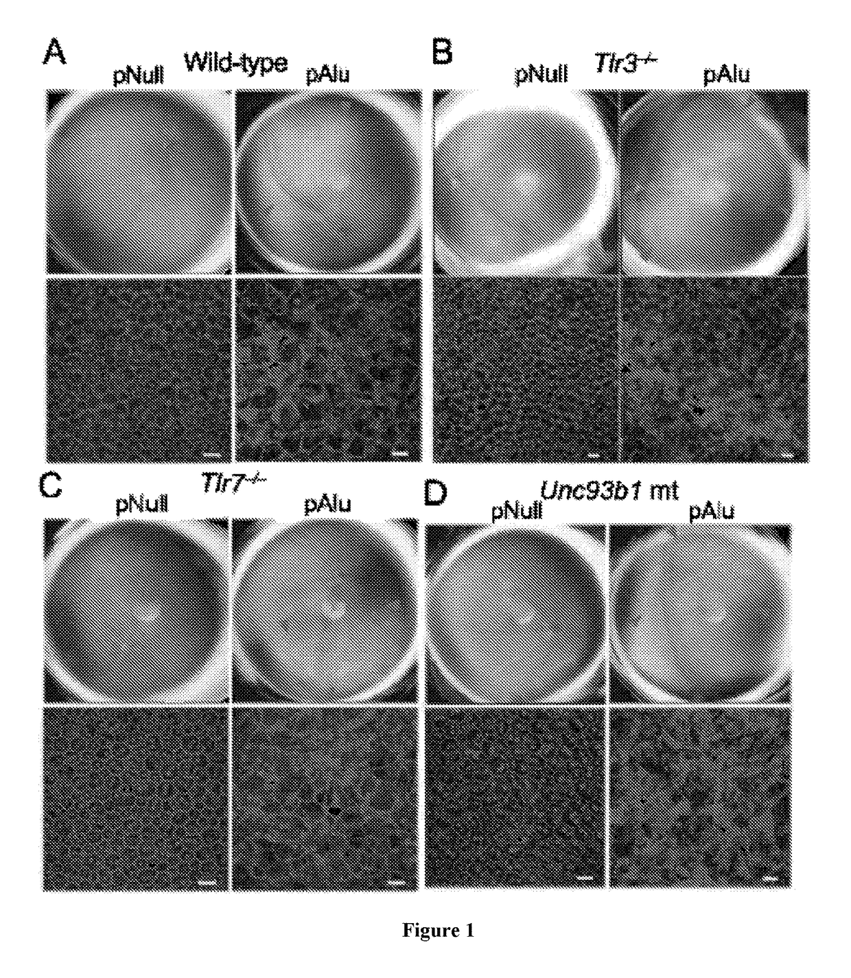
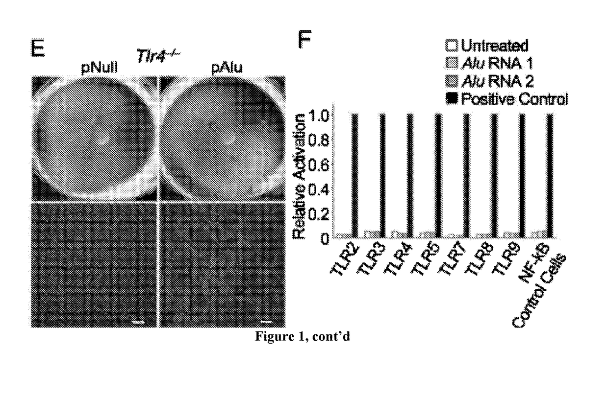
[0146]It has been have shown that Alu RNA induces RPE degeneration via MyD88 signaling, and that Alu RNA increases phosphorylation of IRAK1 and IRAK4, kinases downstream of MyD88. To determine whether IRAK1/IRAK4 phosphorylation is critical to Alu RNA-induced RPE degeneration, N-(2-Morpholinylethyl)-2-(3-nitrobenzoylamido)-benzimidazole, an IRAK/4 inhibitor, was tested. Indeed it was found that this IRAK/4 inhibitor blocked Alu RNA-induced RPE degeneration in wild-type mice in a dose-dependent manner (FIG. 13). With reference to FIG. 13, Alu RNA was injected subretinally in wild-type mice to induce RPE degeneration. Compared to vehicle administration, intravitreous administration of an inhibitor of IRAK1/4 reduced the RPE degeneration induced by Alu RNA in a dose-dependent fashion (1.5-15 μg). With reference to FIG. 14, exposure of human RPE cells to an inhibitor of IRAK1/4 rescued the cell death induced by transfection of pAlu. With reference to FIG. 15, exposure of human RPE cells to an inhibitor of IRAK1/4 rescued the cell death induced by transfection of Alu RNA.
[0147]As noted herein, methods disclosed in the present application can be useful for treating all forms of macular degeneration, including the wet form of macular degeneration (e.g., angiogenesis/blood vessel growth). By way of specific example, a MyD88 inhibitor can be used to treat various forms of macular degeneration, and is specifically contemplated for inhibition of choroidal neovascularization. Relevant data are set forth in FIGS. 16-19. FIG. 16 is a bar graph showing that MyD88−/− mice have reduced laser-induced choroidal neovascularization as compared to wild-type mice. FIG. 17 is a bar graph showing that intravitreous injection of a peptide MyD88 inhibitor reduced laser-induced choroidal neovascularization in wild-type mice compared to a control peptide inhibitor. FIG. 18 is a bar graph showing that intravitreous injection of an siRNA targeting MyD88 (2 μg) reduced laser-induced choroidal neovascularization in wild-type mice at least as well as a neutralizing anti-mouse VEGF antibody, compared to a control sirRNA. FIG. 19 is a bar graph showing that intravitreous injection of an inhibitor of IRAK1/4 reduced laser-induced choroidal neovascularization in wild-type mice compared to vehicle.
[0148]Throughout this document, various references are mentioned. All such references are incorporated herein by reference, including the references set forth in the following list:
REFERENCES
[0149] Adachi, O., Kawai, T., Takeda, K., Matsumoto, M., Tsutsui, H., Sakagami, M., Nakanishi, K., and Akira, S. (1998). Targeted disruption of the MyD88 gene results in loss of IL-1- and IL-18-mediated function. Immunity 9, 143-150. [0150] Akira, S., Uematsu, S., and Takeuchi, O. (2006). Pathogen recognition and innate immunity. Cell 124, 783-801. [0151] Alexander, J. J., and Hauswirth, W. W. (2008). Adeno-associated viral vectors and the retina. Adv Exp Med Biol 613, 121-128 [0152] Alexopoulou, L., Holt, A. C., Medzhitov, R., and Flavell, R. A. (2001). Recognition of double-stranded RNA and activation of NF-kappaB by Toll-like receptor 3. Nature 413, 732-738. [0153] Aliprantis, A. O., Yang, R. B., Weiss, D. S., Godowski, P., and Zychlinsky, A. (2000). The apoptotic signaling pathway activated by Toll-like receptor-2. EMBO J 19, 3325-3336. [0154] Allensworth, J. J., Planck, S. R., Rosenbaum, J. T., and Rosenzweig, H. L. (2011). Investigation of the differential potentials of TLR agonists to elicit uveitis in mice. J Leukoc Biol. [0155] Ambati, J., Ambati, B. K., Yoo, S. H., Ianchulev, S., and Adamis, A. P. (2003). Age-related macular degeneration: etiology, pathogenesis, and therapeutic strategies. Sury Ophthalmol 48, 257-293. [0156] Batzer, M. A., and Deininger, P. L. (2002). Alu repeats and human genomic diversity. Nat Rev Genet 3, 370-379. [0157] Bauernfeind, F., Bartok, E., Rieger, A., Franchi, L., Nunez, G., and Hornung, V. (2011). Cutting edge: reactive oxygen species inhibitors block priming, but not activation, of the NLRP3 inflammasome. J Immunol 187, 613-617. [0158] Bauernfeind, F. G., Horvath, G., Stutz, A., et al. (2009). “Cutting edge: NF-kappaB activating pattern recognition and cytokine receptors license NLRP3 inflammasome activation by regulating NLRP3 expression.”J Immunol 183(2): 787-791. [0159] Bennett, E. A., Keller, H., Mills, R. E., Schmidt, S., Moran, J. V., Weichenrieder, O., and Devine, S. E. (2008). Active Alu retrotransposons in the human genome. Genome Res 18, 1875-1883 [0160] Bernstein, E., Caudy, A. A., Hammond, S. M., and Hannon, G. J. (2001). Role for a bidentate ribonuclease in the initiation step of RNA interference. Nature 409, 363-366. [0161] Blaauwgeers, H. G., Holtkamp, G. M., Rutten, H., Witmer, A. N., Koolwijk, P., Partanen, T. A., Alitalo, K., Kroon, M. E., Kijlstra, A., van Hinsbergh, V. W., et al. (1999). Polarized vascular endothelial growth factor secretion by human retinal pigment epithelium and localization of vascular endothelial growth factor receptors on the inner choriocapillaris. Evidence for a trophic paracrine relation. Am J Pathol 155, 421-428. [0162] Bogdanovich, S., McNally, E. M., and Khurana, T. S. (2008). Myostatin blockade improves function but not histopathology in a murine model of limb-girdle muscular dystrophy 2C. Muscle Nerve 37, 308-316. [0163] Cao, Z., Henzel, W. J., and Gao, X. (1996). IRAK: a kinase associated with the interleukin-1 receptor. Science 271, 1128-1131. [0164] Chandrasekar, B., Vemula, K., Surabhi, R. M., Li-Weber, M., Owen-Schaub, L. B., Jensen, L. E., and Mummidi, S. (2004). Activation of intrinsic and extrinsic proapoptotic signaling pathways in interleukin-18-mediated human cardiac endothelial cell death. J Biol Chem 279, 20221-20233. [0165] Dhellin, O., Maestre, J., and Heidmann, T. (1997). Functional differences between the human LINE retrotransposon and retroviral reverse transcriptases for in vivo mRNA reverse transcription. EMBO J 16, 6590-6602. [0166] Diebold, S. S., Kaisho, T., Hemmi, H., Akira, S., and Reis e Sousa, C. (2004). Innate antiviral responses by means of TLR7-mediated recognition of single-stranded RNA. Science 303, 1529-1531. [0167] Dunaief, J. L., Dentchev, T., Ying, G. S., and Milam, A. H. (2002). The role of apoptosis in age-related macular degeneration. Arch Ophthalmol 120, 1435-1442. [0168] Feldmeyer, L., Keller, M., Niklaus, G., Hohl, D., Werner, S., and Beer, H. D. (2007). The inflammasome mediates UVB-induced activation and secretion of interleukin-1beta by keratinocytes. Curr Biol 17, 1140-1145. [0169] Fernandes-Alnemri, T., Wu, J., Yu, J. W., Datta, P., Miller, B., Jankowski, W., Rosenberg, S., Zhang, J., and Alnemri, E. S. (2007). The pyroptosome: a supramolecular assembly of ASC dimers mediating inflammatory cell death via caspase-1 activation. Cell Death Differ 14, 1590-1604. [0170] Ferrara, N. (2010). Vascular endothelial growth factor and age-related macular degeneration: from basic science to therapy. Nat Med 16, 1107-1111. [0171] Fink, S. L., Bergsbaken, T., and Cookson, B. T. (2008). Anthrax lethal toxin and Salmonella elicit the common cell death pathway of caspase-1-dependent pyroptosis via distinct mechanisms. Proc Natl Acad Sci USA 105, 4312-4317. [0172] Fink, S. L., and Cookson, B. T. (2006). Caspase-1-dependent pore formation during pyroptosis leads to osmotic lysis of infected host macrophages. Cell Microbiol 8, 1812-1825. [0173] Ghayur, T., Banerjee, S., Hugunin, M., Butler, D., Herzog, L., Carter, A., Quintal, L., Sekut, L., Talanian, R., Paskind, M., et al. (1997). Caspase-1 processes IFN-gamma-inducing factor and regulates LPS-induced IFN-gamma production. Nature 386, 619-623. [0174] Gu, Y., Kuida, K., Tsutsui, H., Ku, G., Hsiao, K., Fleming, M. A., Hayashi, N., Higashino, K., Okamura, H., Nakanishi, K., et al. (1997). Activation of interferon-gamma inducing factor mediated by interleukin-1beta converting enzyme. Science 275, 206-209. [0175] Guarda, G., Braun, M., Staehli, F., Tardivel, A., Mattmann, C., Forster, I., Farlik, M., Decker, T., Du Pasquier, R. A., Romero, P., et al. (2011). Type I interferon inhibits interleukin-1 production and inflammasome activation. Immunity 34, 213-223. [0176] Guarda, G., Dostert, C., Staehli, F., Cabalzar, K., Castillo, R., Tardivel, A., Schneider, P., and Tschopp, J. (2009). T cells dampen innate immune responses through inhibition of NLRP1 and NLRP3 inflammasomes. Nature 460, 269-273. [0177] Halle, A., Hornung, V., Petzold, G. C., Stewart, C. R., Monks, B. G., Reinheckel, T., Fitzgerald, K. A., Latz, E., Moore, K. J., and Golenbock, D. T. (2008). The NALP3 inflammasome is involved in the innate immune response to amyloid-beta. Nat Immunol 9, 857-865. [0178] Heil, F., Hemmi, H., Hochrein, H., Ampenberger, F., Kirschning, C., Akira, S., Lipford, G., Wagner, H., and Bauer, S. (2004). Species-specific recognition of single-stranded RNA via toll-like receptor 7 and 8. Science 303, 1526-1529. [0179] Hoebe, K., Du, X., Georgel, P., Janssen, E., Tabeta, K., Kim, S. O., Goode, J., Lin, P., Mann, N., Mudd, S., et al. (2003). Identification of Lps2 as a key transducer of MyD88-independent TIR signalling. Nature 424, 743-748. [0180] Hornung, V., Ellegast, J., Kim, S., Brzozka, K., Jung, A., Kato, H., Poeck, H., Akira, S., Conzelmann, K. K., Schlee, M., et al. (2006). 5′-Triphosphate RNA is the ligand for RIG-I. Science 314, 994-997. [0181] Kanakaraj, P., Ngo, K., Wu, Y., Angulo, A., Ghazal, P., Harris, C. A., Siekierka, J. J., Peterson, P. A., and Fung-Leung, W. P. (1999). Defective interleukin (IL)-18-mediated natural killer and T helper cell type 1 responses in IL-1 receptor-associated kinase (IRAK)-deficient mice. J Exp Med 189, 1129-1138. [0182] Kaneko, H., Dridi, S., Tarallo, V., Gelfand, B. D., Fowler, B. J., Cho, W. G., Kleinman, M. E., Ponicsan, S. L., Hauswirth, W. W., Chiodo, V. A., et al. (2011). DICER1 deficit induces Alu RNA toxicity in age-related macular degeneration. Nature 471, 325-330. [0183] Kanneganti, T. D., Ozoren, N., Body-Malapel, M., Amer, A., Park, J. H., Franchi, L., Whitfield, J., Barchet, W., Colonna, M., Vandenabeele, P., et al. (2006). Bacterial RNA and small antiviral compounds activate caspase-1 through cryopyrin/Nalp3. Nature 440, 233-236. [0184] Kato, H., Takeuchi, O., Sato, S., Yoneyama, M., Yamamoto, M., Matsui, K., Uematsu, S., Jung, A., Kawai, T., Ishii, K. J., et al. (2006). Differential roles of MDA5 and RIG-I helicases in the recognition of RNA viruses. Nature 441, 101-105. [0185] Keller, M., Ruegg, A., Werner, S., and Beer, H. D. (2008). Active caspase-1 is a regulator of unconventional protein secretion. Cell 132, 818-831. [0186] Kleinman, M. E., Kaneko, H., Cho, W. G., Dridi, S., Fowler, B. J., Blandford, A. D., Albuquerque, R. J., Hirano, Y., Terasaki, H., Kondo, M., et al. (2011). Short-interfering RNAs Induce Retinal Degeneration via TLR3 and IRF3. Mol Ther. [0187] Kleinman, M. E., Yamada, K., Takeda, A., Chandrasekaran, V., Nozaki, M., Baffi, J. Z., Albuquerque, R. J., Yamasaki, S., Itaya, M., Pan, Y., et al. (2008). Sequence- and target-independent angiogenesis suppression by siRNA via TLR3. Nature 452, 591-597. [0188] Kumar, H., Kawai, T., Kato, H., Sato, S., Takahashi, K., Coban, C., Yamamoto, M., Uematsu, S., Ishii, K. J., Takeuchi, O., et al. (2006). Essential role of IPS-1 in innate immune responses against RNA viruses. J Exp Med 203, 1795-1803. [0189] Kumar, M. V., Nagineni, C. N., Chin, M. S., Hooks, J. J., and Detrick, B. (2004). Innate immunity in the retina: Toll-like receptor (TLR) signaling in human retinal pigment epithelial cells. J Neuroimmuno1153, 7-15. [0190] Lamkanfi, M., Mueller, J. L., Vitari, A. C., Misaghi, S., Fedorova, A., Deshayes, K., Lee, W. P., Hoffman, H. M., and Dixit, V. M. (2009). Glyburide inhibits the Cryopyrin/Nalp3 inflammasome. J Cell Biol 187, 61-70. [0191] Lander, E. S., Linton, L. M., Birren, B., Nusbaum, C., Zody, M. C., Baldwin, J., Devon, K., Dewar, K., Doyle, M., FitzHugh, W., et al. (2001). Initial sequencing and analysis of the human genome. Nature 409, 860-921. [0192] Loiarro, M., Sette, C., Gallo, G., Ciacci, A., Fanto, N., Mastroianni, D., Carminati, P., and Ruggiero, V. (2005). Peptide-mediated interference of TIR domain dimerization in MyD88 inhibits interleukin-1-dependent activation of NF-κB. J Biol Chem 280, 15809-15814. [0193] Lopez, P. F., Sippy, B. D., Lambert, H. M., Thach, A. B., and Hinton, D. R. (1996). Transdifferentiated retinal pigment epithelial cells are immunoreactive for vascular endothelial growth factor in surgically excised age-related macular degeneration-related choroidal neovascular membranes. Invest Ophthalmol Vis Sci 37, 855-868. [0194] Mariathasan, S., Newton, K., Monack, D. M., Vucic, D., French, D. M., Lee, W. P., Roose-Girma, M., Erickson, S., and Dixit, V. M. (2004). Differential activation of the inflammasome by caspase-1 adaptors ASC and Ipaf. Nature 430, 213-218. [0195] Mariathasan, S., Weiss, D. S., Newton, K., McBride, J., O'Rourke, K., Roose-Girma, M., Lee, W. P., Weinrauch, Y., Monack, D. M., and Dixit, V. M. (2006). Cryopyrin activates the inflammasome in response to toxins and ATP. Nature 440, 228-232. [0196] Maranon, F., Mayor, A., and Tschopp, J. (2009). The inflammasomes: guardians of the body. Annu Rev Immunol 27, 229-265. [0197] Masters, S. L., Dunne, A., Subramanian, S. L., Hull, R. L., Tannahill, G. M., Sharp, F. A., Becker, C., Franchi, L., Yoshihara, E., Chen, Z., et al. (2010). Activation of the NLRP3 inflammasome by islet amyloid polypeptide provides a mechanism for enhanced IL-1beta in type 2 diabetes. Nat Immunol 11, 897-904. [0198] McClure, M. A. (1991). Evolution of retroposons by acquisition or deletion of retrovirus-like genes. Mol Biol Evol 8, 835-856. [0199] McLeod, D. S., Grebe, R., Bhutto, I., Merges, C., Baba, T., and Lutty, G. A. (2009). Relationship between RPE and choriocapillaris in age-related macular degeneration. Invest Ophthalmol Vis Sci 50, 4982-4991. [0200] Miao, E. A., Leaf, I. A., Treuting, P. M., Mao, D. P., Dors, M., Sarkar, A., Warren, S. E., Wewers, M. D., and Aderem, A. (2010). Caspase-1-induced pyroptosis is an innate immune effector mechanism against intracellular bacteria. Nat Immunol 11, 1136-1142. [0201] Muruve, D. A., Petrilli, V., Zaiss, A. K., White, L. R., Clark, S. A., Ross, P. J., Parks, R. J., and Tschopp, J. (2008). The inflammasome recognizes cytosolic microbial and host DNA and triggers an innate immune response. Nature 452, 103-107. [0202] Muzio, M., Ni, J., Feng, P., and Dixit, V. M. (1997). IRAK (Pelle) family member IRAK-2 and MyD88 as proximal mediators of IL-1 signaling. Science 278, 1612-1615. [0203] Nakahira, K., Haspel, J. A., Rathinam, V. A., Lee, S. J., Dolinay, T., Lam, H. C., Englert, J. A., Rabinovitch, M., Cernadas, M., Kim, H. P., et al. (2011). Autophagy proteins regulate innate immune responses by inhibiting the release of mitochondrial DNA mediated by the NALP3 inflammasome. Nat Immunol 12, 222-230. [0204] Ngo, V. N., Young, R. M., Schmitz, R., Jhavar, S., Xiao, W., Lim, K. H., Kohlhammer, H., Xu, W., Yang, Y., Zhao, H., et al. (2011). Oncogenically active MYD88 mutations in human lymphoma. Nature 470, 115-119. [0205] O'Neill, L. A., and Bowie, A. G. (2007). The family of five: TIR-domain-containing adaptors in Toll-like receptor signalling. Nat Rev Immunol 7, 353-364. [0206] Ohtsuki, T., Micallef, M. J., Kohno, K., Tanimoto, T., Ikeda, M., and Kurimoto, M. (1997). Interleukin 18 enhances Fas ligand expression and induces apoptosis in Fas-expressing human myelomonocytic KG-1 cells. Anticancer Res 17, 3253-3258. [0207] Parker, J. S., Roe, S. M., and Barford, D. (2004). Crystal structure of a PIWI protein suggests mechanisms for siRNA recognition and slicer activity. EMBO J 23, 4727-4737. [0208] Picard, C., von Bernuth, H., Ghandil, P., Chrabieh, M., Levy, O., Arkwright, P. D., McDonald, D., Geha, R. S., Takada, H., Krause, J. C., et al. (2010). Clinical features and outcome of patients with IRAK-4 and MyD88 deficiency. Medicine (Baltimore) 89, 403-425. [0209] Pichlmair, A., Lassnig, C., Eberle, C. A., Goma, M. W., Baumann, C. L., Burkard, T. R., Burckstummer, T., Stefanovic, A., Krieger, S., Bennett, K. L., et al. (2011). IFIT1 is an antiviral protein that recognizes 5′-triphosphate RNA. Nat Immunol 12, 624-630. [0210] Puente, X. S., Pinyol, M., Quesada, V., Conde, L., Ordonez, G. R., Villamor, N., Escaramis, G., Jares, P., Bea, S., Gonzalez-Diaz, M., et al. (2011). Whole-genome sequencing identifies recurrent mutations in chronic lymphocytic leukaemia. Nature 475, 101-105. [0211] Qiao, Y., Wang, P., Qi, J., et al. (2012). “TLR-induced NF-kappaB activation regulates NLRP3 expression in murine macrophages.” FEBS Lett 586(7): 1022-1026. [0212] Qureshi, N., Takayama, K., and Kurtz, R. (1991). Diphosphoryl lipid A obtained from the nontoxic lipopolysaccharide of Rhodopseudomonas sphaeroides is an endotoxin antagonist in mice. Infect Immun 59, 441-444. [0213] Rakoff-Nahoum, S., and Medzhitov, R. (2007). Regulation of spontaneous intestinal tumorigenesis through the adaptor protein MyD88. Science 317, 124-127. [0214] Reuter, M., Benninger, P., Chuma, S., Shah, H., Hosokawa, M., Funaya, C., Antony, C., Sachidanandam, R., and Pillai, R. S. (2011). Miwi catalysis is required for piRNA amplification-independent LINE1 transposon silencing. Nature. [0215] Saitoh, T., Fujita, N., Jang, M. H., Uematsu, S., Yang, B. G., Satoh, T., Omori, H., Noda, T., Yamamoto, N., Komatsu, M., et al. (2008). Loss of the autophagy protein Atg16L1 enhances endotoxin-induced IL-1beta production. Nature 456, 264-268. [0216] Schroder, K., and Tschopp, J. (2010). The inflammasomes. Cell 140, 821-832. [0217] Schroder, K., Zhou, R., and Tschopp, J. (2010). The NLRP3 inflammasome: a sensor for metabolic danger? Science 327, 296-300. [0218] Shaikh, T. H., Roy, A. M., Kim, J., Batzer, M. A., and Deininger, P. L. (1997). cDNAs derived from primary and small cytoplasmic Alu (scAlu) transcripts. J Mol Biol 271, 222-234. [0219] Smith, W., Assink, J., Klein, R., Mitchell, P., Klaver, C. C., Klein, B. E., Hofman, A., Jensen, S., Wang, J. J., and de Jong, P. T. (2001). Risk factors for age-related macular degeneration: Pooled findings from three continents. Ophthalmology 108, 697-704. [0220] Stennicke, H. R., Jurgensmeier, J. M., Shin, H., et al. (1998). “Pro-caspase-3 is a major physiologic target of caspase-8.” J Biol Chem 273(42): 27084-27090. [0221] Streilein, J. W. (2003). Ocular immune privilege: therapeutic opportunities from an experiment of nature. Nat Rev Immunol 3, 879-889. [0222] Sun, D., and Ding, A. (2006). MyD88-mediated stabilization of interferon-gamma-induced cytokine and chemokine mRNA. Nat Immunol 7, 375-381. [0223] Sun, Q., Sun, L., Liu, H. H., Chen, X., Seth, R. B., Forman, J., and Chen, Z. J. (2006). The specific and essential role of MAVS in antiviral innate immune responses. Immunity 24, 633-642. [0224] Suzuki, N., Chen, N. J., Millar, D. G., Suzuki, S., Horacek, T., Hara, H., Bouchard, D., Nakanishi, K., Penninger, J. M., Ohashi, P. S., et al. (2003). IL-1 receptor-associated kinase 4 is essential for IL-18-mediated NK and Thl cell responses. J Immunol 170, 4031-4035. [0225] Suzuki, N., Suzuki, S., Duncan, G. S., Millar, D. G., Wada, T., Mirtsos, C., Takada, H., Wakeham, A., Itie, A., Li, S., et al. (2002). Severe impairment of interleukin-1 and Toll-like receptor signalling in mice lacking IRAK-4. Nature 416, 750-756. [0226] Tabeta, K., Hoebe, K., Janssen, E. M., Du, X., Georgel, P., Crozat, K., Mudd, S., Mann, N., Sovath, S., Goode, J., et al. (2006). The Unc93b1 mutation 3d disrupts exogenous antigen presentation and signaling via Toll-like receptors 3, 7 and 9. Nat Immunol 7, 156-164. [0227] Takeda, A., Baffi, J. Z., Kleinman, M. E., Cho, W. G., Nozaki, M., Yamada, K., Kaneko, H., Albuquerque, R. J., Dridi, S., Saito, K., et al. (2009). CCR3 is a target for age-related macular degeneration diagnosis and therapy. Nature 460, 225-230. [0228] Thornberry, N. A., Bull, H. G., Calaycay, J. R., Chapman, K. T., Howard, A. D., Kostura, M. J., Miller, D. K., Molineaux, S. M., Weidner, J. R., Aunins, J., et al. (1992). A novel heterodimeric cysteine protease is required for interleukin-1 beta processing in monocytes. Nature 356, 768-774. [0229] Tschopp, J., Maranon, F., and Burns, K. (2003). NALPs: a novel protein family involved in inflammation. Nat Rev Mol Cell Biol 4, 95-104. [0230] Vandanmagsar, B., Youm, Y. H., Ravussin, A., Galgani, J. E., Stadler, K., Mynatt, R. L., Ravussin, E., Stephens, J. M., and Dixit, V. D. (2011). The NLRP3 inflammasome instigates obesity-induced inflammation and insulin resistance. Nat Med 17, 179-188. [0231] Vandenabeele, P., Vanden Berghe, T., and Festjens, N. (2006). Caspase inhibitors promote alternative cell death pathways. Sci STKE 2006, pe44. [0232] Verhoef, P. A., Kertesy, S. B., Lundberg, K., Kahlenberg, J. M., and Dubyak, G. R. (2005). Inhibitory effects of chloride on the activation of caspase-1, IL-1beta secretion, and cytolysis by the P2X7 receptor. J Immunol 175, 7623-7634. [0233] Vogt, S. D., Curcio, C. A., Wang, L., Li, C. M., McGwin, G., Jr., Medeiros, N. E., Philp, N. J., Kimble, J. A., and Read, R. W. (2011). Retinal pigment epithelial expression of complement regulator CD46 is altered early in the course of geographic atrophy. Exp Eye Res. [0234] von Bernuth, H., Picard, C., Jin, Z., Pankla, R., Xiao, H., Ku, C. L., Chrabieh, M., Mustapha, I. B., Ghandil, P., Camcioglu, Y., et al. (2008). Pyogenic bacterial infections in humans with MyD88 deficiency. Science 321, 691-696. [0235] Wen, H., Orris, D., Lei, Y., Jha, S., Zhang, L., Huang, M. T., Brickey, W. J., and Ting, J. P. (2011). Fatty acid-induced NLRP3-ASC inflammasome activation interferes with insulin signaling. Nat Immunol 12, 408-415. [0236] Yamamoto, M., Sato, S., Hemmi, H., Hoshino, K., Kaisho, T., Sanjo, H., Takeuchi, O., Sugiyama, M., Okabe, M., Takeda, K., et al. (2003). Role of adaptor TRIF in the MyD88-independent toll-like receptor signaling pathway. Science 301, 640-643. [0237] Yang, P., Tyrrell, J., Han, I., and Jaffe, G. J. (2009). Expression and modulation of RPE cell membrane complement regulatory proteins. Invest Ophthalmol Vis Sci 50, 3473-3481. [0238] Yang, Y. L., Reis, L. F., Pavlovic, J., Aguzzi, A., Schafer, R., Kumar, A., Williams, B. R., Aguet, M., and Weissmann, C. (1995). Deficient signaling in mice devoid of double-stranded RNA-dependent protein kinase. EMBO J 14, 6095-6106. [0239] Yang, Z., Stratton, C., Francis, P. J., Kleinman, M. E., Tan, P. L., Gibbs, D., Tong, Z., Chen, H., Constantine, R., Yang, X., et al. (2008). Toll-like receptor 3 and geographic atrophy in age-related macular degeneration. N Engl J Med 359, 1456-1463. [0240] Zaki, M. H., Boyd, K. L., Vogel, P., Kastan, M. B., Lamkanfi, M., and Kanneganti, T. D. (2010). The NLRP3 inflammasome protects against loss of epithelial integrity and mortality during experimental colitis. Immunity 32, 379-391.
Login to view more PUM


Description & Claims & Application Information
We can also present the details of the Description, Claims and Application information to help users get a comprehensive understanding of the technical details of the patent, such as background art, summary of invention, brief description of drawings, description of embodiments, and other original content. On the other hand, users can also determine the specific scope of protection of the technology through the list of claims; as well as understand the changes in the life cycle of the technology with the presentation of the patent timeline. Login to view more.
Login to view more Similar technology patents
Compositions and methods for modulating expression of frataxin
InactiveUS20160201064A1Increase levelDelay or reduce riskSugar derivativesMuscular disorderCancer researchFrataxin
Owner:RANA THERAPEUTICS INC
Cultured dairy products and methods of manufacture
InactiveUS20060286209A1Cost efficientIncrease levelMilk preparationOther dairy technologyCheese creamMilk products
Owner:KRAFT FOODS GLOBAL BRANDS LLC
Methods, systems, and software for providing a blocked sewer alert
Owner:WASTE INNOVATION CONCEPTS LLC
Combinatorial therapy with an acetylcholinesterase inhibitor and (3aR)-1,3a,8-trimethyl-1,2,3,3a,8,8a-hexahydropyrrolo[2,3,-b]indol-5-yl phenylcarbamate
InactiveUS20050182044A1Reduce riskIncrease levelBiocideNervous disorderAcetylcholinesterase inhibitorΒ amyloid peptide
Owner:AXONYX INC
Method of controlling a power plant
InactiveUS20140142776A1Increase levelMechanical power/torque controlBatteries circuit arrangementsPower storageNuclear engineering
Owner:SIEMENS AG
Classification and recommendation of technical efficacy words
- Increase level
- Inhibit activation
Inferring object status based on detected watermark data
Owner:DIGIMARC CORP (FORMERLY DMRC CORP) +1
Continuous process for producing ethanol using raw starch
InactiveUS20050239181A1High levelIncrease levelBiofuelsAnimal feeding stuffChemistryStarch production
Owner:BROIN & ASSOC
Application of a system parameter as a method and mechanism for controlling eye chamber stability
Owner:JOHNSON & JOHNSON SURGICAL VISION INC
Increased Seed Oil and Abiotic Stress Tolerance Mediated by HSI2
InactiveUS20120066794A1Increase toleranceIncrease levelOther foreign material introduction processesPlant peptidesDrought resistanceOil content
Owner:NAT RES COUNCIL OF CANADA
Presence awareness agent
InactiveUS20070230681A1Increase levelContinue operationCustomer communicationsManual exchangesPresence awarenessHuman–computer interaction
Owner:AVAYA TECH LLC
Thiazole and thiadiazole compounds for inflammation and immune-related uses
InactiveUS20070254925A1Suppress immune systemInhibit activationBiocideSenses disorderHypersensitivity DiseasesImmune disorder
Owner:SYNTA PHARMA
Use of specific T2R taste receptors to identify compounds that block bitter taste
ActiveUS20030170608A1Block bitter tasteInhibit activationCompound screeningApoptosis detectionDrug6-nitrosaccharin
Owner:SENOMYX INC
Vehicle AC Ground Fault Detection System
InactiveUS20090091868A1Prevent shockInhibit activationElectric devicesShort-circuit testingElectrical switchingElectricity
Owner:LEAR CORP
Uses of inhibitors for the activation of CXCR4 receptor by SDF-1 in treating rheumatoid arthritis
InactiveUS20050202005A1Inhibit activationOrganic active ingredientsAntibody ingredientsInflammatory arthropathyMedicine
Owner:THE TRUSTEES OF COLUMBIA UNIV IN THE CITY OF NEW YORK
NF-κB inhibitor containing substituted benzoic acid derivative as active ingredient
InactiveUS7064124B2Inhibit activationAntibacterial agentsBiocideMedicinal chemistryActive ingredient
Owner:ASUBIO PHARMA