Thawing method for frozen embryonic stem cell
A technology of human embryonic stem cells and resuscitation solution, which is applied in the field of resuscitation of frozen embryonic stem cells, which can solve the problems of time-consuming and labor-intensive operations, and complicated operation of vitrification cryopreservation
- Summary
- Abstract
- Description
- Claims
- Application Information
AI Technical Summary
Problems solved by technology
Method used
Image
Examples
Embodiment 1
[0049] Cell cryopreservation
[0050] Cryopreservation solution: mix knock-out serum replacement (SR) and dimethyl sulfoxide (DMSO) in a ratio of 9:1, place on ice, and pre-cool.
[0051] Take hES cells of different passage numbers (20-25 ES cell clones each) for cryopreservation, and the experiment was repeated 3 times (see Table 1). Methods as below:
[0052] Take well-grown hES cells, digest with 1mg / ml type IV collagenase, transfer the clones to an eppendorf tube, add culture medium dropwise, gently pipette into small cell clumps, centrifuge at 1000rpm / min for 3 minutes, discard the supernatant Add the cryopreservation solution to the tube slowly drop by drop, and shake it up constantly. Quickly transfer the cell suspension to the cryopreservation tube, put it into the program cooling box, and cool down at 1°C / min until -80°C. -80°C overnight, transfer it to a liquid nitrogen tank the next day, and store it in the refrigerator.
Embodiment 2
[0055] Take out the cryopreservation tube prepared in Example 1 from the liquid nitrogen, quickly place it in a 37°C water bath, shake it until it dissolves, and scrub the tube wall with 75% alcohol cotton (heating speed is about 100°C / min). Then slowly add the cells to the resuscitation solution (the resuscitation solution is hES culture solution containing 0.2 mol / L trehalose), centrifuge at 1000 rpm / min for 3 minutes, and discard the supernatant. The ES cells were transferred to a pre-prepared feeder layer, cultured in hES culture medium containing 0.1 mol / L trehalose, and replaced with conventional hES culture medium after 1 hour of culture.
Embodiment 3
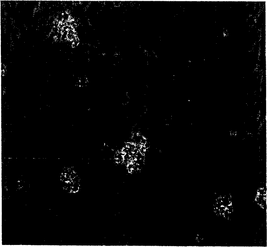
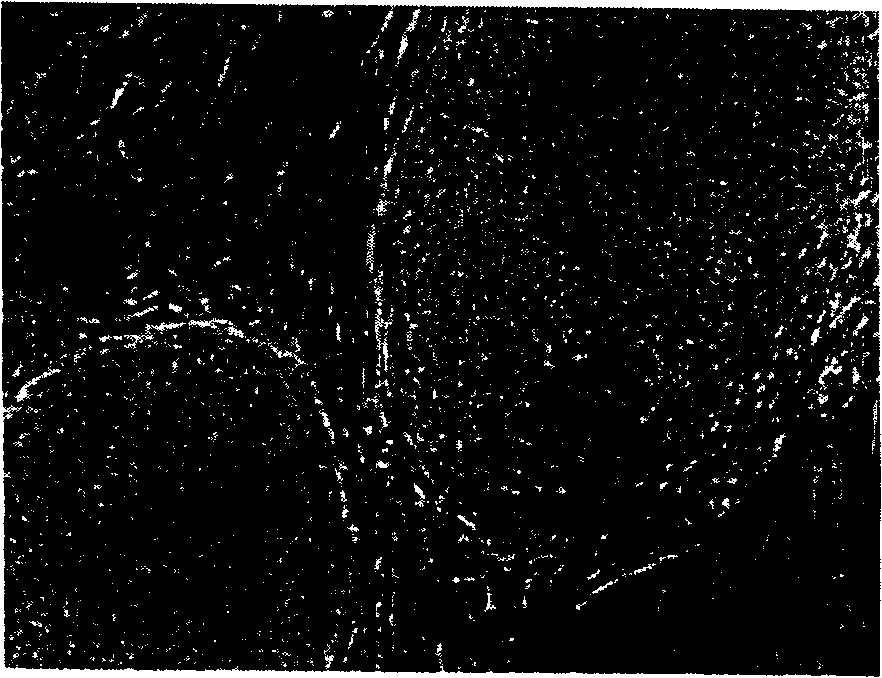
[0057] Morphological observation and characterization of human ES cells after resuscitation
[0058] 1. Method
[0059] (a) Morphological observation of human ES cells after resuscitation
[0060] Use a phase contrast microscope to observe the survival and differentiation of cells after resuscitation. The clone formation rate was calculated after 8 days of culture.
[0061] Clone formation rate=number of undifferentiated ES clones after recovery / total number of ES clones before cryopreservation×100%
[0062] (b) Characterization of human ES cells after resuscitation
[0063] (1) Alkaline phosphatase (AKP) staining
[0064] According to the method of Xu et al. (Xu X, Tsung HC, Yan Yc. Establishment and differential of murine EG cell lines derived from primordial germcells. Acta Biologiae Exper Sinica 1999; 32: 251-63), NBT and BCIP (Sigma) were used as Substrate and staining agent for AKP activity identification.
[0065] (2) Immunocytochemistry
[0066] SSEA-4 and Oct-4 monoclonal...
PUM
 Abstract
Description
Claims
Application Information
 - R&D
- Intellectual Property
- Life Sciences
- Materials
- Tech Scout
- Unparalleled Data Quality
- Higher Quality Content
- 60% Fewer Hallucinations
Browse by: Latest US Patents, China's latest patents, Technical Efficacy Thesaurus, Application Domain, Technology Topic, Popular Technical Reports.
© 2025 PatSnap. All rights reserved.Legal|Privacy policy|Modern Slavery Act Transparency Statement|Sitemap|About US| Contact US: help@patsnap.com


