Electrocardiograph with three-dimensional image and method for implementing same
An electrocardiograph and stereoscopic image technology, applied in the fields of equipment, image data processing, medical science, etc., can solve the problems of different lead systems, VCG value not being properly recognized, popularized, and complicated operations
- Summary
- Abstract
- Description
- Claims
- Application Information
AI Technical Summary
Problems solved by technology
Method used
Image
Examples
Embodiment 1
[0062] 1. The formation of space center vector ring:
[0063] The heart is a three-dimensional, hollow, solid organ. The generation of cardiac bioelectricity comes from the conduction system of the heart and the electro-chemical diffusion of the myocardium. The countless instantaneous heart vectors formed by it are distributed in the three-dimensional space field. If the farthest ends of each instant vector are connected to each other, a three-dimensional ring of space trajectory is formed, which is called a space heart vector ring or a three-dimensional heart vector ring ( attached Figure 13 ). Figure 13 The middle ring body in the figure represents the three-dimensional heart vector ring, F, H, and S represent the frontal, transverse, and lateral plane heart vector rings generated after the first projection, and V1 and V5 represent the two plane heart vector rings generated after the second projection. ECG with a lead axis.
[0064] 2. Calculation formula of spatial EC...
Embodiment 2
[0078] A method for realizing a three-dimensional image electrocardiogram. The steps include: establishing a three-dimensional coordinate liner, a cross plate composed of lines, flat plates, colors, light, etc. as a "three-dimensional liner"; The heart bushing is placed on the annulus of the three-dimensional space vector. Synchronous or selective sampling, display, observation, editing and tracing are used for 1-78 leads. "Three-dimensional lining" is a prior art, and the patent No. 98117316.0 is titled "Imaging method and imager for three-dimensional electrocardiogram", and this patent has a characteristic description of "three-dimensional lining".
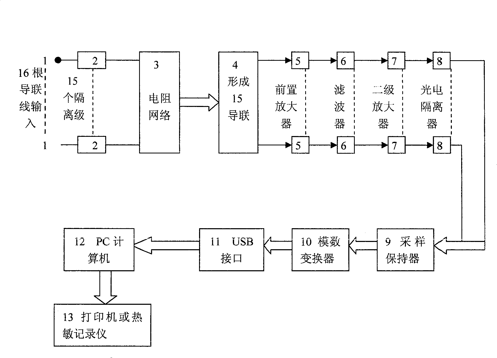
[0079] A method for realizing a three-dimensional image electrocardiogram, the steps include providing a set of leads, that is, 16 electrode wires (15 channels) input lead system including Frank's 7-electrode system and traditional 12-lead ECG Figure 10 An electrode line, in which Frank's positive Y-axis is connected in parall...
Embodiment 3
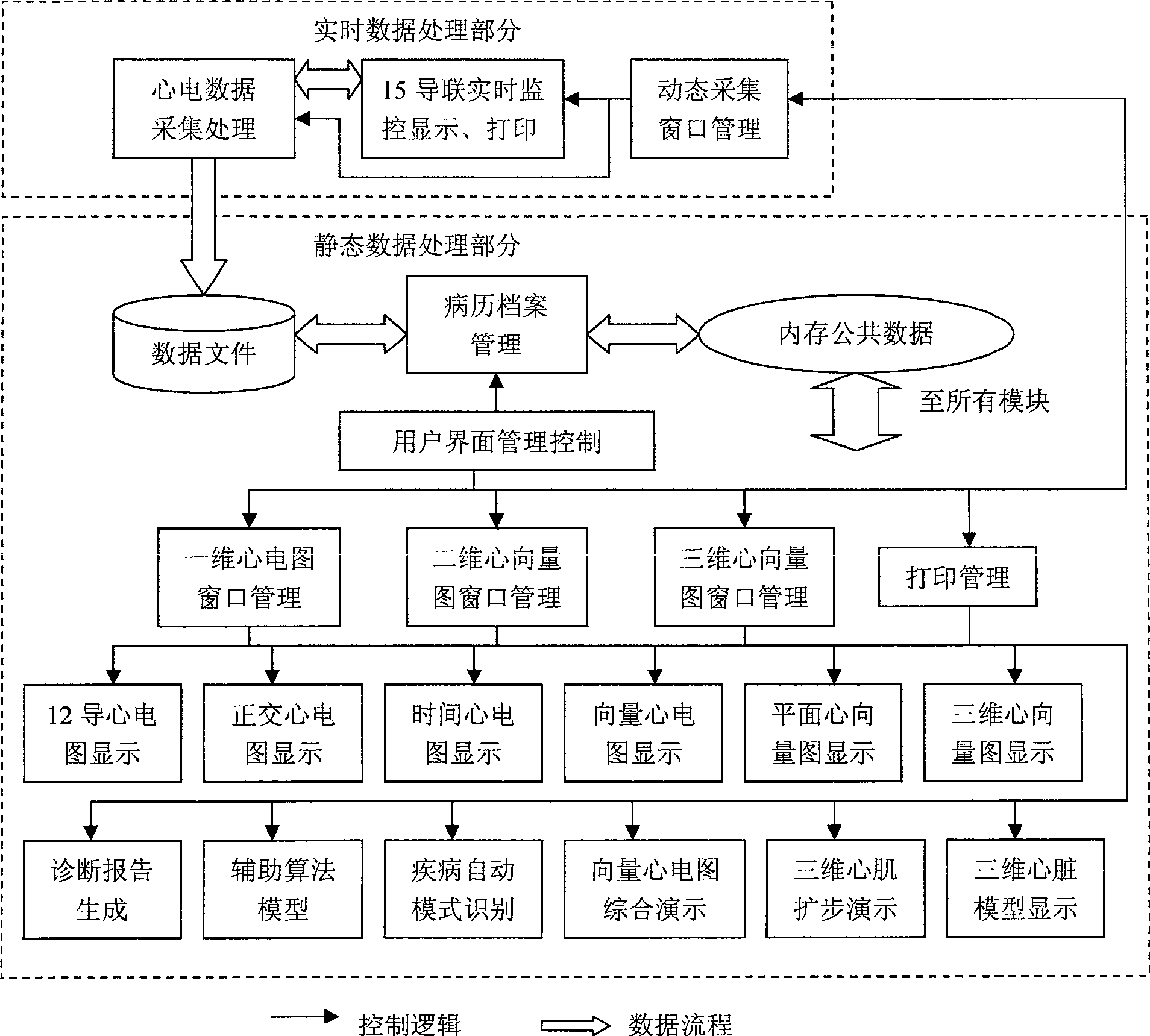
[0090] Such as figure 1 As shown, in the past, the anatomical morphology and electrocardiology of the heart belonged to two separate research fields, such as B-ultrasound and electrophysiology. The object of the present invention is to restore the three-dimensional anatomical structure of the heart and the three-dimensional electrocardiogram into one, closely combine the three-dimensional electrocardiogram in three-dimensional space and the anatomical structure of the virtual heart in three-dimensional space, and observe and diagnose with images and / or images. heart disease. The so-called visualization, intelligence, graphics, imaging, and automatic diagnosis of the structure and function of the heart. Its significance lies in that it is clear at a glance, and a picture is worth a thousand words. That is, three-dimensional, all-round, and intuitively reflect the phase relationship between cardiac bioelectrical activity and actual heart position; the forward and reverse time...
PUM
 Abstract
Description
Claims
Application Information
 - R&D
- Intellectual Property
- Life Sciences
- Materials
- Tech Scout
- Unparalleled Data Quality
- Higher Quality Content
- 60% Fewer Hallucinations
Browse by: Latest US Patents, China's latest patents, Technical Efficacy Thesaurus, Application Domain, Technology Topic, Popular Technical Reports.
© 2025 PatSnap. All rights reserved.Legal|Privacy policy|Modern Slavery Act Transparency Statement|Sitemap|About US| Contact US: help@patsnap.com


